Nitric oxide (NO) and neuronal nitric oxide synthase (nNOS) have been localized in every cell type in the retina, and the NO/cyclic guanosine monophosphate (cGMP) signaling pathway is involved at several levels of visual processing and in several retinal pathologies (Goldstein et al., Reference Goldstein, Ostwald and Roth1996; Eldred, Reference Eldred, Steinbusch, De Vente and Vincent2000). The NO/cGMP pathway is predominantly activated by an increase in intracellular Ca2+, which activates nNOS to produce NO. The primary downstream target of NO is the heterodimeric enzyme soluble guanylate cyclase (sGC), which is composed of α (sGCα) and β (sGCβ) subunits. NO binds to heme in sGC, activating sGC to catalyze the conversion of GTP to cGMP (see review, Sitaramayya, Reference Sitaramayya2002). Thus, NO can act through the second messenger cGMP to target phosphodiesterases (PDEs), cyclic nucleotide-gated (CNG) channels, and cGMP-dependent protein kinase (PKG) (Zhang & Cote, Reference Zhang and Cote2005).
Some components of the NO/cGMP pathway have been previously examined in the salamander retina. Nitric oxide synthase (NOS) has been anatomically localized in the salamander retina in photoreceptor ellipsoids, amacrine cells, and distal regions of both Müller and bipolar cells using both nicotinamide adenine dinucleotide phosphate-diaphorase (NADPH-d) histochemistry and nNOS immunocytochemistry (Liepe et al., Reference Liepe, Stone, Koistinaho and Copenhagen1994; Kurenni et al., Reference Kurenni, Thurlow, Turner, Moroz, Sharkey and Barnes1995). However, there were no detailed descriptions of the specific cell types involved. There is also physiological evidence that NO can activate CNG Ca2+ channels in photoreceptors in the salamander retina through the second messenger cGMP (Kurenny et al., Reference Kurenny, Moroz, Turner, Sharkey and Barnes1994). CNG channels have been physiologically localized in isolated retinal ganglion cells, where an NO stimulus can increase levels of cGMP, resulting in the opening of Ca2+ channels through a mechanism that involves sGC and PKG (Hirooka et al., Reference Hirooka, Kourennyi and Barnes2000). Additionally, the NO/cGMP pathway has been shown to be involved in the structural plasticity of isolated salamander photoreceptor somata through a cGMP-dependent mechanism (Zhang et al., Reference Zhang, Beuve and Townes-Anderson2005).
The goal of this study was to functionally activate and anatomically localize several aspects of the NO/cGMP signal transduction pathway in the tiger salamander retina. The detailed characterization of the NO/cGMP pathway in this model system will serve as a foundation for determining the role of NO in normal visual processing and retinopathies.
Using immunocytochemistry, we localized nNOS in photoreceptor ellipsoids, four amacrine cell types, Müller and bipolar cells, and in somata in the ganglion cell layer (GCL). Nitric oxide imaging with the NO-sensitive dye, diaminofluorescein (DAF), indicated light-stimulated NO production in specific retinal cell types. In order to identify the cell types that responded to NO, cyclic guanosine monophosphate–like immunoreactivity (cGMP-LI) was used to functionally localize NO-activated sGC in four different amacrine cell types, three different bipolar cell types, and in somata within the GCL. cGMP-LI was also immunocytochemically colocalized with its downstream target PKGII in processes in the outer retina and in somata in the inner retina. These results showed that the NO/cGMP pathways were present and functional in all nerve cell types within the salamander retina. Nitric oxide was produced by all cell types in response to different light stimuli, and its downstream target sGC was activated and synthesized the second messenger cGMP in specific cell types in the inner retina.
Experimental procedures
Tissue preparation
Larval tiger salamanders (Ambystoma tigrinum) were maintained on a 12-h light/12-h dark cycle. Near the middle of the light cycle, animals were decapitated using a method that was approved by the Boston University Charles River Campus Institutional Animal Care and Use Committee, their eyes were enucleated, and the anterior chambers and lenses were removed. Following the pharmacological incubations, the salamander eyecups were fixed with 4% paraformaldehyde in 0.1 m sodium phosphate buffer (PB, pH 7.4) for 30–90 min for control tissue and for 4 h for all experiments using stimulation with NO donor. Eyecups were cryoprotected using a sucrose gradient (5–30%), embedded in optimal cutting temperature media (OCT; Tissue-Tek; Miles Inc., Elkhart, IN), frozen, cut into 14-μm-thick cross sections using a cryostat, and then collected on Superfrost® Plus slides (Fisher Scientific, Waltham, MA). Sections were stored at −20°C before they were used for immunocytochemistry.
In vitro stimulation with NO donor
Following the dissections described above, the isolated salamander eyecups were allowed to recover for 45 min at room temperature in aerated HEPES-buffered amphibian balanced salt solution (BSS) (110 mm NaCl, 2.5 mm KCl, 3 mm CaCl2, 2 mm MgCl2, 10 mm glucose, and 5 mm HEPES, pH 7.8). Eyecups were then preincubated for 20 min at room temperature in aerated BSS with the following PDE inhibitor cocktail: the nonspecific PDE inhibitor 3-isobutyl-1-methylxanthine (1 mm; Sigma Aldrich, St. Louis, MO); a selective inhibitor for PDE 3, trequinsin (0.25 μ m; Tocris Cookson Inc., Ellisville, MO); a selective inhibitor for PDE 1-5, zaprinast (250 μ m; Tocris Cookson Inc.); and a selective inhibitor for PDE 5, dipyridamole (50 μ m; BIOMOL Research Laboratories Inc., Plymouth Meeting, PA). Following the preincubation with PDE inhibitors, the NO donor (+/−)-S-nitroso-N-acetylpenicillamine (SNAP) (100 μ m; Molecular Probes, Eugene, OR) was added to the BSS. After 30 min of stimulation, eyecups were fixed for 4 h and then prepared for immunocytochemical analysis. The following controls were also tested: PDE inhibitor cocktail only, depleted NO donor with PDE inhibitor cocktail (the SNAP stock solution was left open at room temperature for 1 week), and control BSS (no PDE inhibitor cocktail). All incubations were done in normal room light.
Immunocytochemical staining
Sections on slides were washed with 0.1 m PB to remove excess OCT. All antiserum solutions were diluted in 0.1 m PB with 0.3% Triton X-100 (PBtx). Nonspecific labeling was blocked by incubation in 5% normal goat, donkey, or rabbit serum (Jackson ImmunoResearch Labs, West Grove, PA) as appropriate, in PBtx, for 1 h. Sections were then incubated in primary antiserum overnight at 4°C. The following primary antisera were used: a rabbit polyclonal antiserum directed against the C-terminus (amino acids 1409–1429) of nNOS, diluted 1:1000 (N7280; Sigma Aldrich), and a rabbit polyclonal antiserum directed against the C-terminus (amino acids 1–120) of PKGII, diluted 1:100 (sc-25430; Santa Cruz Biotechnology Inc., Santa Cruz, CA). The rabbit and sheep antisera against cGMP were the generous gifts of Dr. Jan de Vente (Department of Pharmacology, Faculty of Medicine, Free University, Amsterdam, The Netherlands) and were diluted 1:10,000.
Following the incubation in primary antiserum, the sections were washed in PB and incubated for 2 h in one of the following appropriate fluorescent secondary antisera diluted 1:500 in PBtx: Alexa 488–conjugated donkey anti-goat immunoglobulin G (IgG), Alexa 488–conjugated donkey anti-sheep IgG, Alexa 488–conjugated goat anti-rabbit IgG (Molecular Probes, Carlsbad, CA), or Cy5-conjugated donkey anti-rabbit IgG (Jackson ImmunoResearch Labs). To control for nonspecific secondary antiserum staining, some slides were incubated in normal serum without primary antiserum before the secondary antiserum was applied. The slides were then washed in PB and cover slipped with glycerol before they were visualized using a Fluoview™ 300 confocal microscope (Olympus Corporation, Melville, NY) with a 40× oil immersion objective with a numerical aperture of 1.00. For double-labeling experiments, each secondary antiserum was excited by a separate laser (488 or 647 nm) and recorded in a separate channel. Images that were used for colocalization analysis were captured separately as described above, and then, the red and green channels were analyzed using the RG2B colocalization plugin as developed by Christopher Philip Mauer for Image J (http://rsb.info.nih.gov/ij/plugins/rg2bcolocalization.html). The colocalization figures shown in this report are shown as a “collapsed” stack of sixteen 1-μm sections in order to show more details of the anatomy than were visible in a single optical section. However, the colocalizations were confirmed by comparing the same single optical sections for each imaging channel.
The digital stacks of twelve to fourteen 1-μm-thick optical sections were collected and then collapsed and converted to 8-bit grayscale images using Image J software (Wayne Rasband, National Institute of Mental Health, Bethesda, MD) and then imported into Corel Draw 11 (Corel Corporation, Ontario, Canada) for brightness and contrast adjustment, arrangement, and labeling. For cell tracings (Figs. 4 and 9), Image Pro Plus (Media Cybernetics Inc., Bethesda, MD) was used. This software allowed for the accurate tracing of cells and their processes through a stack of confocal z-sections.
Criteria for the characterization of cell types
The following parameters were used to characterize specific cell types. The inner nuclear layer (INL) was anatomically divided as follows: near the INL–inner plexiform layer (IPL) border, the central INL, and near the outer plexiform layer (OPL)–INL border. The locations within the IPL were described as percentages ranging from S0 to S100, with S0 being the INL–IPL border and S100 being the IPL–GCL border. The distal IPL (S0–S40), containing synapses of OFF cells, is referred to as the OFF sublamina, and the proximal sublamina (S60–S100), containing synapses of ON cells, is referred to as the ON sublamina (Wu et al., Reference Wu, Gao and Maple2000; Pang et al., Reference Pang, Gao and Wu2002, Reference Pang, Gao and Wu2004). Additionally, cells were classified based on how their processes stratified within the IPL: cells were described as being “narrowly monostratified” (cells with dendrites ramifying in only 10% of the IPL), “broadly monostratified” (cells with dendrites ramifying in 20–30% of the IPL), or “diffuse” (cells ramifying in 80–100% of the IPL).
NO imaging
Animals used for NO imaging were dark adapted overnight and maintained in total darkness during tissue preparation and processing using infrared imaging equipment throughout the dissection, retinal slice preparation, dye loading, and pharmacological stimulation treatments of the retinal slices. Retinal slices were prepared as described previously (Lukasiewicz et al., Reference Lukasiewicz, Maple and Werblin1994; Eldred & Blute, Reference Eldred and Blute2005). Briefly, the eyes were enucleated, and the anterior segments of the eyes and lenses were removed, and then, the eyecups were hemisected, placed vitreous side down on nitrocellulose filters, and sectioned into 250-μm-thick slices. Slices were allowed to recover from slicing for at least 30 min and then were incubated for 60 min in BSS containing 10 μ m of the NO-sensitive dye 4-amino-5-methylamino-2′,7′-difluorofluorescein diacetate (DAF-2DA) (Molecular Probes, Eugene, OR). The slices were washed for 45 min with fresh aerated BSS and placed into lighttight boxes that were fitted with a fiber optic attached to a light-emitting diode. Some slices were stimulated for 10 min with the light-emitting diode to produce either flashing (660 nm, 3 Hz, 20 μW, 25% duty cycle) or steady light (660 nm, 20 μW) stimulation, while a matched set of slices were kept in the dark for comparison. Following light stimulation, the slices were fixed in the dark for 2 h with 4% paraformaldehyde in PB and then washed in PB. The nitric oxide–induced fluorescence (NO-IF) was imaged using the Fluoview 300 confocal microscope and its associated Fluoview software (Olympus Corporation). The images of NO-IF are presented in reverse contrast such that the NO-IF is dark, that is, a darker signal indicates more NO.
Western blot analysis
Retinas from enucleated eyecups were removed and homogenized by ultrasonication in homogenization lysis buffer (50 mm Tris–Cl, pH 7.5), 250 mm sucrose, 1 mm ethylenediaminetetraacetic acid, 1% Nonidet phenoxylpolyethoxylethanol-40, 0.1% sodium dodecyl sulfate, 0.5% deoxycholate acid, 10 mm l-phenylmethylsulfonyl fluoride, and 1% protease inhibitor cocktail) (Complete Mini®; Roche, Indianapolis, IN). Retinal homogenates were spun down at 500 × g for 5 min to clean up the samples, and the supernatants were run on western blots. Protein concentrations were determined by a modified Bradford assay. Forty micrograms of protein were run on an 8% Tris–glycine PAGEr Duramide Precast Gel (Cambrex Bio Science Rockland Inc., Rockland, ME) at 110 V for 90 min and transferred overnight (30 V) to a polyvinylidene fluoride membrane (Millipore, Billerica, MA). Following washes in Tris-buffered saline with Tween 20 (TBST: 50 mm Tris, 150 mm sodium chloride, 1 mm magnesium chloride, and 0.25% Tween 20, pH 7.4), the membranes were blocked with 5% casein in TBST for 1.5 h at room temperature and incubated in primary antiserum diluted in 2% bovine serum albumin in Tris-buffered saline (TBS) (50 mm Tris, 150 mm sodium chloride, and 1 mm magnesium chloride, pH 7.4) for 2 h. After washing in TBST, the secondary anti-rabbit IgG antiserum conjugated to horseradish peroxidase (1:300,000; Jackson ImmunoResearch Labs) was applied for 1 h. After several washes in TBST, chemiluminescent detection using Immobilon Western Chemiluminescent HRP Substrate (Millipore) and Kodak Biomax Light Film (Kodak, Rochester, NY) was used to detect labeled proteins. Molecular weight determinations were done using Multi-Analyst (Bio-Rad, Hercules, CA) software.
Results
Characterization of the antisera used for immunocytochemistry
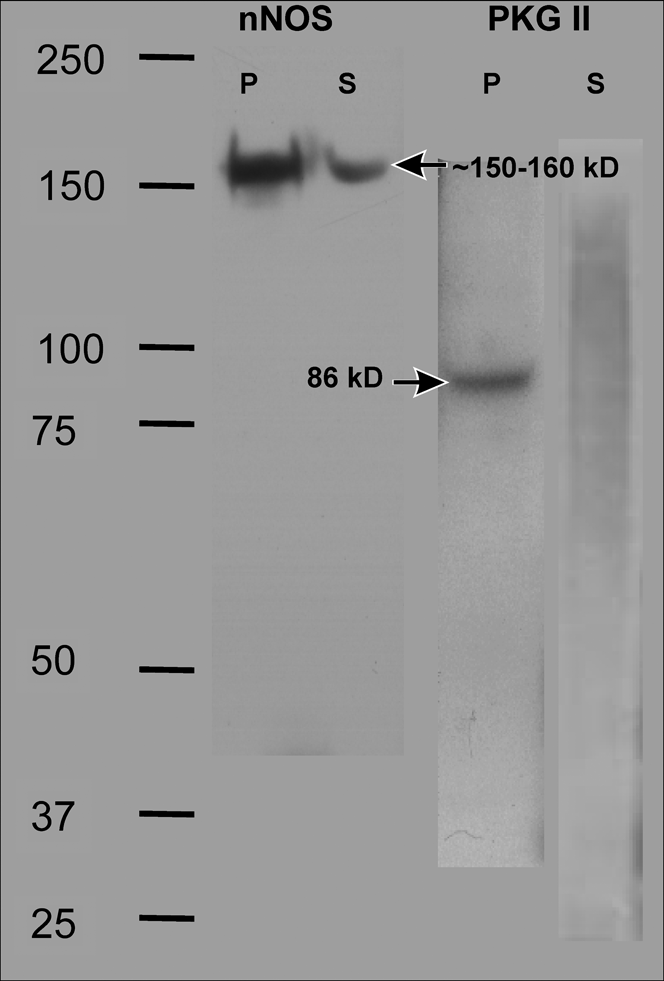
Western blots were used to confirm that the antisera used in this study specifically recognized the correct antigens in salamander retinal tissue. Several differential centrifugation steps were done on retinal homogenates in order to separate soluble and particulate fractions. The presence of a single band of the correct molecular weight was confirmed for each of the protein antisera used in this study, nNOS (~150–160 kDa) and PKGII (86 kDa) (Fig. 1). The production, characterization, and specificity of the cGMP antisera used in this study have been previously described (de Vente et al., Reference de Vente, Steinbusch and Schipper1987) and therefore were not repeated.
Characterization of nNOS and PKGII antisera. Salamander retinal homogenates were spun down and separated into soluble (S) and particulate (P) fractions and then run on a western blot and probed with antisera specific for nNOS or PKGII. A single band with the correct molecular weight was identified using each antiserum (black arrows): nNOS ~150–160 kDa and PKGII 86 kDa.

Localization of NOS: Neuronal nitric oxide synthase–like immunoreactivity
In order to specifically localize the neuronal isoform of NOS (nNOS), immunocytochemistry was done using an antiserum specifically directed against the C-terminus of nNOS. Neuronal nitric oxide synthase–like immunoreactivity (nNOS-LI) was present in both the outer and the inner retina (Fig. 2A). The strongest nNOS-LI was observed in a band in the OPL, in somata in the INL, in the IPL, and in somata in the GCL. There was no immunoreactivity when the primary antiserum was omitted (Fig. 2B).
Localization of NOS: nNOS-LI. (A) nNOS-LI. An antiserum specific for the C-terminus of nNOS was used to localize nNOS-LI. In the outer retina, there was faint nNOS-LI adjacent to photoreceptor somata in the ONL (horizontal black arrowhead) and intense nNOS-LI activity in processes in the OPL (single asterisk). In the inner retina, nNOS-LI was in bipolar cells (B), Müller cell somata (horizontal black arrow), select populations of amacrine cells (A) in the INL, and in isolated somata in the GCL (G). Strong nNOS-LI was also observed in the form of puncta and fibrous banding within S40–S60 of the IPL (double asterisks). (B) Negative control. There was no immunoreactivity when no primary antiserum was used. Scale bars = 25 μm.

In the outer retina, there was faint nNOS-LI in Müller cell processes adjacent to the somata in the outer nuclear layer (ONL) and in Müller cell somata in the INL. Intense nNOS-LI was observed in many puncta and in processes within the OPL (Fig. 2A). Given the size and orientation of the labeled processes in the OPL, it is likely that some of the nNOS-LI was in processes of horizontal and bipolar cells.
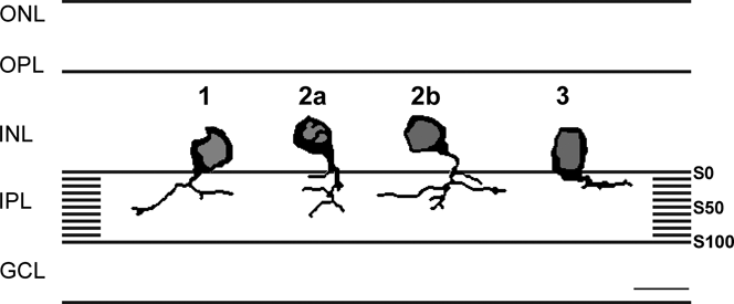
In the inner retina, nNOS-LI was present in select bipolar and amacrine cell somata, as well as in somata in the GCL in both the central and the peripheral retina (Fig. 2A). These amacrine cells had very strong nNOS-LI in their processes and in their perinuclear cytoplasm. Cell counts were done to determine the percentages of these cells within the salamander retina. Of all the total somata that were counted in the INL, ~6.76% of them were positive for nNOS-LI. Of all the cells in the INL, ~2.3% were able to be classified as being one of the four amacrine cell types: nNOS-LI 1, nNOS-LI 2a, nNOS-LI 2b, and nNOS-LI 3 (Fig. 3).
Characterization of nNOS-LI amacrine cells. In this figure and subsequent figures, the borders of the IPL are defined by black lines on the left. (A) Type 1: nNOS-LI Type 1 cells had large rounded somata (19 μm wide and 16 μm high) that were located in the INL adjacent to the IPL (black horizontal arrow). These somata gave rise to single tapered primary processes that branched in S10–S20 of the IPL (diagonal black arrow) and had terminal arborizations in S40–S60 of the IPL (vertical arrows). (B) Type 2a: nNOS-LI Type 2a cells had large ovoid-shaped somata (18 μm wide and 14 μm high) that were located in the middle of the INL (horizontal black arrow). These nNOS-LI Type 2 cells had a large primary process that tapered (diagonal black arrow) and extended to the S40–S80 region of the IPL where it arborized (vertical black arrow). (C) Type 2b: nNOS-LI Type 2b cells had rounded somata (16 μm wide and 14 μm high) that were located in the middle of the INL (top horizontal black arrow). These nNOS-LI Type 2a cells had thin primary processes that branched at S0–S10 (bottom horizontal black arrow) to form terminal arborizations in S40–S80 of the IPL (vertical black arrow). (D) Type 3: nNOS-LI Type 3 cells were large and had somata that ranged from ovoid to rectangular (13 μm wide and 16 μm high) that were located in the INL adjacent to the IPL (horizontal black arrow). The thick primary process was heavily stained, and these cells arborized in S0–S20 of the IPL (vertical black arrow). Scale bars = 25 μm.

The nNOS-LI 1 cells (Fig. 3A) were the most common in retinal cross sections (0.69% of all cells in the INL), and their somata were located in the INL adjacent to the IPL. The cytoplasmic staining in their large (19 μm wide and 16 μm high) rounded somata and in their processes was very intense. A distinguishing characteristic of these nNOS-LI 1 cells was their initial arborization in S10–S20 of the IPL. Their subsequent processes were broadly monostratified and extended into S40–S60 of the IPL.
The nNOS-LI 2 cells were common and were split into two types: nNOS-LI 2a (Fig. 3B) and nNOS-LI 2b (Fig. 3C), 0.56 and 0.43% of all cells in the INL, respectively. Their somata were relatively large (nNOS-LI 2a, 18 μm wide and 14 μm high; nNOS-LI 2b, 16 μm wide and 14 μm high), and they had strong cytoplasmic staining in their somata and in their processes. These cells were distinguished from the nNOS-LI 1 cells by the fact that the somata of both the nNOS-LI 2a and the nNOS-LI 2b cells were located in the middle of the INL. The two types of nNOS-LI 2 cells were similar in where their processes terminated; however, their morphology was slightly different. The nNOS-LI 2a cells had irregularly shaped ovoid somata with a thick primary process that extended to the S40–S80 region of the IPL where it arborized. The nNOS-LI 2b cells had more rounded somata with a thinner primary process that initially arborized in S0–S10 of the IPL. Their terminal processes of both the nNOS-LI 2a and the nNOS-LI 2b arborized in S40–S80 of the IPL.
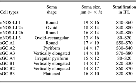
The nNOS-LI 3 cells (Fig. 3D) were relatively less common (0.61%) in retinal cross sections. Their large somata (13 μm wide and 16 μm high) were located on the border of the INL and IPL, and their shape ranged from ovoid to rectangular. Their somatic cytoplasm and their thick primary process were heavily stained and tapered into thinner processes. Their dendritic processes were broadly monostratified and ran in S0–S20 of the IPL. The amacrine cell types with nNOS-LI are summarized in Table 1 and Fig. 4.
nNOS-LI summary diagram. Diagrammatic summary of all four nNOS-LI amacrine cell types observed in the salamander retina.

Summary of nNOS and sGC containing cell types

A small percentage (0.5%) of the somata within the GCL had positive staining for nNOS-LI. It was difficult to determine whether or not these cells were ganglion cells or displaced amacrine cells because few, if any, of their processes were labeled.
Our localizations of nNOS-LI results support and extend previous descriptions (Liepe et al., Reference Liepe, Stone, Koistinaho and Copenhagen1994; Kurenni et al., Reference Kurenni, Thurlow, Turner, Moroz, Sharkey and Barnes1995) and suggest that NO can be produced in photoreceptor ellipsoids, four distinct classes of amacrine cells, Müller cells, somata in the GCL, and in processes within the OPL and IPL in the salamander retina.
Localization of NO
In order to identify where NO could actually be produced in response to different light stimuli, the NO-sensitive fluorescent dye, DAF-2DA, was loaded into dark-adapted retinal slices. These slices were either left in the dark or stimulated with a steady or flashing light stimulus, fixed, and then the NO-IF was imaged. This method has been previously used and perfected to visualize NO production in retinal slices (Eldred & Blute, Reference Eldred and Blute2005). In dark-adapted retinal slices (Fig. 5A), there was strong NO-IF present in some horizontal and amacrine cells and in some puncta in the IPL. Stimulation with the two different light stimuli yielded different patterns of NO-IF. When stimulated with steady light (Fig. 5B), NO-IF was present in horizontal cells and in their processes in the OPL, in some amacrine cell somata, and in some puncta in the IPL. In response to flashing light (Fig. 5C), NO-IF was present in photoreceptors, some apparent bipolar cell somata, and horizontal cells. There was also a robust increase in the number of labeled amacrine cells in the INL and labeled puncta in the IPL. Most dramatically, there was strong NO-IF in many somata in the GCL.
Nitric oxide imaging. NO imaging in response to light stimuli in the salamander retina. Retinal slices were dark adapted (A) and then stimulated with a steady (B) or flashing (C) light stimulus, fixed, and the NO-IF was imaged. Images are contrast inverted, with NO-IF as black. (A) In dark-adapted retinal slices, some NO-IF was present in the inner and outer retina. In the INL, there was strong NO-IF present in horizontal (H) and amacrine (A) cells. There were also some puncta of NO-IF in the IPL. (B) When dark-adapted retinal slices were stimulated with steady light, there was an increase in NO-IF in horizontal (H) and amacrine (A) cells and in puncta in the IPL. There was also some weak NO-IF in bipolar cells (B). (C) When dark-adapted retinal slices were stimulated with flashing light, there was strong NO-IF in the outer retina in photoreceptors (P). There was an increase in NO-IF in amacrine (A) cell somata, horizontal cells (H), and bipolar cells (B). There were also many more and larger puncta with NO-IF in the IPL and in presumptive ganglion cell somata (G). The amacrine cell (A, horizontal arrow) closely resembles the nNOS-LI 3 amacrine cell type. Scale bars = 25 μm.

Activation of sGC with an NO donor to produce cGMP-LI
In order to localize functional sGC, retinal eyecup preparations were stimulated with an NO donor (100 μ m SNAP) and probed for cGMP-LI using a cGMP-specific antiserum. In order to visualize and retain maximum levels of cGMP-LI, a cocktail of specific and nonspecific PDE inhibitors was used to inhibit the degradation of cGMP. When the primary antiserum was omitted, all staining was eliminated (not shown).
There were very low basal levels of cGMP-LI in unstimulated retinas in the absence of PDE inhibitors. cGMP-LI was predominantly localized in one type of monostratified bipolar cell (Fig. 6A) with processes that arborized in S20–S30 of the IPL. In the presence of the PDE inhibitor cocktail (Fig. 6B), there was an increase in cGMP-LI in processes in the OPL and in bipolar somata in the INL, with their processes arborizing in S20–S30 or S60–S70 of the IPL. To control for any nonspecific activity of the NO donor, the effects of depleted NO donor were also examined. The cGMP-LI seen in response to the depleted NO donor with PDE inhibitor cocktail (Fig. 6C) was similar to the unstimulated eyecups (Fig. 6B).
Activation of sGC with an NO donor. Eyecup preparations were stimulated with an NO donor and were then labeled with cGMP antiserum to functionally localize activated sGC. (A) Unstimulated control retina without PDE inhibitors. cGMP-LI in unstimulated control retinas without PDE inhibitors was confined to monostratified OFF bipolar cells (B) with processes that arborized near S20–S30 of the IPL (asterisk). (B) Unstimulated control retina with PDE inhibitors. In the presence of the PDE inhibitors, there was an increase in cGMP-LI in processes within the OPL (black arrowhead) and in somata of ON and OFF bipolar cells (B) that arborized in S20–S30 (single asterisk) and S60–S70 (double asterisks) of the IPL, respectively. (C) Stimulation with a depleted NO donor with PDE inhibitors. To control for the potential artifactual activity of the NO donor, depleted NO donor was used for the stimulation. The cGMP-LI seen in response to depleted NO donor was localized in processes within the OPL (black arrowhead) and in somata of ON and OFF bipolar cells (B) that arborized in S20–S30 (single asterisk) and S60–S70 (double asterisks) of the IPL, respectively. This was similar to that seen in unstimulated eyecups with PDE inhibitor cocktail. (D) Stimulation with NO donor with PDE inhibitors. When stimulated with the NO donor (100 μ m SNAP), there was a dramatic increase in cGMP-LI in amacrine (A) and bipolar (B) cell somata, in processes in the OPL (vertical arrowhead), and in somata in the GCL (G). There was a robust increase in labeled puncta in the IPL, with two strong bands of cGMP-LI at S20–S30 (single asterisk) and S60–S70 (double asterisks). Scale bars = 25 μm.

Eyecup preparations that were stimulated with the NO donor (100 μ m SNAP) with PDE inhibitor cocktail (Fig. 6D) demonstrated a dramatic overall increase in cGMP-LI in the inner retina. There was an increase in the labeling intensity of bipolar cell somata; however, there did not seem to be an increase in the types of labeled bipolar cells (Fig. 6D). There was also an increase in labeled amacrine cell somata within the INL and in somata in the GCL (Fig. 6D). Although these cells in the GCL could have been displaced amacrine cells or ganglion cells, it is likely that some of them were ganglion cells due to the presence of axons with strong cGMP-LI in the GCL. Finally, there was an overall increase in cGMP-LI within the IPL, especially in two bands: one at S20–S30 and one at S60–S70 (Fig. 6D).
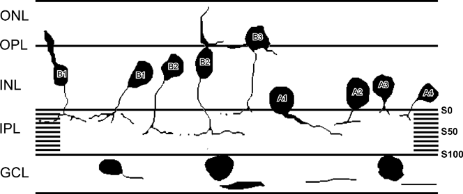
In the presence of exogenous NO, there was a large increase in cGMP-LI in a variety of retinal neurons within the inner retina (Figs. 7 and 8). All the cells will be referred to as sGC for soluble guanylate cyclase, followed by a letter A for amacrine or B for bipolar cell and a number that corresponds to the relative frequency in which these cells were identified in retinal sections. For example, sGC A1 was the most common amacrine cell type, while sGC A4 was rarely seen in retinal cross sections. The relative percentages of each amacrine cell type with cGMP-LI were not able to be determined due to the high levels of cGMP-LI in the IPL, which often prohibited detailed analysis of each cell’s dendritic arborization. These cells are summarized in Table 1 and Fig. 9.
Characterization of amacrine cells with NO-stimulated cGMP-LI. (A) sGC A1 cells had large rounded somata (17 μm wide and 19 μm high) adjacent to the IPL (black horizontal arrow). These somata gave rise to large primary process that tapered (vertical black arrow) and arborized in S50–S70 of the IPL (asterisk). (B) sGC A2 cells had relatively small pyriform-shaped somata (14 μm wide and 17 μm high) that were located adjacent to the IPL (vertical black arrow). These somata gave rise to a single primary process (horizontal black arrow) that arborized in S30–S40 of the IPL (asterisk). (C) sGC A3 cells had large vertically elongated somata (14 μm wide and 18 μm high) that were located adjacent to the IPL (black horizontal arrow). These somata quickly branched in S10–S30 of the IPL (diagonal black arrow) into secondary processes that arborized in S70–S80 of the IPL (asterisk). (D) sGC A4 cells had relatively small irregularly shaped somata (15 μm wide and 12 μm high) that were located in the middle of the INL (black horizontal arrow). These somata gave rise to a thin primary process (diagonal black arrow) that terminated in S0–S20 of the IPL (asterisk).

Characterization of bipolar cells with cGMP-LI. (A) sGC B1 cells had large somata (14 μm wide and 17 μm high) that were located adjacent to the OPL in the INL (black horizontal arrows). Some of these somata had Landolt’s clubs (horizontal black arrowhead). These cells gave rise to thin single primary process that branched at S0–S10 (diagonal black arrow) to give rise to their final arborizations in S20–S30 of the IPL (asterisk). (B) sGC B2 cells had large vertically elongated somata (14 μm wide and 17 μm high) that were located adjacent to the OPL in the INL (black horizontal arrow). Some of these somata had Landolt’s clubs (horizontal black arrowhead). These somata gave rise to a thin primary process (top diagonal black arrow) that arborized in S50–S60 of the IPL (bottom diagonal black arrow) and terminated in S60–S70 of the IPL (asterisk). (C) sGC B3 cells had large flattened somata (16 μm wide and 10 μm high) that were displaced and located in the ONL (horizontal black arrowhead). These somata had a primary process (horizontal black arrow) that branched at S0–S15 (diagonal black arrow) and had terminal arborizations in the S20–S30 of the IPL (asterisk). These cells also had well-labeled lateral processes that extended 20–30 μm in the OPL (vertical black arrow).

Summary of cGMP-LI. Diagrammatic summary of all cGMP-LI cell types observed with SNAP stimulation (100 μ m) in the presence of PDE inhibitors. The numbers reflect relative frequency of cell types with one being most common. A, amacrine cell; B, bipolar cell.

Amacrine cells with cGMP-LI
There were many putative amacrine cells located in both the central and the peripheral regions of the retina. These amacrine cells had strong cGMP-LI in both their somata and their processes. Additionally, there was an increase in cGMP-LI in somata in the GCL. Although it is possible that at least some of these cells in the GCL were displaced amacrine cells, at least some of these somata could be ganglion cells due to the labeled axons in the GCL.
Although the strong and extensive cGMP-LI in the IPL made it difficult to morphologically identify, quantify, and characterize many of the labeled amacrine cells, it was possible to characterize four different amacrine cell types. sGC A1 cells (Fig. 7A) were most common, and their large rounded somata (19 μm high and 17 μm wide) were located in the INL adjacent to the IPL. These cells had a single thick primary process that projected to one side of the somata that tapered as it projected to arborize in S50–S70 of the IPL. sGC A2 cells (Fig. 7B) had pyriform-shaped somata (17 μm high and 14 μm wide) in the INL adjacent to the IPL. These cells gave rise to a single thin primary process that went straight down into the IPL where it arborized in S30–S40. sGC A3 cells (Fig. 7C) had large vertically elongated somata (18 μm high and 14 μm wide) that were located in the INL adjacent to the IPL. A single short stout primary process came from the bottom of the cell and quickly branched in S10–S30 of the IPL into secondary processes that continued to S60–S70, where they had their terminal arborizations. sGC A4 cells (Fig. 7D) had irregularly shaped somata (12 μm high and 15 μm wide) that were located in the middle of the INL. These cells gave rise to a single delicate primary process that had terminal arborizations around S20–S30 of the IPL.
Bipolar cells with cGMP-LI
Many bipolar cell somata were labeled, but often, their processes and their dendritic arborizations were not clearly labeled. Although the characterization of bipolar cell types was often limited by the absence of distinctly labeled process and/or the extensive cGMP-LI in the IPL, at least three different bipolar cell types were identified. The lack of well-labeled processes raises the possibility that additional bipolar cell types may contain sGC.
sGC B1 cells (Fig. 8A) were relatively common, and they were the only bipolar cells identified in unstimulated control retinas without PDE inhibitors (BSS only) (Fig. 6A), and they were present in all retinas and stimulus conditions. Their somata (16 μm high and 14 μm wide) were observed both with and without Landolt’s clubs that extended into the ONL. These cells had relatively few processes that were obvious in the OPL. They had single thin primary processes that branched at S0–S10 to give rise to their final arborizations in S20–S30 of the IPL. Due to the location of their processes in the OFF sublamina of the IPL, it is likely that these are physiologically OFF bipolar cells.
sGC B2 cells (Fig. 8B) were apparent only in the presence of the PDE inhibitor cocktail and were not present in BSS only, suggesting that these cells have high endogenous levels of PDE activity under basal conditions. These cells had vertically elongated somata (16 μm high and 14 μm wide) and were also seen both with and without Landolt’s clubs. These cells had clearly apparent processes in the OPL, and each had a single primary process that gave rise to an initial arborization in the IPL at S50–S60 and processes that terminated in S60–S70.
sGC B3 cells (Fig. 8C) were displaced bipolar cells that had relatively large flattened somata (10 μm high and 16 μm wide) with clearly labeled prominent processes in the OPL. These cells had a single primary process that branched in S0–S15 of the IPL and terminated in S20–S30 of the IPL. These cells were also seen only in the presence of PDE inhibitor cocktail, and thus, they also have high endogenous levels of PDE activity under basal conditions. A diagrammatic summary of all the cell types with NO-stimulated cGMP-LI is shown in Fig. 9.
Colocalization analysis of cGMP-LI and PKG-like immunoreactivity (PKG-LI)
One of the downstream targets of cGMP is PKG. PKG has both soluble (PKGIα and PKGIβ) and particulate (PKGII) isoforms (Hofmann et al., Reference Hofmann, Feil, Kleppisch and Schlossmann2006). Although we tested antisera directed against PKGIα, PKGIβ, and PKGII, only the antiserum directed against the first 120 amino acids of PKGII produced a single band of the correct molecular weight on western blots (Fig. 1). Double labeling of cGMP-LI and PKGII-LI was done on retinal cross sections that were stimulated with the NO donor SNAP (Fig. 10A–10C). PKGII-LI (Fig. 10B) was present in many processes in the OPL, in sporadic amacrine cell somata, in puncta in the IPL, and in select somata in the GCL. PKGII-LI was colocalized with cGMP-LI (Fig. 10C) in many processes in the OPL, in select amacrine cell somata, in puncta in the IPL, and in select somata in the GCL. It was often difficult to identify specific amacrine cell types that contained both PKGII-LI and cGMP-LI because their processes were not strongly filled with PKGII-LI; however, the sGC A1 and sGC A2 amacrine cell types were double labeled (not shown).
Colocalization analysis of cGMP-LI with PKGII-LI. Colocalization analysis of cGMP-LI with its downstream target, PKGII-LI, was done in tissue that was stimulated with the NO donor SNAP (100 μ m). (A) cGMP-LI levels were high after stimulation with the NO donor (100 μ m SNAP). There were well-labeled amacrine (horizontal arrow) and bipolar (vertical arrow) cell somata within the INL, some labeled processes in the OPL, ganglion cell axons (vertical arrowhead), as well as somata in the GCL (horizontal arrowhead). There was also an increase in cGMP-LI within the IPL (asterisk). (B) PKGII-LI was found in processes in the OPL (vertical black arrow), some amacrine cells (horizontal black arrow), select somata in the GCL (horizontal black arrowhead), in axons in the ganglion cell axon layer (vertical arrowhead), and in puncta in the IPL (asterisk). (C) PKGII-LI was colocalized with cGMP-LI in processes in the OPL (vertical black arrow), isolated amacrine cells (horizontal arrow), in somata in the GCL (horizontal arrowhead), in the ganglion cell axon layer (vertical arrowhead), and in puncta in the IPL (asterisk). Scale bars = 25 μm.

Discussion
Localization of NOS
NOS has been previously localized in the salamander retina in photoreceptor ellipsoids, amacrine cells, and the distal regions of both Müller and bipolar cells using both NADPH-d histochemistry and nNOS immunocytochemistry (Liepe et al., Reference Liepe, Stone, Koistinaho and Copenhagen1994; Kurenni et al., Reference Kurenni, Thurlow, Turner, Moroz, Sharkey and Barnes1995), but there were no detailed descriptions of the specific cell types involved. The staining seen in our experiments was very similar to those previous studies, with the exception of the previously reported strong staining of Müller cells in the INL. The difference in this staining could reflect that the previous study used antisera that might also recognize inducible NOS (iNOS), as suggested by Kurenni et al. (Reference Kurenni, Thurlow, Turner, Moroz, Sharkey and Barnes1995). Our results support these original studies, and we were able to additionally characterize the subset of four specific amacrine cell types.
nNOS-LI in amacrine cells
The present report confirms and expands on the previous localizations of nNOS in salamander retina by identifying four distinct amacrine cell types with nNOS-LI: nNOS-LI 1, nNOS-LI 2a, nNOS-LI 2b, and nNOS-LI 3 (Fig. 3). The characteristics of these cell types are summarized in Fig. 4 and Table 1. Twenty subtypes of amacrine cells have been previously identified based on their cell morphology and light responses in salamander retina (Pang et al., Reference Pang, Gao and Wu2002). These cell types were intracellularly filled and morphologically characterized as having dendrites that were narrowly monostratified, broadly monostratified, multistratified, or diffusely stratified in the IPL. The strong and extensive nNOS-LI in the IPL made it difficult to conclusively correlate the four nNOS amacrine cell types described here to cell types that have been previously described (Pang et al., Reference Pang, Gao and Wu2002). However, based on the localizations and stratification of their dendritic arborizations, it is possible to make some comparisons.
nNOS-LI 1 cells were consistent with Type 11 cells that are described as being ON/OFF cells that are broadly monostratified in S30 (ON stratum) and S40 (OFF stratum) (Pang et al., Reference Pang, Gao and Wu2002). nNOS-LI Type 2a and Type 2b cells were more diffusely stratified with processes that terminated in S40–S80 of the IPL, and thus, they would be consistent with the Type 5 or Type 6 cells that arborize in S50 and S60, respectively, classifying them as ON/OFF cells. nNOS-LI 3 cells were consistent with Type 13 cells as they had broadly monostratified processes in S0–S20 (OFF stratum) of the IPL. The NO-IF that was seen in amacrine cells in both dark and light stimulations (Fig. 5) was consistent with the fact that the amacrine cells with nNOS-LI can ramify in either ON or OFF regions of the IPL.
nNOS-LI and NO-IF
In retinal slices, the NO-sensitive dye DAF is able to localize NO production within specific retinal cell types, and it had been shown that NO-IF can be produced in all cell types within the retina (Eldred & Blute, Reference Eldred and Blute2005). Previous reports (Wood & Garthwaite, Reference Wood and Garthwaite1994) have suggested that NO is freely diffusible. NO imaging in this report (Fig. 5) and in turtle retina (Blute et al., Reference Blute, Lee and Eldred2000b; Eldred & Blute, Reference Eldred and Blute2005) does not support this theory. NO-IF is rigidly localized within cell somata, processes, and synaptic boutons in the inner and outer retina, and there was no evidence of diffusion. There are several reasons why NO would not be freely diffusible. One explanation is that NO is bound to thiols such as glutathione to form nitrosothiols (Upchurch et al., Reference Upchurch, Welch and Loscalzo1995; Singh et al., Reference Singh, Hogg, Joseph and Kalyanaraman1996; Hogg, Reference Hogg2002). Very high levels of glutathione have been localized in specific retinal cell types such as Müller and horizontal cells (Pow & Crook, Reference Pow and Crook1995). Such a cellular sink for NO has been suggested to be involved in shaping low-level NO signals (Griffiths & Garthwaite, Reference Griffiths and Garthwaite2001). Additionally, protein disulfide isomerase (PDI) is able to catalyze thiol-disulfide exchange reactions to the intracellular compartments (Zai et al., Reference Zai, Rudd, Scribner and Loscalzo1999). Enzymes like PDI could serve to transport any NO that leaves the cell back into the cell using a mechanism that involves transnitrosation.
In general, there was good correlation between nNOS-LI and NO-IF in that amacrine, bipolar, and ganglion cell somata had both. In salamander, it was difficult to correlate specific nNOS-LI cell types with light-stimulated NO-IF because although many somata had clear NO-IF, the processes of these cells did not have strong NO-IF, and if it was present, it tended to be punctate. However, some comparisons could be made. There was a common amacrine cell type with NO-IF that had a vertically elongated soma that quickly arborized in the IPL (Fig. 5C) that closely resembled the nNOS-LI 3 amacrine cell type. The other nNOS amacrine cell types were not as clearly identifiable and may have high levels of endogenous synaptic inhibition that prevented their light-stimulated production of NO. Synaptic inhibition plays a critical role in NO modulating production in retina (Yu & Eldred, Reference Yu and Eldred2003, Reference Yu and Eldred2005a,Reference Yu and Eldredb).
Some apparent bipolar cell somata also had light-stimulated NO-IF, and this correlated well with the nNOS-LI in bipolar cell somata in the INL and in processes in the OPL. However, there were no bipolar cells that had sufficient NO-IF in their processes to make any precise anatomical correlations. There was also a significant population of horizontal cells that had strong NO-IF, and therefore, it was likely that some of the nNOS-LI located in the OPL was in horizontal cell processes.
Putative Müller cell processes in the ONL and their somata in the INL had light nNOS-LI; however, NO-IF was not present in Müller cells retinal slices that were dark adapted or stimulated with steady or flashing light. Although nNOS was present in Müller cells, it did not actively produce NO in the dark or in response to a light stimulus.
There were very few somata in the GCL that were positive for nNOS-LI; however, in the presence of a flashing light stimulus, there were relatively more somata that were positive for NO-IF. It is possible that the high levels of NO-IF in the GCL are due to another calcium-activated isoform of NOS, eNOS. It was unlikely that iNOS was involved, given that the time required to induce the synthesis of active iNOS would be considerably longer than the flashing light stimulus.
Functional activation of sGC with an NO donor
The addition of exogenous NO produced elevated levels of cGMP-LI in multiple amacrine and bipolar cell types and in somata in the GCL. In control retinas that were not stimulated, there were very low basal levels of cGMP-LI within the inner retina. The comparison of unstimulated control retinas (Fig. 6A) and unstimulated control retinas with PDE inhibitors (Fig. 6B) indicated the presence of high levels of endogenous PDE activity. These results were similar to those in turtle, where eight amacrine and three bipolar cell types with functional sGC are identified in the presence of PDE inhibitor, and cGMP-LI is low in the absence of PDE inhibitor (Blute et al., Reference Blute, Velasco and Eldred1998). The other interesting similarity was that in both turtle and salamander, the stratification of cGMP-LI in the IPL was very precise with only certain strata being labeled in both the ON and the OFF regions of the IPL. This suggests that cGMP plays a role in very specific circuits in both the ON and the OFF signaling pathways.
cGMP-gated ion channels are located in photoreceptors (Kurenny et al., Reference Kurenny, Moroz, Turner, Sharkey and Barnes1994), ganglion (Ahmad et al., Reference Ahmad, Leinders-Zufall, Kocsis, Shepherd, Zufall and Barnstable1994), bipolar (Nawy & Jahr, Reference Nawy and Jahr1991; Shiells & Falk, Reference Shiells and Falk1992; de la Villa et al., Reference de la Villa, Kurahashi and Kaneko1995), and Müller cells (Kusaka et al., Reference Kusaka, Dabin, Barnstable and Puro1996). The present localization of NO-IF, soluble guanylate cyclase–like immunoreactivity, and cGMP-LI in somata in the INL and GCL suggested that NO may function as an intracellular messenger in the regulation of these CNG channels.
Amacrine cells
Twenty subtypes of amacrine cells (Pang et al., Reference Pang, Gao and Wu2002) and 20 bipolar cell types (Pang et al., Reference Pang, Gao and Wu2004) are identified in salamander retina based on their cell morphology and light responses. In our study, there were four amacrine and three bipolar cell types with cGMP-LI that were identified. In the presence of exogenous NO, the very high levels of cGMP-LI in the IPL made it very difficult to trace the dendritic arborizations of individual amacrine cells for long distances. However, it was possible to make some speculations as to the physiology of these cells with cGMP-LI.
sGC A3 cells (Fig. 7C) were consistent with the Type 7 cells described by Pang et al. (Reference Pang, Gao and Wu2002). These cells were distinct because they branched in the S10–S30 layer of the IPL into two processes that formed terminal arborizations in S70–S80 (ON stratum) of the IPL. NO produced in response to a light stimulus would activate sGC in this cell type to produce cGMP. It is possible that these sGC A3 cells could have CNG channels that would contribute to their ON responses. Due to the high levels of cGMP-LI and their diffuse stratifications in the IPL, sGC A1 cells (Fig. 7A), sGC A2 cells (Fig. 7B), and sGC A4 cells (Fig. 7D) could not be correlated to the cell types described by Pang et al. (Reference Pang, Gao and Wu2002). It was possible that these cells were novel amacrine cell types that were different from the 20 cell types previously described. Similar results were seen in turtle where NO donor–stimulated cGMP-LI is found in more than 10 previously undescribed amacrine cell types (Blute et al., Reference Blute, Velasco and Eldred1998). However, based on the physiological results of Pang et al. (Reference Pang, Gao and Wu2002), it was possible to speculate on the physiological responses of these sGC amacrine cells based on their stratification in the IPL. The sGC A1 cells were likely to be primarily ON cells because they have terminal processes in both ON/OFF (S50–60) and ON (S70) strata. sGC A2 cells had terminal processes primarily in OFF (S30–S40) strata and were most likely OFF cells. Finally, the sGC A4 cells had terminal processes in the OFF (S0–S20) strata and would certainly be OFF cells.
Although it was difficult to say with certainty, the sGC A4 cells had some anatomical similarity, in terms of their somata shape and location, with nNOS-LI 2a cells. This raises the possibility that this cell type can make NO and respond to it by making its own cGMP. A similar situation occurs in turtle where there is an amacrine cell with nNOS-LI that also has NO donor–stimulated increases in cGMP-LI (Blute et al., Reference Blute, Velasco and Eldred1998). This would suggest that NO can serve as an intracellular messenger in these amacrine cells. Another similarity to turtle (Blute et al., Reference Blute, Velasco and Eldred1998) is the close correlation of nNOS-LI and cGMP-LI in strata within the IPL. This would suggest that if NO is functioning as an intercellular messenger, it is doing so over relatively short distances.
Bipolar cells
In the salamander, 20 bipolar cell types are identified based on their cell morphology and responses to light (Pang et al., Reference Pang, Gao and Wu2004). Some speculations about the physiological function of the sGC bipolar cells can be made based on the physiological and anatomical criteria for bipolar cells that are defined in that paper.
sGC B1 cells (Fig. 8A) terminated in S20–S30 of the IPL, which correlated with Type 3 or Type 4 OFF bipolar cells that are described by Pang et al. (Reference Pang, Gao and Wu2004). sGC B3 cells (Fig. 8C) terminated in S20–S30 of the IPL, as well, and thus could be characterized as OFF bipolar cells (Maple et al., Reference Maple, Zhang, Pang, Gao and Wu2005). sGC B1 cells had cGMP-LI both in the unstimulated control conditions and in the presence of exogenous NO. It was likely that the cGMP levels in these cells were not regulated by NO, given that NOS inhibitors do not decrease the cGMP-LI in these bipolar cells in salamander (Cimini et al., Reference Cimini, Strang, Wotring, Keyser and Eldred2008). It was possible that the cGMP-LI in these bipolar cells was related to the activation of particulate guanylate cyclases by natriuretic peptides that are described in turtle retina (Blute et al., Reference Blute, Lee, Huffmaster, Haverkamp and Eldred2000a). The sGC B3 cells were seen in the presence of PDE inhibitors in response to NO donor, suggesting that these cells had high levels of PDE activity that limited their response to basal levels of NO and that they were only activated by higher levels of NO. sGC B2 cells (Fig. 8B) were consistent with the Type 6 or Type 7 ON bipolar cells of Pang et al. (Reference Pang, Gao and Wu2004).
Colocalization analysis of cGMP with PKG
Functional studies of the NO/cGMP pathways have shown that the downstream target of cGMP, PKG, can play a role at several levels of visual processing. Colocalization analysis of cGMP-LI and PKGII-LI demonstrated that PKGII-LI colocalized with cGMP-LI in processes in the OPL, in amacrine cells in the INL, as well as in somata in the GCL (Fig. 10).
The NO/cGMP pathway has been shown to play a specific role in closing gap junctions between horizontal cells. The intracellular injection of arginine (a precursor to NO) (Miyachi et al., Reference Miyachi, Murakami and Nakaki1990) or cGMP (Miyachi et al., Reference Miyachi, Miyakawa and Murakami1991) in turtle horizontal cells blocks gap junctional coupling, and intracellular injections of cGMP uncouple horizontal cells in catfish (DeVries & Schwartz, Reference De Vries and Schwartz1989). In our study, we showed that NO-IF was present in horizontal cells and that NO-activated sGC increased cGMP-LI in the OPL. There was also a colocalization of cGMP-LI with PKGII-LI in these processes within the OPL. All these results were consistent with studies of horizontal cells in the hybrid bass, which indicate that NO, cGMP, and PKG are involved in uncoupling the gap junctions between the cells (Lu & McMahon, Reference Lu and McMahon1997).
In conclusion, our results showed that the NO/cGMP pathway was present and well developed in all nerve cell types within the salamander retina. This study supported previous studies localizing NOS in the salamander retina, and we additionally characterized the specific amacrine cell types that contained nNOS. Using NO imaging, we provided evidence that NO was produced by all neuronal cell types in the salamander retina and that exogenous NO activated sGC to synthesize cGMP in select amacrine cells, bipolar cells, and somata in the GCL. Colocalization analysis of cGMP-LI and its downstream target, PKGII-LI, provided anatomical support for previous functional studies done in retina. This work supported that NO/cGMP pathways have the ability to influence the function of all the retinal neuron types in the salamander.
Acknowledgments
This research was supported by NIH-NEI EY04785 to W.D.E. We would like to thank Felicitas B. Eldred for her technical assistance and Dr. Jan de Vente for his generous gifts of the cGMP antiserum.