INTRODUCTION
Fascioliasis is a parasitic disease caused by two trematode species, Fasciola hepatica of almost worldwide distribution, and Fasciola gigantica restricted to given regions of Africa and Asia (Mas-Coma et al. Reference Mas-Coma, Valero and Bargues2009a). Despite its importance in animal husbandry due to the economic losses it causes, human infection by Fasciola has historically been of secondary importance (Chen and Mott, Reference Chen and Mott1990). However, the description of human fascioliasis endemic areas in many countries in the last two decades has changed the concept of this food-borne zoonosis (Mas-Coma, Reference Mas-Coma2005). Moreover, experimental studies on the long-term infection indicate that fascioliasis pathogenicity is not only important in the invasive or acute phase, as previously considered, but also in the biliary and advanced chronic periods of the disease, mainly in human fascioliasis endemic areas (Valero et al. Reference Valero, Santana, Morales, Hernandez and Mas-Coma2003, Reference Valero, Navarro, Garcia-Bodelon, Marcilla, Morales, Garcia, Hernandez and Mas-Coma2006a, Reference Valero, Girones, García-Bodelon, Periago, Chico-Calero, Khoubbane, Fresno and Mas-Coma2008). In the human infection by Fasciola, high pathogenicity is now recognized in several clinical situations previously neglected. Consequently, their worrying impact in human endemic areas of developing countries is now being emphasized (Mas-Coma et al. Reference Mas-Coma, Agramunt and Valero2014). Additionally, immune-modulation by fasciolids in the acute infection phase (Brady et al. Reference Brady, O'Neill, Dalton and Mills1999; Dalton et al. Reference Dalton, Robinson, Mulcahy, O'Neill and Donnelly2013), and their immune suppressive effect in the biliary and advanced chronic phases (Girones et al. Reference Girones, Valero, Garcia-Bodelon, Chico-Calero, Punzon, Fresno and Mas-Coma2007) underlie common co-infections with other parasitic and infectious diseases (Esteban et al. Reference Esteban, Flores, Aguirre, Strauss, Angles and Mas-Coma1997a, Reference Esteban, Flores, Angles, Strauss, Aguirre and Mas-Comab, Reference Esteban, Gonzalez, Bargues, Sanchez, Naquira and Mas-Coma2002, Reference Esteban, Gonzalez, Curtale, Muñoz-Antoli, Valero, Bargues, El-Sayed, El Wakeel, Abdel-Wahab, Montresor, Engels, Savioli and Mas-Coma2003; Gonzalez et al. Reference Gonzalez, Esteban, Bargues, Valero, Ortiz, Naquira and Mas-Coma2011; Zumaquero-Rios et al. Reference Zumaquero-Rios, Sarracent-Perez, Rojas-Garcia, Rojas-Rivero, Martinez-Tovilla, Valero and Mas-Coma2013).
The transmission characteristics of this freshwater snail-borne disease in human endemic areas are highly dependent on abiotic factors such as those inherent to climate and environment (Fuentes et al. Reference Fuentes, Valero, Bargues, Esteban, Angles and Mas-Coma1999, Reference Fuentes, Malone and Mas-Coma2001). This underlies the pronounced impact of climate and global changes on fascioliasis and has been related to the increasing number of human infection reports in numerous countries (Mas-Coma et al. Reference Mas-Coma, Valero and Bargues2008, Reference Mas-Coma, Valero and Bargues2009b). The impact of climate change and anthropogenic modifications of the environment on the transmission risk, long-term trend and seasonality of human fascioliasis has been recently examined in southern Asia (Afshan et al. Reference Afshan, Fortes-Lima, Artigas, Valero, Qayyum and Mas-Coma2014). The spreading capacity of both lymnaeid snail vectors and fasciolid flukes by means of human movements and import/export of livestock have also been highlighted (Mas-Coma et al. Reference Mas-Coma, Valero and Bargues2009a; Bargues et al. Reference Bargues, Artigas, Khoubbane, Flores, Glöer, Rojas-Garcia, Ashrafi, Falkner and Mas-Coma2011).
Considering this scenario of increasing concern, the World Health Organization (WHO) decided to launch a worldwide initiative against this disease, with the aim to decrease morbidity, mainly focusing on children (WHO, 2007, 2008). Human endemic areas of different epidemiological characteristics and transmission patterns were initially selected for a pilot initiative. Specific strategies were designed for each one of the four countries involved (Bolivia, Peru, Egypt, Vietnam), including differing (i) diagnostic techniques, (ii) control measures and (iii) monitoring methods for the evaluation of the efficacy of the activities performed (Villegas et al. Reference Villegas, Angles, Barrientos, Barrios, Valero, Hamed, Grueningr, Ault, Montresor, Engels, Mas-Coma and Gabrielli2012). Therefore, bespoke country-specific strategies were applied and evaluated (Curtale et al. Reference Curtale, Hassanein, El Wakeel, Mas-Coma and Montresor2003b; Valero et al. Reference Valero, Periago, Perez-Crespo, Angles, Villegas, Aguirre, Strauss, Espinoza, Herrera, Terashima, Tamayo, Engels, Gabrielli and Mas-Coma2012a).
The aforementioned WHO initiative highlighted the need for diagnostic techniques more accurate than those traditionally used for the diagnosis of human fascioliasis previously (Chen and Mott, Reference Chen and Mott1990; Hillyer, Reference Hillyer and Dalton1999; Mas-Coma et al. Reference Mas-Coma, Esteban and Bargues1999b) and for techniques and methods useful at large scale for both mass screening and monitoring. The present study reviews the diagnosis of human fascioliasis, emphasizing (i) the challenges posed by the new worldwide scenario and (ii) analysing the need to deal appropriately with the different epidemiological situations and transmission patterns in both individual diagnosis and community surveys, as well as (iii) the advantages and problems posed by the many diagnostic techniques presently available. Focus is given to diagnosis of fascioliasis by means of stool and blood samples, based on parasitological and immunological techniques. Older techniques no longer or only rarely used and other complementary techniques are only briefly discussed. This review focuses on human fascioliasis, so that reference to diagnosis of fascioliasis in animals is only included where necessary; specific literature regarding livestock should be sought elsewhere if required (e.g. Charlier et al. Reference Charlier, Vercruysse, Morgan, van Dijk and Williams2013). Molecular techniques are not included in the present review, due to their specific characteristics, complexity and the length its analysis would need. This is the most comprehensive approach to review faecal- and blood-based diagnosis of human fascioliasis in light of the current worldwide scenario.
The purposes of this review are mainly (i) to highlight the problems which the diagnostic methods and techniques may face in the present new global scenario (instead of a review of all techniques available in the past and present), (ii) to help physicians in hospitals and health centres and those with responsibility for health in endemic countries to better select appropriate diagnostic strategies depending on their situations (individual patient, patient treatment follow-up, community surveys, control campaign monitoring, etc.), (iii) to highlight diagnostic techniques and tests which have been adequately evaluated or at least sufficiently applied as to obtain significant conclusions (there is a very large literature about similar tests developed in different countries which are afterwards never or almost never applied) and (iv) to state an approach from the point of view of the praxis problems posed by human fascioliasis diagnosis and their solutions (technological details of the different techniques and tests are only considered secondarily and noted when needed).
OLD AND NEW SCENARIOS
Before the 1990s, diagnosis of fascioliasis focused on individual patients in hospitals or health centres. Case reports were mainly from developed countries (Arjona et al. Reference Arjona, Riancho, Aguado, Salesa and Gonzalez-Macias1995) and usually concerned isolated human infection in animal fascioliasis endemic areas (Mas-Coma et al. Reference Mas-Coma, Bargues, Esteban and Dalton1999a). In such cases, blood eosinophilia was an element of significant diagnostic guidance, given that co-infections with other helminthiases were rare in autochthonous people, although care should be taken with immigrants and travellers to tropical/subtropical areas. Infrastructures and diagnostic capacities available were thus usually at a high level. Consequently, although diagnostic methods and techniques available were not very sensitive and specific, they were sufficient for the needs in such individual patient situations. Adult subjects were the most frequently involved, usually without gender differences. Diagnosis was usually made shortly after beginning of symptoms by passive detection of patients who attended a health centre.
These patients were most frequently in the invasive phase of the disease, so that serological techniques were mainly applied due to Fasciola eggs not yet appearing in stools. Serological techniques were also solving the usual problem of low egg shedding, related to low infection burdens, and even absence of eggs in stools during the biliary phase due to (i) incapacity of Fasciola to produce eggs related to the lack of adaptation of the fluke to the human host or (ii) encapsulation of eggs in liver abscesses or granulomas due to being laid in the liver parenchyma. This explains why intensity by egg counts was almost never assessed.
In cases of isolated patients or situations of short series of patients in developed countries, valuable information regarding anamnesis was relatively easy to obtain, including previous ingestion of watercress (rarely other metacercariae-carrying freshwater vegetables). Finally, verification of successful treatment was also easily performed because individual patient follow-up is affordable, thus allowing for the assessment of symptom and egg disappearance.
However, from the mid-1990s onwards, due to the progressive description of human fascioliasis endemic areas and human infection reports in developing countries, new situations, hitherto neglected, entered into the global scenario. Field studies have recently demonstrated that human fascioliasis is pronouncedly more heterogeneous than previously thought. The different transmission patterns and epidemiological situations give rise to different scenarios (Mas-Coma et al. Reference Mas-Coma, Bargues and Valero2005, Reference Mas-Coma, Valero and Bargues2009a).
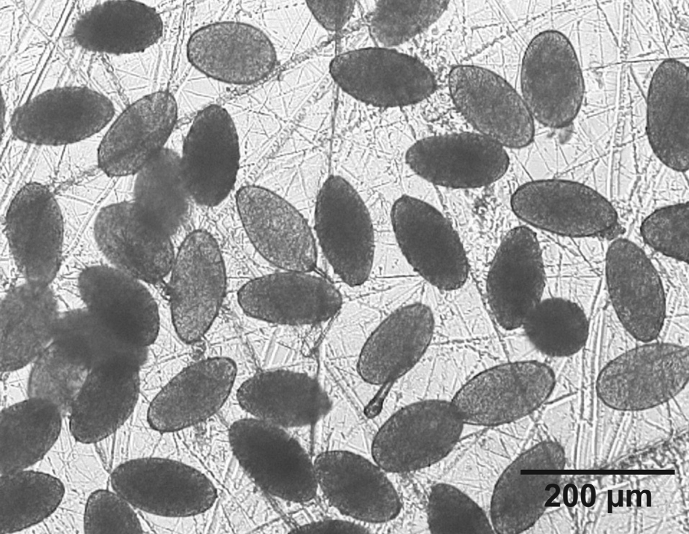
There is a need for community surveys, as situations with very high prevalences (up to 72 and 100% prevalence by coprology and serology, respectively), and very high intensities (up to more than 8000 eggs per gram (epg) in children) have been described in human fascioliasis endemic areas of developing countries (Fig. 1) (Esteban et al. Reference Esteban, Flores, Aguirre, Strauss, Angles and Mas-Coma1997a, Reference Esteban, Flores, Angles, Strauss, Aguirre and Mas-Comab, Reference Esteban, Flores, Angles and Mas-Coma1999, Reference Esteban, Gonzalez, Bargues, Sanchez, Naquira and Mas-Coma2002, Reference Esteban, Gonzalez, Curtale, Muñoz-Antoli, Valero, Bargues, El-Sayed, El Wakeel, Abdel-Wahab, Montresor, Engels, Savioli and Mas-Coma2003; Gonzalez et al. Reference Gonzalez, Esteban, Bargues, Valero, Ortiz, Naquira and Mas-Coma2011; Zumaquero-Rios et al. Reference Zumaquero-Rios, Sarracent-Perez, Rojas-Garcia, Rojas-Rivero, Martinez-Tovilla, Valero and Mas-Coma2013). In poor-income rural areas, blood eosinophilia loses its usefulness as an element of diagnostic guidance, because co-infections with other helminthiases appear frequently in autochthonous people. Insufficient health infrastructures and diagnostic capacities, mainly in remote rural areas, add another aspect of concern. Moreover, patients are now increasingly diagnosed in urban areas neighbouring rural endemic areas. This includes (i) patients from rural areas attending health centres in the city for diagnosis or (ii) people living in the cities becoming infected after visiting the rural areas or (iii) after ingestion of contaminated vegetables transported from the endemic rural areas and sold in uncontrolled city food markets.
Massive shedding of Fasciola eggs in a faecal sample of a female child from the human fascioliasis endemic area of the Nile Delta, Egypt.

The diagnostic methods and techniques needed for community surveys should be very sensitive to also detect subjects infected by low burdens, and highly specific to avoid cross-reactions with other co-existing parasitoses. In contrast to developed countries, the disease shows relationships with age and gender: children and females appearing to be the most frequently involved and coprological prevalences in adult subjects usually decreasing by around 50% (Esteban et al. Reference Esteban, Flores, Aguirre, Strauss, Angles and Mas-Coma1997a, Reference Esteban, Flores, Angles, Strauss, Aguirre and Mas-Comab, Reference Esteban, Flores, Angles and Mas-Coma1999; Mas-Coma, Reference Mas-Coma, Cotruvo, Dufour, Rees, Bartram, Carr, Cliver, Craun, Fayer and Gannon2004).
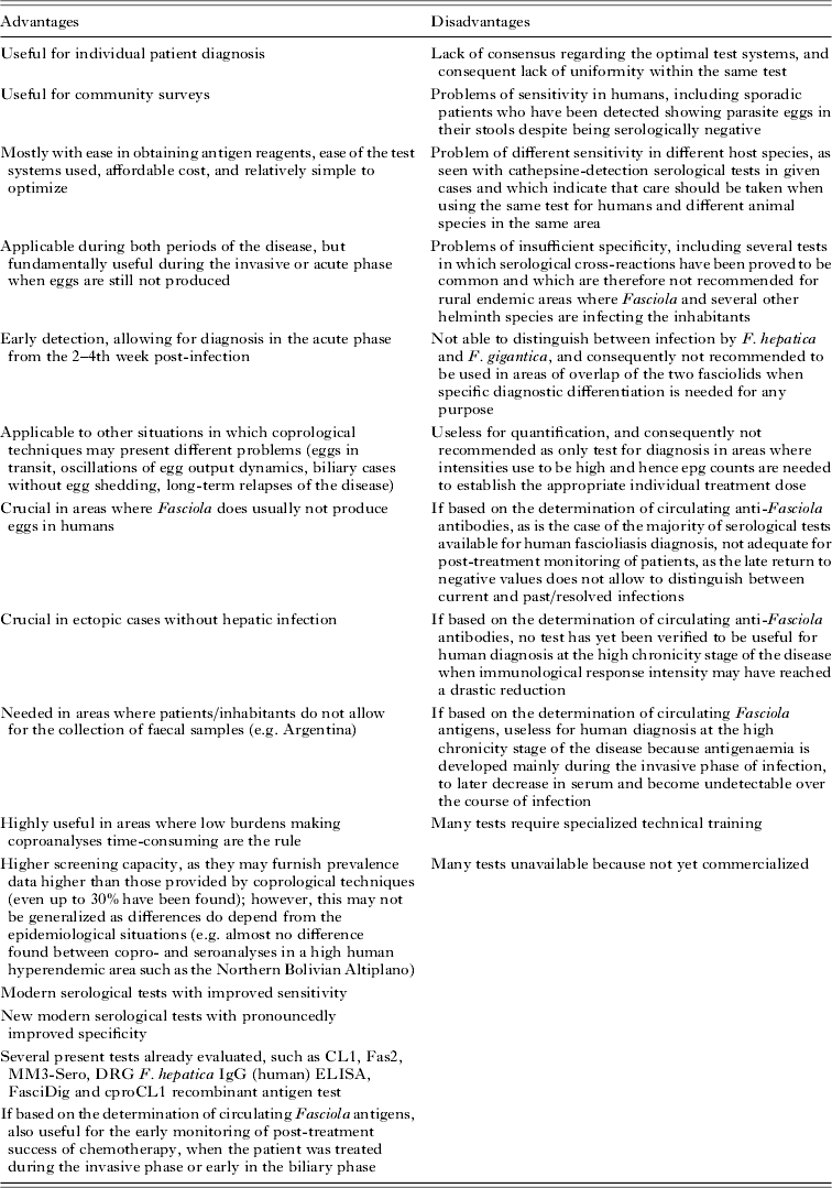
In human fascioliasis endemic rural areas, active detection of patients is needed, because diagnosis in the invasive phase is rarely performed given that children with symptoms of the acute phase do not attend health centres for diagnosis. In such situations, a successful diagnosis could only be performed during the biliary phase, as diagnosis usually relies on faecal egg detection, as serological techniques are usually not available for diagnosis in the invasive phase in rural health centres.
Many aspects, which were never given importance in human fascioliasis diagnosis before, may now be considered crucial. For example, in several human fascioliasis endemic areas blood or stool samples cannot be obtained due to ethnic/religious aspects of the inhabitants (for example, in Bolivia and Argentina). In many areas, disease transmission is seasonal (monoseasonal or biseasonal), thus prevalences and intensities may differ according to the season in which the survey is made, as in the valley pattern in Andean countries (Mas-Coma, Reference Mas-Coma2005) or in Pakistan (Afshan et al. Reference Afshan, Fortes-Lima, Artigas, Valero, Qayyum and Mas-Coma2014). In other areas transmission is permanent throughout the year, as for instance in the altiplano pattern in Andean countries where lymnaeid populations are linked to permanent water bodies due to the high evapotranspiration rates at very high altitude (Mas-Coma et al. Reference Mas-Coma, Angles, Esteban, Bargues, Buchon, Franken and Strauss1999c).
Other aspects to be considered when undertaking surveys in rural endemic areas include the need for methods and techniques enabling appropriate collection of high numbers of stool and/or blood samples and subsequent transport to and preservation/archiving in the laboratory. This means looking for different fixation/conservation methods, analysing problems of distances between the rural area and the laboratory, and considering the potential problem posed by high temperatures and consequently foreseeing the use of high-capacity refrigerators and freezers.
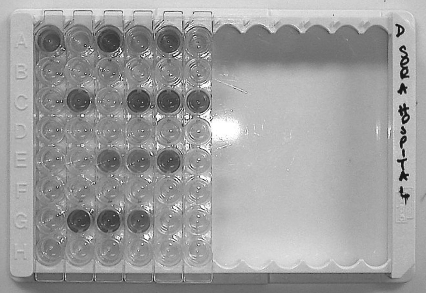
The need for rapid techniques and expert teams able to diagnose high numbers of samples in a short time arises when surveys are intended to cover large areas or to be performed in highly populated communities (Fig. 2). The use of coprological techniques enabling the simultaneous detection of Fasciola and other protozoan and helminth parasites is needed, mainly in community surveys of poor rural areas. Moreover, at least one quantitative diagnostic technique should be used to allow for the establishment of the appropriate treatment dose, because infection burdens in such areas may be high (50–400 epg) or very high (400–8000 epg). A threshold of 400 epg was established to decide on a mid-dose to avoid the risk of colic in massively infected subjects (WHO, 2007; Villegas et al. Reference Villegas, Angles, Barrientos, Barrios, Valero, Hamed, Grueningr, Ault, Montresor, Engels, Mas-Coma and Gabrielli2012; Valero et al. Reference Valero, Periago, Perez-Crespo, Angles, Villegas, Aguirre, Strauss, Espinoza, Herrera, Terashima, Tamayo, Engels, Gabrielli and Mas-Coma2012a). Unfortunately, intensity by egg count is usually not assessed in fascioliasis surveys.
ELISA plate showing positive results obtained with a specific and rapid serological test applied to samples from many patients simultaneously diagnosed in the regional hospital of Quy Nhon, Vietnam.
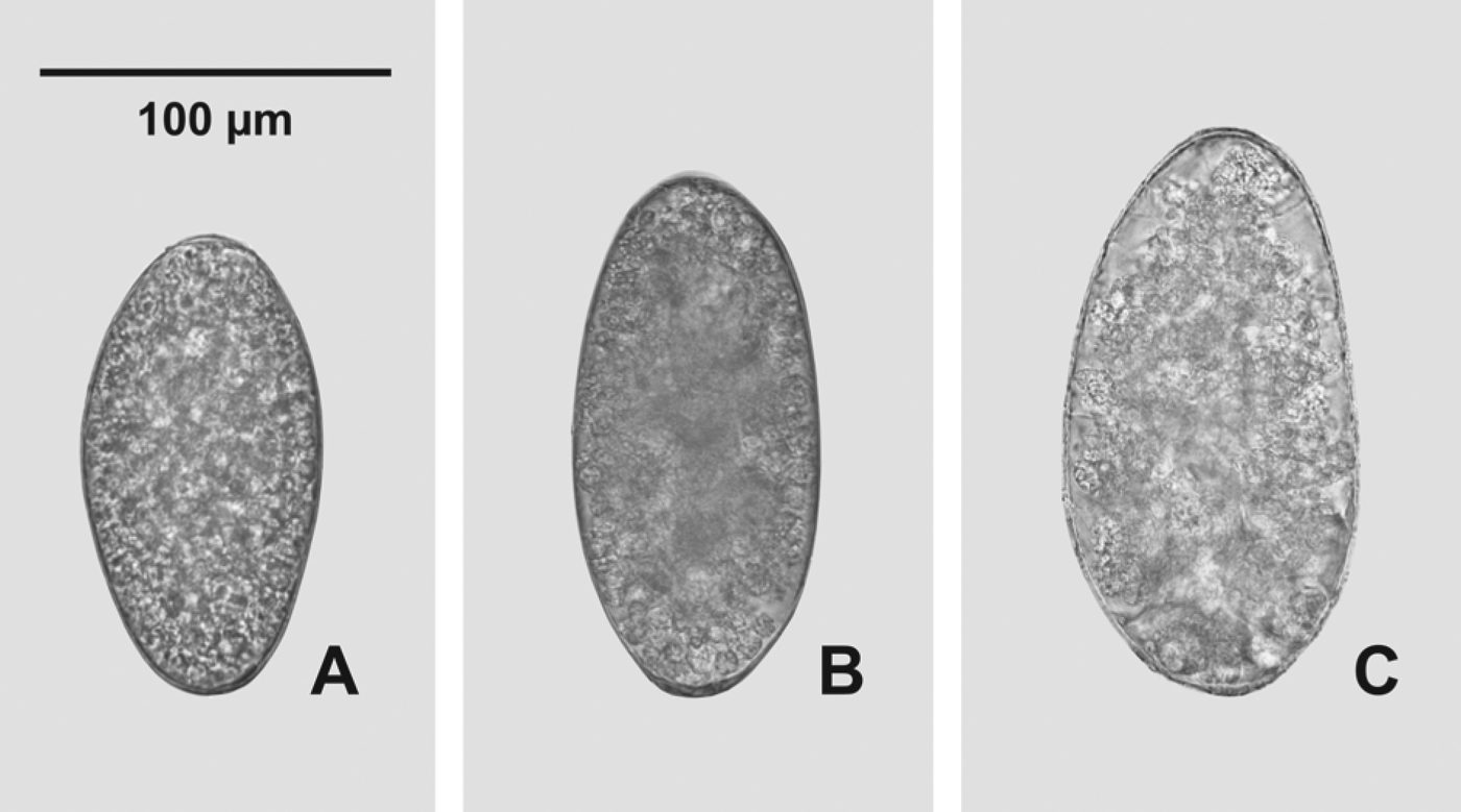
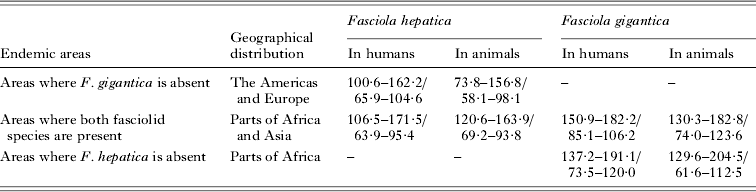
In several regions of Africa and Asia it should be considered that F. hepatica and F. gigantica coexist (Mas-Coma et al. Reference Mas-Coma, Valero and Bargues2009a) and that the differential specific diagnosis is of interest because of their different pathology, transmission, epidemiology and control measures. Although egg size may be helpful (Valero et al. Reference Valero, Perez-Crespo, Periago, Khoubbane and Mas-Coma2009a; Mas-Coma et al. Reference Mas-Coma, Agramunt and Valero2014), unfortunately all indirect immunological techniques have proved to be useless for differentiation between both fasciolid species, so that nowadays only certain molecular techniques are able to do so due to the problem of hybrids/intermediate forms (Mas-Coma et al. Reference Mas-Coma, Valero and Bargues2009a).
Anamnesis appears to be rarely useful in rural endemic areas because of the usual ingestion of different freshwater vegetables, local foods and beverages (Mas-Coma et al. Reference Mas-Coma, Angles, Esteban, Oviedo and Buchón1995; Mas-Coma, Reference Mas-Coma, Cotruvo, Dufour, Rees, Bartram, Carr, Cliver, Craun, Fayer and Gannon2004; Ashrafi et al. Reference Ashrafi, Valero, Massoud, Sobhani, Solaymani-Mohammadi, Conde, Khoubbane, Bargues and Mas-Coma2006a). Results of questionnaires on diet and food traditions are of low credibility when dealing with children, but may give interesting general information when obtained from respective parents (Zumaquero-Rios et al. Reference Zumaquero-Rios, Sarracent-Perez, Rojas-Garcia, Rojas-Rivero, Martinez-Tovilla, Valero and Mas-Coma2013).
Finally, verification of successful treatment in community surveys performed in rural endemic areas is often difficult. The need for highly sensitive techniques allowing for appropriate monitoring in post-survey treatments and control campaigns, and subsequent surveillance has been highlighted in recent years (Valero et al. Reference Valero, Periago, Perez-Crespo, Angles, Villegas, Aguirre, Strauss, Espinoza, Herrera, Terashima, Tamayo, Engels, Gabrielli and Mas-Coma2012a). Additional problems in rural areas include the ‘crowding effect’ (Valero et al. Reference Valero, De Renzi, Panova, Garcia-Bodelon, Periago, Ordoñez and Mas-Coma2006b), more- or less-frequent re-infections due to the high disease transmission risk, and the invasive phase repeatedly overlapping the biliary chronic phase observed mainly in children (Valero et al. Reference Valero, Santana, Morales, Hernandez and Mas-Coma2003, Reference Valero, Navarro, Garcia-Bodelon, Marcilla, Morales, Garcia, Hernandez and Mas-Coma2006a, Reference Valero, Girones, García-Bodelon, Periago, Chico-Calero, Khoubbane, Fresno and Mas-Coma2008).
DIAGNOSIS IN DIFFERENT SITUATIONS
For both the selection of the appropriate diagnostic technique and the correct interpretation of the results obtained from the technique applied, the following aspects should be considered in the diagnosis: (i) the intra-organic migration followed by the juvenile fasciolid after infection by metacercariae ingestion until reaching the definitive hepatic microhabitat of the biliary canals and gall bladder where fluke maturation will occur and egg shedding will start and (ii) the related different clinical pictures of the disease periods (Mas-Coma and Bargues, Reference Mas-Coma and Bargues1997; Mas-Coma et al. Reference Mas-Coma, Agramunt and Valero2014).
Similarly, the heterogeneity of fascioliasis regarding transmission patterns and epidemiological situations throughout (Mas-Coma, Reference Mas-Coma2005; Mas-Coma et al. Reference Mas-Coma, Bargues and Valero2005, Reference Mas-Coma, Valero and Bargues2009a) should also be taken into account in both the diagnosis of individual patients and the design of community surveys and interpretation of their results.
Individual diagnosis in different clinical situations
Several clinical situations may be taken into account when deciding which method and technique might be most appropriate for the diagnosis of a patient:
Eggs in transit
People who have ingested infected domestic animal livers (mainly cattle, sheep and pig) a short time before testing may reflect ‘false’ fascioliasis when the fluke eggs are found in their stools (Stork et al. Reference Stork, Venables, Jennings, Beesley, Bendezu and Capron1973; Ragab and Farag, Reference Ragab and Farag1978; Campo et al. Reference Campo, Marcen, Gonzalez, Milazzo, Pascual and Ponce1980). Such spurious infection may give rise to false positives. Although an expert microscopist may differentiate the somewhat degenerated aspect of ‘eggs in transit’ from ‘normal eggs’, to avoid confusion in such cases diagnosis requires placing the patient on a liver-free diet and performing repeated follow-up stool examinations.
Invasive and biliary periods of the disease
In humans, the incubation phase (from a few days to 2–3 months) is shorter than the prepatent period (at least 3–4 months), meaning that infected subjects may present clinical findings long before eggs could be found in the stools (Mas-Coma and Bargues, Reference Mas-Coma and Bargues1997), making egg finding in faeces as an early diagnosis tool impossible. Thus, egg finding techniques become useful only 3–4 months post-infection (p.i.). Serological tests allow for an earlier detection of infection and are therefore the appropriate techniques for patient diagnosis in the invasive phase. A serological test may allow for the detection of anti-Fasciola antibodies as early as 2 weeks p.i. although it does not differentiate between current or past infection, whereas a positive antigen-detecting test indicates an active infection although only from the 8th week p.i., that is around 2 months before beginning of egg shedding (see below).
Asymptomatic cases
Patients without symptoms have been reported, sometimes even in as relatively high percentages as 15% (Haseeb et al. Reference Haseeb, El-Shazly, Arafa and Morsy2003a). Several of these patients do moreover not present eggs in faeces (Arjona et al. Reference Arjona, Riancho, Aguado, Salesa and Gonzalez-Macias1995; Adachi et al. Reference Adachi, Kotani, Shimizu, Tanaka and Okada2005; Demir et al. Reference Demir, Ellidokuz, Degirmenci, Yücel and Gökçe2005). Eosinophilia, invasive techniques such as laparoscopy with liver biopsy, and non-invasive ones such as ultrasonography may be useful for guiding towards a confirmative diagnosis of such patients by means of a serological test and/or coproantigen test.
Egg output dynamics
In livestock the absence of a direct relationship between number of flukes present and number of eggs shed is well known. Egg production oscillations in trematodes in general and Fasciola in particular throughout their lifespan is also well known (Valero et al. Reference Valero, Panova, Comes, Fons and Mas-Coma2002). In humans, Fasciola egg output number and dynamics are unfortunately unknown. Different possibilities such as intermittent and very low egg shedding, especially in infection burdens of only one or a few fluke adults as well as in old infections, may not be ruled out and may be overlooked when analysing only one stool sample or when applying insufficiently sensitive techniques. The stimulation of the contraction of the gall bladder, where eggs accumulate, by administration of appropriate food (cholagogues) increases the appearance of eggs in stools thereafter. This has been useful in surveys of schoolchildren (R. Angles, La Paz, unpublished data). Significant intra-specimen variations in six Kato–Katz slides from a single stool sample were found, while the inter-specimen variation was almost negligible in daily exams for 5 consecutive days (El-Morshedy et al. Reference El-Morshedy, Shehab, Zaki and Farag2002). However, pronounced epg variations were found in children who were followed-up during a longer period in which no drug for treatment was available (unpublished data). Consequently, multiple and repeated follow-up stool examinations may be needed in such cases.
Biliary cases without egg shedding due to immature flukes or liver abscesses
Except in human fascioliasis endemic regions, humans are generally believed to be a non-suitable host for Fasciola. Consequently, sporadic or isolated case reports in animal endemic areas (Mas-Coma et al. Reference Mas-Coma, Bargues, Esteban and Dalton1999a) highlight the possibility of hepatic infections by flukes unable to attain maturity in human subjects. This may explain patients in whom eggs could never be found despite being in the biliary phase of the disease.
In many human infections, eggs are often not found in stools even after multiple faecal examinations (Hillyer, Reference Hillyer and Dalton1999). In Europe for instance, the diagnosis of human fascioliasis is frequently established using serological tests, because the detection of F. hepatica eggs in stools is not always possible. Thus, in the Limousin region, central France, egg detection in stools was positive in only 27·6% of a total of 711 subjects with fascioliasis (Rondelaud et al. Reference Rondelaud, Dreyfuss and Vignoles2006). In Asia, a similar situation is found in Vietnam, where cases have been ascribed to F. gigantica (De et al. Reference De, Murrell, Cong, Cam, Chau, Toan and Dalsgaard2003) and egg detection in stools was positive in only 16·9% of a total of 249 infected patients (De et al. Reference De, Nguyen, Dang, Nguyen, Le and Trieu2005). In a human fascioliasis mesoendemic area recently described in Mexico, where individual intensities in children were low (24–384 epg), eggs were only found in 14 out of a total of 50 children positive by coproantigen test (Zumaquero-Rios et al. Reference Zumaquero-Rios, Sarracent-Perez, Rojas-Garcia, Rojas-Rivero, Martinez-Tovilla, Valero and Mas-Coma2013).
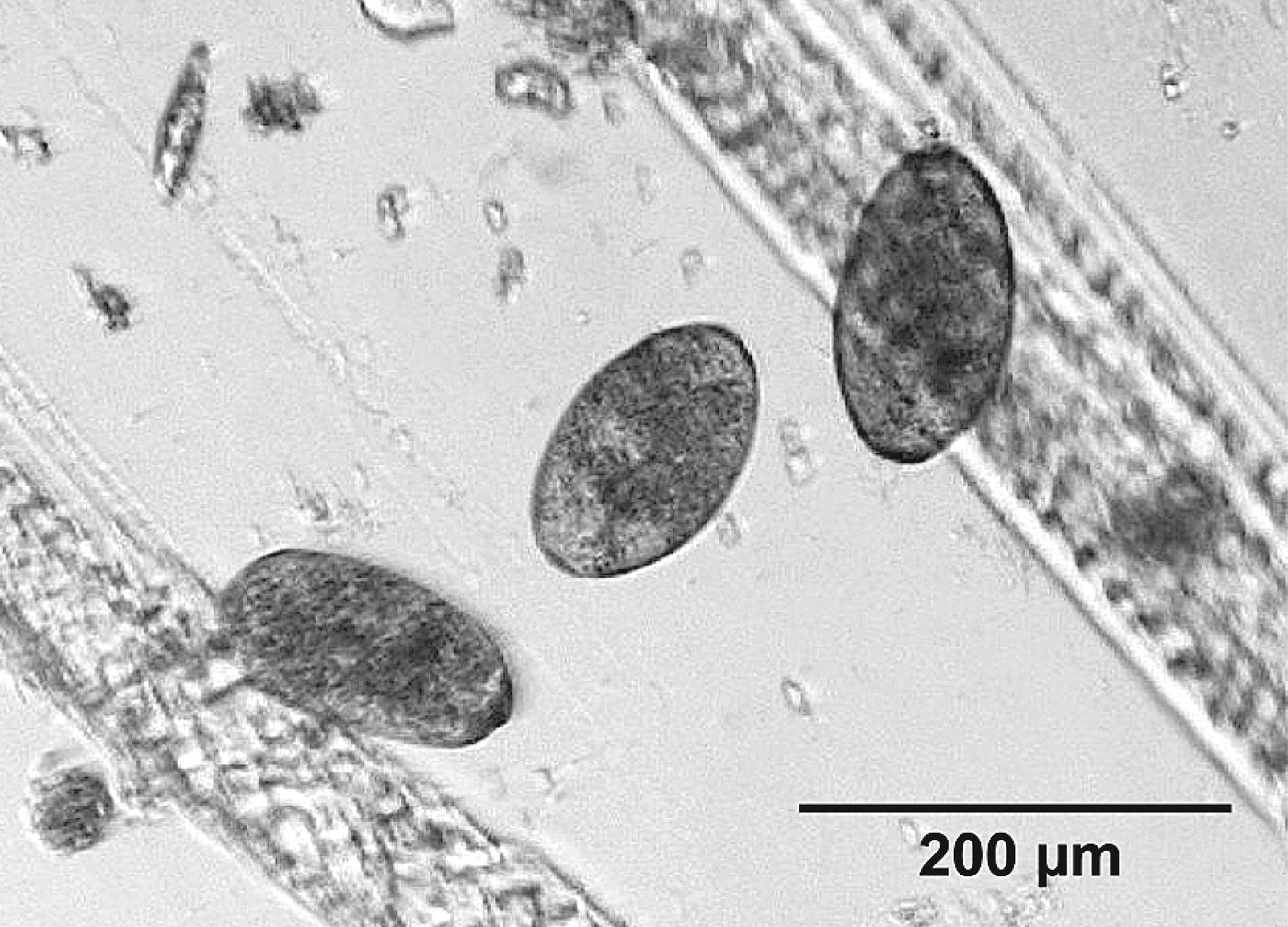
Rare cases have also been reported in which a fluke reached maturity in the liver but its ‘encapsulation’ in a hepatic abscess did not allow eggs to reach the biliary canals and be shed with faeces (Fig. 3).
Eggs of Fasciola hepatica found in a liver abscess of a male adult Italian patient who was shedding no eggs in stools.
Re-infections and overlap of acute and chronic phases
Mainly in human fascioliasis endemic areas where the infection risk is high, but also in individual patients whose infection is related to the habit of usually consuming freshwater vegetables in animal fascioliasis endemic areas, subjects may be re-infected once already in the biliary period due to previous infection. In such cases, a relapse of the clinical characteristics of the invasive phase may appear and overlap with symptoms of the chronic period, as infected hosts do not develop resistance due to the rapid and potent ability of fasciolids to suppress the immune response (Girones et al. Reference Girones, Valero, Garcia-Bodelon, Chico-Calero, Punzon, Fresno and Mas-Coma2007; Dalton et al. Reference Dalton, Robinson, Mulcahy, O'Neill and Donnelly2013). In this group of patients, an increase of egg shedding may occur once the migrating juveniles reach the biliary canals, mature and start egg production.
Long-term relapses of the disease
Fascioliasis may have been overlooked due to milder symptomatology or misdiagnosis with patients going on to suffer a relapse including impressive manifestations and even death after many years of chronic infection (Ruggieri et al. Reference Ruggieri, Correa and Martínez1967; Correa et al. Reference Correa, Ruggieri and Martinez1969; Auer et al. Reference Auer, Kristoferitsch, Picher and Wessely1982; Kristoferitsch et al. Reference Kristoferitsch, Wessely, Auer and Picher1982). It should be considered that flukes may survive up to 9–13·5 years in the human host (Mas-Coma et al. Reference Mas-Coma, Bargues and Valero2005).
In Argentina, the long time elapsed between the appearance of symptoms and confirmation of infection by appropriate diagnosis has been emphasized in many patients: nearly four years in average, including patients having suffered from symptoms for 10 or more years without diagnosis (Mera y Sierra et al. Reference Mera y Sierra, Agramunt, Cuervo and Mas-Coma2011). This suggests either infected subjects had not sought professional diagnosis due to mild symptoms caused by low fluke burdens and/or misdiagnosis of patients due to the non-pathognomonic clinical picture, easily confused with other diseases when the patient attends a health centre not used to dealing with fascioliasis (La Pook et al. Reference La Pook, Magun, Nickerson and Meltzer2000).
The breakage of multiple old liver abscesses, aberrant specimens surviving as immature flukes for long periods, and hypersensitivity of the Arthus reaction type appear to underlie in these cases (Mas-Coma et al. Reference Mas-Coma, Agramunt and Valero2014). In such cases, eggs may be found in stools, but sometimes not when the flukes involved in the pathogenic picture are ectopic migrants. Neither do eggs appear in stools from patients presenting clinical disorders and biological abnormalities after years from the last date of fascioliasis treatment. Such symptoms appearing just after treatment and persisting afterwards up to 2–18 years were suggested to be due to the presence of persistent hepatic lesions following fascioliasis treatment (Rondelaud et al. Reference Rondelaud, Dreyfuss and Vignoles2006).
Ectopic infections
In fascioliasis, juvenile flukes sometimes deviate during migration from the intestine to the liver and enter other organs. Ectopic localization of the parasite may cause a confusing clinical presentation in both the acute and chronic infections. Ectopic lesions most frequently reported are in the gastrointestinal tract and subcutaneous tissue. Other ectopic locations described include: heart, blood vessels, the lung and pleural cavity; brain; dorsal spine; orbit; abdominal wall; stomach, caecum and appendix; pancreas; spleen; peritoneum; inguinal nodes; cervical node; blood vessels; skeletal muscle; and epididymis. Ectopic manifestations usually appear shortly after infection, although exceptions have been reported. Such ectopic flukes achieve maturity only very rarely (Mas-Coma et al. Reference Mas-Coma, Agramunt and Valero2014). No eggs in stools may be found when such ectopic lesions appear during the invasive phase, which is usually the case, but positive faecal samples may be found when in the biliary phase. Serological tests are thus the techniques of choice for such cases. Imaging techniques which may help guide towards fascioliasis diagnosis in several of the aforementioned localizations are unfortunately not usually available in remote poor rural areas of developing countries.
Diagnosis in different epidemiological situations
Human fascioliasis is very heterogeneous due to the different epidemiological situations and transmission patterns known in different countries (Mas-Coma et al. Reference Mas-Coma, Bargues, Esteban and Dalton1999a, Reference Mas-Coma, Esteban and Barguesb, Reference Mas-Coma, Bargues and Valero2005, Reference Mas-Coma, Valero and Bargues2009a; Mas-Coma, Reference Mas-Coma2005). This heterogeneity should be considered when deciding which diagnostic method(s) and technique(s) would be more appropriate depending on the area the patient(s) comes from or where the surveys should be implemented. A method and a technique may be useful in a given area but not in another. Unfortunately, only a few have been evaluated for different epidemiological situations and transmission patterns (Valero et al. Reference Valero, Periago, Perez-Crespo, Rodriguez, Perteguer, Garate, Gonzalez-Barbera and Mas-Coma2012b). Moreover, the use of only one technique may not always be sufficient and an appropriate combination of two or more techniques should be employed.
Different fasciolids
In areas of Europe, the Americas, Australia and the African Maghreb only F. hepatica is present and in western Africa there is only F. gigantica. The presence of only one of the two fasciolids is due to the lymnaeid species present in the aforementioned regions, only belonging to vector species specific for F. hepatica or F. gigantica, respectively (see details of this selective geographical distribution in Mas-Coma et al. Reference Mas-Coma, Valero and Bargues2009a). There is therefore no need for differential diagnosis when dealing with subjects inhabiting those regions, except in cases of immigrants from and travellers to endemic regions where the distribution of the two fasciolid species overlap.
However, there is an overlap of the geographical distribution of the two Fasciola species in eastern and southern Africa, as well as in many regions of Asia. In these regions, two different overlap situations can be distinguished (Mas-Coma et al. Reference Mas-Coma, Valero and Bargues2009a): (i) local overlap, in places where climatic characteristics throughout the year enable the coexistence of Galba and Radix species in the same locality, nearby water bodies and even sometimes the same water body. This allows transmission and consequent infection of livestock and humans living sedentarily in the locality by both F. hepatica and F. gigantica; (ii) Zonal overlap, in areas with different altitudes so that highlands offer the necessary cold-mild weather conditions for Galba truncatula and F. hepatica, and lowlands offer the warm-hot climate necessary for Radix species and F. gigantica. Thus, definitive hosts become co-infected by both fasciolids when moving from lowlands to neighbouring highlands and vice versa (animals because of interzonal transhumance, transportation and trade; humans when moving around for various reasons). Egypt is an example of local overlap, whereas Iran and Pakistan are examples of zonal overlap (see more details on local/zonal overlaps in Mas-Coma et al. Reference Mas-Coma, Valero and Bargues2009a).
In both kinds of overlaps, hybrid intermediate adult and egg forms are found, with higher and lower frequency in local and zonal overlap areas, respectively. Thus, techniques allowing for differential diagnosis are recommended for both local and zonal overlap areas given the different transmission characteristics, pathogenicity and control measures for one or other fasciolid species (Mas-Coma et al. Reference Mas-Coma, Valero and Bargues2009a). It should be considered here that immunological techniques (serological and coproantigen tests) do not differentiate between the two species, and that egg-finding coprological techniques may sometimes not be sufficient (Valero et al. Reference Valero, Perez-Crespo, Periago, Khoubbane and Mas-Coma2009a; Mas-Coma et al. Reference Mas-Coma, Agramunt and Valero2014). Only specific molecular techniques allow for such a differential diagnosis between F. hepatica, F. gigantica and hybrid intermediate forms. Morphological phenotyping of adult flukes may also be applied if parasites can be obtained with invasive methods, although this is also prone to difficulty and time-consuming, and may sometimes furnish inconclusive results if specimens are intermediate forms and their number too few (Mas-Coma et al. Reference Mas-Coma, Valero and Bargues2009a).
Situations in human fascioliasis endemic areas
Most of these situations are in developing countries, although they have also been infrequently described in impoverished remote rural areas of developed countries. Different prevalences and intensities are characteristic features in the human hyper-, meso- and hypoendemic areas (Mas-Coma et al. Reference Mas-Coma, Bargues, Esteban and Dalton1999a, Reference Mas-Coma, Esteban and Barguesb, Reference Mas-Coma, Bargues and Valero2005, Reference Mas-Coma, Valero and Bargues2009a). Whereas none or only very low differences may be obtained between prevalence results from immunological and direct coprological techniques in human hyperendemic areas with very high prevalences such as the Northern Bolivian Altiplano (Valero et al. Reference Valero, Periago, Perez-Crespo, Angles, Villegas, Aguirre, Strauss, Espinoza, Herrera, Terashima, Tamayo, Engels, Gabrielli and Mas-Coma2012a; Esteban et al. unpublished data), there may be differences between results from immunological and direct coprological techniques when in meso- and hypoendemic areas, and also hyperendemic areas with not very high prevalences. In the latter areas, immunological techniques, whether serological or coproantigen tests, furnish more accurate prevalence results (Espinoza et al. Reference Espinoza, Maco, Marcos, Saez, Neyra, Terashima, Samalvides, Gotuzzo, Chavarry, Huaman, Bargues, Valero and Mas-Coma2007; Valero et al. Reference Valero, Periago, Perez-Crespo, Angles, Villegas, Aguirre, Strauss, Espinoza, Herrera, Terashima, Tamayo, Engels, Gabrielli and Mas-Coma2012a; Zumaquero-Rios et al. Reference Zumaquero-Rios, Sarracent-Perez, Rojas-Garcia, Rojas-Rivero, Martinez-Tovilla, Valero and Mas-Coma2013).
Moreover, highly sensitive diagnostic techniques are needed for areas presenting low prevalences and intensities. Additionally, in human fascioliasis endemic areas there are adult subjects who may have been infected for many years (Esteban et al. Reference Esteban, Flores, Aguirre, Strauss, Angles and Mas-Coma1997a, Reference Esteban, Flores, Angles, Strauss, Aguirre and Mas-Comab, Reference Esteban, Flores, Angles and Mas-Coma1999). The adult flukes infecting them may be very old and consequently no longer shedding eggs whilst even eosinophilia may have become negligible.
In human fascioliasis endemic areas in general, children presenting first symptoms of the invasive phase do not usually attend a health centre for diagnosis, and if they do they are usually misdiagnosed because of absence of eggs in stools and lack of serological diagnostics, especially in rural health centres. Thus, children are the main target for epidemiological assessment and coprological methods should be used to avoid community disturbance due to blood extraction procedures. Inhabitants of remote rural endemic areas are usually reluctant to give blood samples, and if successful in convincing them for one occasion, this usually poses problems for subsequent studies.
Epidemics of human fascioliasis have also been described in human fascioliasis endemic areas. In such outbreaks involving a prevalence increase, epidemics are usually related to previous climatic conditions having favoured both the parasite and the snail life cycles (Mas-Coma et al. Reference Mas-Coma, Valero and Bargues2008, Reference Mas-Coma, Valero and Bargues2009b), and also to anthropogenic modifications of the environment (Afshan et al. Reference Afshan, Fortes-Lima, Artigas, Valero, Qayyum and Mas-Coma2014). Such epidemics can take place in hypoendemic, mesoendemic and hyperendemic areas (Mas-Coma et al. Reference Mas-Coma, Bargues, Esteban and Dalton1999a, Reference Mas-Coma, Esteban and Barguesb, Reference Mas-Coma, Bargues and Valero2005, Reference Mas-Coma, Valero and Bargues2009a) and may show a parallelism with outbreaks in livestock, early detection of which may help in forecasting the human epidemic.
Situations in animal fascioliasis endemic areas
This is the usual situation in developed countries, but can also appear in developing countries where human fascoliasis does not appear to be a serious public health problem. People usually present for diagnosis in hospitals and health centres as soon as the first symptoms appear. Therefore, patients are usually diagnosed in the invasive period or beginning of the biliary period. Consequently, there are usually no eggs in stools, but anamnesis and eosinophilia provide valuable evidence and the diagnosis should preferentially rely on serodiagnosis. An immunological test is also better due to the fact that there may be fluke strains not adapted to humans and therefore not able to reach maturity and produce eggs in the patients in given areas.
Patients are usually isolated and sporadic cases in animal fascioliasis endemic areas (Mas-Coma et al. Reference Mas-Coma, Bargues, Esteban and Dalton1999a, Reference Mas-Coma, Esteban and Barguesb, Reference Mas-Coma, Bargues and Valero2005, Reference Mas-Coma, Valero and Bargues2009a). However, infection reports of immigrants from endemic areas (Auer et al. Reference Auer, Kristoferitsch, Picher and Wessely1982; Pelletier et al. Reference Pelletier, Chosidow, Rogeaux, Lenoir, Piette, Frances and Herson1995; Graham et al. Reference Graham, Brodie and Weller2001; MacLean et al. Reference Mac Lean, Varghese and Graeme-Cook2002; Noyer et al. Reference Noyer, Coyle, Werner, Dupouy-Camet, Tanowitz and Wittner2002; Turrientes et al. Reference Turrientes, Saenz de Sta. Maria, Ceballos, Diaz, Bareno, Muro, Pardo and Lopez-Velez2004; Cheung et al. Reference Cheung, Enns, Romney, Reynolds and Amar2005; Channabasappa et al. Reference Channabasappa, Vettiankal, Riles and Attar2006; Fullerton et al. Reference Fullerton, Vitale and Vitale2006; Morse et al. Reference Morse, McLean and Bedi2006; Alatoom et al. Reference Alatoom, Sheffield, Gander, Shaw and Cavuoti2008; Chand et al. Reference Chand, Herman, Partridge, Hewitt and Chiodini2009; Novo-Veleiro et al. Reference Novo-Veleiro, Martín-Sánchez, Elcuaz-Romano, Muro, Afonso-Rodríguez, García-Bardeci, Bordes-Benítez, Carranza-Rodríguez, Hernández-Cabrera, Alvela-Suárez and Pérez-Arellano2012), and travellers (professionals, business people, tourists) and military personnel having visited endemic areas are not uncommon in developed countries.
The rarity of Fasciola infection in those areas makes diagnosis challenging and often unrecognized. It may also lead to misdiagnosis or late diagnosis when local physicians are not aware of fascioliasis entering into the diagnostic probabilities related to the clinical picture presented (La Pook et al. Reference La Pook, Magun, Nickerson and Meltzer2000; Mera y Sierra et al. Reference Mera y Sierra, Agramunt, Cuervo and Mas-Coma2011).
Human epidemics have also been described in animal fascioliasis endemic areas. These outbreaks appear in zones where previous human reports have always been isolated and sporadic. Such outbreaks usually concern a very few subjects infected from the same contaminated source (family or small group reports; contaminated wild, home-grown or commercially grown watercress or other metacercariae-carrying vegetables) (Mas-Coma et al. Reference Mas-Coma, Bargues, Esteban and Dalton1999a, Reference Mas-Coma, Esteban and Barguesb, Reference Mas-Coma, Bargues and Valero2005, Reference Mas-Coma, Valero and Bargues2009a). Outbreaks of this type usually appear following heavy rainfall in the previous years or seasons (Carme et al. Reference Carme, Niel, Danis and Gentilini1978; Perez Rodríguez et al. Reference Perez Rodríguez, Casero Valladares, Martinez Rodriguez and Perez Martin1986; Ripert et al. Reference Ripert, Tribouley, Luong Dinh Giap, Combe and Laborde1987; Cadel et al. Reference Cadel, Barbier, Duhamel and Georges1996). Sometimes seasonality is related to ingestion of infected plants. For example, in France most human cases occurred during the October–April watercress season (Ripert et al. Reference Ripert, Tribouley, Luong Dinh Giap, Combe and Laborde1987), with a maximum from November to February (Rondelaud et al. Reference Rondelaud, Amat-Frut and Pestre-Alexandre1982). Unexpected problems of contamination of watercress cultures due to, for example, disease spread by an introduced sylvatic reservoir animal such as the nutria (Houin et al. Reference Houin, Moquet, Czeher, Vallois, Mas-Coma, Bargues, Esteban and Valero2004) appear related to the recent emergence of human fascioliasis in particular areas (Mailles et al. Reference Mailles, Vaillant, Schepens, Ajana, Capek, Ilef, Fillebeen, Lefort, Therouanne, Volant, Flavigny and De Valk2003, Reference Mailles, Capek, Ajana, Schepens, Ilef and Vaillant2006). In rural areas of developing countries, the best method to differentiate between an outbreak in an animal endemic area and a human endemic situation is mass diagnostic screening of schoolchildren.
Different transmission patterns
Different disease seasonalities have been described in human and also in animal fascioliasis endemic areas. Mono- and bi-seasonalities and permanent transmission may be distinguished. Diagnostic results are unavoidably influenced by these different local transmission patterns, mainly when working with direct coprological techniques allowing for egg finding (Valero et al. Reference Valero, Periago, Perez-Crespo, Angles, Villegas, Aguirre, Strauss, Espinoza, Herrera, Terashima, Tamayo, Engels, Gabrielli and Mas-Coma2012a). Intensities (epg) are higher in seasons when human subjects are at the beginning of the egg-shedding period. Consequently these seasons should be preferred when implementing mass screenings (Esteban et al. Reference Esteban, Flores, Angles and Mas-Coma1999; Afshan et al. Reference Afshan, Fortes-Lima, Artigas, Valero, Qayyum and Mas-Coma2014) to avoid overlooking cases with very low burdens. Serological antibody-detecting tests may be less influenced by seasonality, because of the perseverance of antibodies. Antigen-detecting techniques may also be influenced by seasonality, as antigen amounts, both faecal and circulating, are related to the burden. Seasonal infection data in livestock inhabiting the same area may be useful in establishing the best season for human surveys (Mas-Coma et al. Reference Mas-Coma, Angles, Esteban, Bargues, Buchon, Franken and Strauss1999c).
In areas of zonal overlap where hybridization is rare (Mas-Coma et al. Reference Mas-Coma, Valero and Bargues2009a), transmission by pure F. hepatica may occur in different months than transmission of pure F. gigantica, due to their different temperature thresholds, F. hepatica preferring milder months and F. gigantica warmer months (Afshan et al. Reference Afshan, Fortes-Lima, Artigas, Valero, Qayyum and Mas-Coma2014). In areas of local overlap where hybridization is common (Mas-Coma et al. Reference Mas-Coma, Valero and Bargues2009a), the existence of two transmission peaks, each corresponding to one Fasciola species, may become diluted and human infection and subsequent beginning of egg shedding after the prepatent period appear continuous throughout more months.
Social habits and cultural traditions
Human diet and behaviour related to livestock (local animal management traditions) have to be considered in prevalence surveys as well as ethnic aspects facilitating the participation (or not) in surveys of the inhabitants of the endemic area. In the Northern Bolivian Altiplano, the highest human hyperendemic area known, the Aymara inhabitants do not allow blood sampling due to religious customs. Thus all surveys in these areas have been made with coprological techniques (Esteban et al. Reference Esteban, Flores, Angles and Mas-Coma1999; Mas-Coma et al. Reference Mas-Coma, Angles, Esteban, Bargues, Buchon, Franken and Strauss1999c). In initial studies, the acceptance of inhabitants of given localities to furnish blood samples was obtained (Hillyer et al. Reference Hillyer, Soler de Galanes, Rodriguez-Perez, Bjorland, Silva de Lagrava, Guzman and Bryan1992) although very few additional ones could later be further obtained (Strauss et al. Reference Strauss, Angles, Esteban and Mas-Coma1997; O'Neill et al. Reference O'Neill, Parkinson, Strauss, Angles and Dalton1998). Since then, Aymaras of the endemic area have remained reluctant to give blood, and as a result all subsequent serological studies were repeatedly using the same old, few samples obtained for the aforementioned interventions (ignoring this fact has given rise to several misunderstandings in recent reviews). Thus, the present WHO control initiative is based only on coprological techniques (Villegas et al. Reference Villegas, Angles, Barrientos, Barrios, Valero, Hamed, Grueningr, Ault, Montresor, Engels, Mas-Coma and Gabrielli2012; Valero et al. Reference Valero, Periago, Perez-Crespo, Angles, Villegas, Aguirre, Strauss, Espinoza, Herrera, Terashima, Tamayo, Engels, Gabrielli and Mas-Coma2012a).
A contrasting situation, however, is described in a human fascioliasis endemic area recently described in Argentina (Carnevale et al. Reference Carnevale, Cabrera, Cucher, di Risio, Malandrini, Kamenetzky, Alazraqui, Etchart, Pantano and Velasquez2013), where inhabitants are reluctant to give stool samples, so that diagnostic studies relied only on serological tests (Malandrini, personal communication). This situation will also, in turn, pose problems for the verification of treatment success.
Surveys
The detection of human fascioliasis endemic areas from the 1990s posed new challenges for human fascioliasis diagnosis which had never been asked before. This included requests for easy and quick techniques able to be performed in the field and applied to a high number of subjects. In human fascioliasis endemic areas, a strategy is needed to facilitate the implementation of quantitative techniques allowing for epg counts and subsequent decision of appropriate treatment dose for each patient to avoid colic risks (WHO, 2007).
The need to look for co-infections and associations of fascioliasis with other protozoan and helminth infections indicates the convenience of selecting a diagnostic technique with the capacity to detect fascioliasis, protozoosis and other helminthiasis at the same time, in order to avoid including an additional diagnostic technique to the intervention design. Such a multiparasite diagnostic technique should be a coprological one, as no immunological technique available at present (serological, coproantigen) furnishes such an advantage (Esteban et al. Reference Esteban, Flores, Aguirre, Strauss, Angles and Mas-Coma1997a, Reference Esteban, Flores, Angles, Strauss, Aguirre and Mas-Comab, Reference Esteban, Gonzalez, Bargues, Sanchez, Naquira and Mas-Coma2002, Reference Esteban, Gonzalez, Curtale, Muñoz-Antoli, Valero, Bargues, El-Sayed, El Wakeel, Abdel-Wahab, Montresor, Engels, Savioli and Mas-Coma2003; Gonzalez et al. Reference Gonzalez, Esteban, Bargues, Valero, Ortiz, Naquira and Mas-Coma2011). Moreover, if the volume (weight) of stool sample used for the coprological analysis is measured from the beginning, it may additionally be useful for the quantification of eggs of Fasciola and other helminths. Such a combined method for surveying becomes crucial to assess the morbidity of human fascioliasis situations in endemic areas, mainly in children, given the immunosuppression-immunomodulation capacity of Fasciola and its pathogenic synergism with other pathogenic parasites (Mas-Coma, Reference Mas-Coma, Cotruvo, Dufour, Rees, Bartram, Carr, Cliver, Craun, Fayer and Gannon2004; Dalton et al. Reference Dalton, Robinson, Mulcahy, O'Neill and Donnelly2013).
Given the heterogeneity of human fascioliasis, diagnosis methods should suit the specific characteristics of each endemic area (simple extrapolation of measures successful in one country to another country, as suggested in recent reviews, should not be made). In Egypt, active detection of infected subjects is made by means of surveys in schoolchildren and locality inhabitants (Curtale et al. Reference Curtale, Hammoud, El Wakeel, Mas-Coma and Savioli2000, Reference Curtale, Mas-Coma, Hassanein, Barduagni, Pezzotti and Savioli2003a, Reference Curtale, Hassanein, Barduagni, Yousef, El Wakeel, Hallaj and Mas-Coma2007; Esteban et al. Reference Esteban, Gonzalez, Curtale, Muñoz-Antoli, Valero, Bargues, El-Sayed, El Wakeel, Abdel-Wahab, Montresor, Engels, Savioli and Mas-Coma2003) with selective treatment and follow-up applied to subjects diagnosed as positive (Curtale et al. Reference Curtale, Hassanein, El Wakeel, Mas-Coma and Montresor2003b). In Vietnam, passive detection of infected subjects is organized through the national network of health centres distributed throughout all endemic areas, so that patients are diagnosed, treated and followed-up in each hospital (WHO, 2008). In Bolivia, active detection of infected subjects is made by coprological surveys and control measures include yearly mass treatments with triclabendazole (Villegas et al. Reference Villegas, Angles, Barrientos, Barrios, Valero, Hamed, Grueningr, Ault, Montresor, Engels, Mas-Coma and Gabrielli2012). In Peru, active detection of infected subjects is made by means of surveys with both serological and coprological tests (Valero et al. Reference Valero, Periago, Perez-Crespo, Angles, Villegas, Aguirre, Strauss, Espinoza, Herrera, Terashima, Tamayo, Engels, Gabrielli and Mas-Coma2012a) with control measures including similar periodic mass treatments.
Qualitative and quantitative diagnosis
In human fascioliasis, qualitative diagnostic techniques may be of interest not only for the detection of patient infection by the liver fluke, but also for the differential diagnosis between F. hepatica, F. gigantica and hybrid intermediate forms. For such purpose, only coprological and invasive techniques providing eggs and/or adult flukes which may subsequently be appropriately analysed phenotypically and genotypically are useful (Mas-Coma et al. Reference Mas-Coma, Valero and Bargues2009a). The aforementioned techniques may also be useful when providing living materials of the fasciolids (viable eggs, living adult flukes) which may be used to obtain laboratory isolates to assess resistance to drugs. Unfortunately, present knowledge on drug resistance in fascioliasis is not sufficient to allow development of the necessary molecular tests for such a screening, which means that drug resistance has still to be established by means of experimental assays.
In contrast to serological and coproantigen techniques, direct coprological assays have the added value of allowing for burden quantification by epg counts. Although it is well known that in animal fascioliasis a direct relationship between adult fluke number and epg counts does not appear to be robustly accurate, at present there is no other non-invasive method enabling quantification. Therefore, epg is the only measure used to indirectly assess burden, intensity-related pathogenicity and post-treatment colic risk. Unfortunately, epg counts are lacking in the majority of publications on case reports and community surveys. Although epg counts were considered unnecessary in the past, because patients affected by a massive infection had been reported very rarely, we know today that very high intensities may be found in human endemic areas, mainly in children and females. Hence, quantification should be applied to subjects from these areas to verify post-treatment colic risk by comparing with the 400 epg threshold (WHO, 2007). Moreover, epg counts may provide valuable information about individual re-infections and family risks, as well as the status of infection risk in the field around given localities, and are thus useful indications for prevention and control.
Although all coprological techniques may be implemented in a quantitative way, by simply starting from a known quantity of stool sample, only a very few have been developed to essentially be used for quantification. Unfortunately these quantitative techniques show a low sensitivity which requires repetitive analyses to reach sufficient sensitivity levels.
Post-treatment monitoring and post-control surveillance
Diagnosis for monitoring and surveillance requires techniques fulfilling specific capacities. The first requirement is the ability to confirm parasite eradication/removal of infection shortly following successful treatment. In patients in the biliary period who are shedding eggs, the post-treatment disappearance of eggs in stools is the most usual feature looked for when using coprological diagnostic techniques. If the patient was in the invasive period and therefore no eggs were found before treatment, disappearance of eosinophilia and return of hepatic function tests to normal values, besides recovery from symptoms and signs, are results on which to rely (Mas-Coma et al. Reference Mas-Coma, Agramunt and Valero2014).
Worth mentioning here is the fact that serological techniques are not recommended for post-treatment monitoring of patients, given the relatively long time they need to return to negative values (usually at least 4–5 months). There are, however, a few reports on shorter times depending on the antigen. In one study, the IgG ELISA results became negative by the second month of triclabendazole treatment in 40% of the cured cases (Apt et al. Reference Apt, Aguilera, Vega, Miranda, Zulantay, Perez, Gabor and Apt1995). Other studies showed that after the first month of treatment, IgG ELISA became negative in 83·3% of cases, reaching 95% after 4 months (Tawfeer and Hussein, Reference Tawfeer and Hussein2000). In contrast, coproantigen tests appear to be very useful for such an endeavour when the patient was treated in the biliary period of the disease, because specific antigens disappear rapidly in stools following successful treatment (Valero et al. Reference Valero, Ubeira, Khoubbane, Artigas, Muiño, Mezo, Perez-Crespo, Periago and Mas-Coma2009b, Reference Valero, Periago, Perez-Crespo, Angles, Villegas, Aguirre, Strauss, Espinoza, Herrera, Terashima, Tamayo, Engels, Gabrielli and Mas-Coma2012a; Zumaquero-Ríos et al. Reference Zumaquero-Rios, Sarracent-Perez, Rojas-Garcia, Rojas-Rivero, Martinez-Tovilla, Valero and Mas-Coma2013).
A second desirable attribute of a diagnostic technique useful for post-treatment monitoring would be the capacity to detect infection at very low burdens, especially in patients for whom the drug dose applied was not sufficient to kill all flukes present. In coprological procedures, the solution to such a situation is implementing repeated stool analyses over several days immediately after treatment. Coproantigen tests may simplify that task, as they are able to detect infection in subjects shedding very few or no eggs in stools (Valero et al. Reference Valero, Periago, Perez-Crespo, Angles, Villegas, Aguirre, Strauss, Espinoza, Herrera, Terashima, Tamayo, Engels, Gabrielli and Mas-Coma2012a; Zumaquero-Ríos et al. Reference Zumaquero-Rios, Sarracent-Perez, Rojas-Garcia, Rojas-Rivero, Martinez-Tovilla, Valero and Mas-Coma2013). This does not mean, unfortunately, that coproantigen tests may not sometimes fail, for unknown reasons (Valero et al. Reference Valero, Periago, Perez-Crespo, Angles, Villegas, Aguirre, Strauss, Espinoza, Herrera, Terashima, Tamayo, Engels, Gabrielli and Mas-Coma2012a), so confirmation by referring to eosinophilia, hepatic function tests, symptoms and signs is always recommended.
For the monitoring of mass treatments after or during community control initiatives, diagnostic techniques must fulfil not only the aforementioned two requirements, but also be useful for fieldwork in surveys and fast to perform, to allow for the screening of large numbers of subjects in a short time. Similar requirements are needed for general surveillance in human fascioliasis endemic areas where different control measures may be systematically applied:
– for short-term impact indicators, a quantitative method such as Kato–Katz may be applied to assess a decrease of intensity and a coproantigen test to assess the decrease of prevalence;
– for medium-term impact indicators, new infections in children (or another designated cohort in countries where fascioliasis does not concentrate in children, such as in Europe, Iran or Vietnam) may be determined by Kato–Katz and coproantigen tests, to which a serological test may be added to assess the decrease of antigenaemia (as for instance in European countries).
Annual data may be consulted to avoid bias related to seasonality, so that seasonal infection risk may be taken into account to define which month(s) are more appropriate for surveys in sentinel schools.
SAMPLE MANAGEMENT: COLLECTION, FIXATION, TRANSPORT AND PRESERVATION
Up to the 1980s, almost all patients affected by fascioliasis in developed countries attended a hospital or health centre for diagnosis after symptom appearance. In these health centres, the stool or blood sample could be directly and easily obtained. In the new scenario from the 1990s, aspects not considered for fascioliasis diagnosis previously should now be taken into account. Such aspects may include (i) long distances between the locality inhabited by the patient in a remote rural area and the laboratory where the sample may be analysed, (ii) field characteristics of the area to be surveyed, or (iii) time needed to analyse a large number of samples obtained in a survey.
Thus, decisions should be taken on sample procedures, depending on (i) situations of individual patient diagnosis or surveys, including (ii) sample collection procedures, (iii) processing of fresh faecal samples or immediate fixation after biological sample collection, (iv) adequate transport of the samples to ensure arrival in good condition to the laboratory and (v) appropriate preservation of the samples if it is not possible to process and analyse immediately after arrival in the laboratory or when the large number of samples arrived will give rise to several days’ work of analysis.
A number of textbooks and laboratory manuals cover these crucial aspects of sample collection, fixation, transport and preservation for diagnosis of parasitic diseases in detail (e.g. Ash and Orihel, Reference Ash and Orihel1987; WHO, 1991). However, several characteristics particular to stool and blood samples for human fascioliasis diagnosis should be emphasized.
Procedures for stool samples
Although fascioliasis does not cause diarrhoea by itself, several of the diseases usually associated with fascioliasis do, such as giardiasis. Liquid specimens should be examined within 30 min of passage, or the material should be placed in an appropriate preservative. Formed specimens may be kept at room temperature for several hours before examination, but should be examined the same day. If the specimens are to be sent to another laboratory for examination, they must be fixed (if sending will take time) or appropriately preserved whether refrigerated or frozen in portable equipment (if arrival at the laboratory will only take a relatively short time) to keep the parasites in good condition.
To optimize individual diagnosis of human fascioliasis, multiple specimens collected over a period of several days or weeks may sometimes be necessary (Espino et al. Reference Espino, Diaz, Perez and Finlay1998).
In surveys, faecal samples should be transported to a laboratory close to the endemic area within 1–5 h of collection if there is a need to keep them fresh. Hermetic containers of appropriate size are needed to avoid problems in transport. Similarly, the storage capacity (refrigerators, freezers) of the laboratory should have been prepared in advance, if the number of samples is high.
The relevance of also looking for concomitant protozoans and other helminths should be considered. If required, several options may be followed for the examination of fresh faecal samples, and many fixatives are available for the preservation of the stools, including commercial kit systems (Ash and Orihel, Reference Ash and Orihel1987; WHO, 1991). One or other fixative, or applying more than one fixative to the sample of each individual surveyed, may be needed depending on the diagnostic methods to be used according to the parasite species looked for.
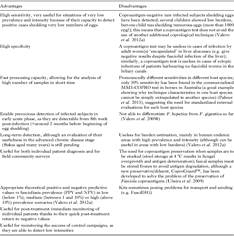
For coproantigen tests, the samples should be refrigerated (3−5 °C) for short-term preservation or frozen (−20 °C) for long-term conservation. In fascioliasis hyperendemic areas, the number of subjects who participate in surveys of this kind is very large. This can produce problems in transporting and preserving the faecal samples, as in many techniques the coproantigen degrades at ambient temperature within a few days and the faecal material cannot be treated with any classical fixative. The alternative of freezing the samples poses the additional problems of (i) the difficulty and cost of transporting frozen samples to the laboratory where the examination is to take place and (ii) the unsuitability of frozen samples for the diagnosis of several co-infecting parasites. Another solution is the use of a new faecal preservative/diluent, CoproGuard™, which has been demonstrated to be convenient for sample preservation in these kinds of surveys including the application of a coproantigen detection test (Ubeira et al. Reference Ubeira, Muiño, Valero, Periago, Perez-Crespo, Mezo, Gonzalez-Warleta, Romaris, Paniagua, Cortizo, Llovo and Mas-Coma2009).
The usefulness of this product, specifically developed to prevent degradation of the MM3-recognized Fasciola coproantigens in stool samples over time, has recently been evaluated. This preservative enhanced coproantigen extraction without affecting the detection limit of the assay, and the antigenicity of Fasciola coproantigens in faecal samples stored at 37 °C was retained throughout the entire observation period (120 days). The use of CoproGuard™ combined with MM3-Copro ELISA may be a very useful tool for the diagnosis of human fascioliasis, mainly in field community surveys in developing countries (Ubeira et al. Reference Ubeira, Muiño, Valero, Periago, Perez-Crespo, Mezo, Gonzalez-Warleta, Romaris, Paniagua, Cortizo, Llovo and Mas-Coma2009)
In areas of low prevalence and intensity, for burden estimation to establish treatment dose, and for post-treatment follow-up, diagnostic concentration methods are needed. Of the two most commonly used concentration procedures, the zinc sulphate flotation and the formalin-ether sedimentation methods, the latter is the most useful. Formalin–ether sedimentation or rapid sedimentation are useful for both fresh and formalin-preserved material and can be used with polyvinyl alcohol solution (PVA) material, but with some decreased efficiency (Lumbreras et al. Reference Lumbreras, Cantella and Burga1962; Ash and Orihel, Reference Ash and Orihel1987). Problems associated with the safety of keeping diethyl ether in laboratories have been addressed through substitution with ethyl acetate (Ash and Orihel, Reference Ash and Orihel1987). Samples fixed in merthiolate-iodine-formaldehyde (MIF) fixative (1:3 stool amount/final faecal solution) are processed by the direct MIF (Sapero and Lawless, Reference Sapero and Lawless1953) and MIF concentration (MIFC) (Blagg et al. Reference Blagg, Schloegel, Mansour and Khalaf1955) techniques.
After many years of diagnostic use in different epidemiological situations and transmission patterns of fascioliasis, a standardized easy and low-cost coprodiagnostic procedure for human fascioliasis has been shown to work well for both individual diagnosis and surveys (Esteban et al. Reference Esteban, Flores, Aguirre, Strauss, Angles and Mas-Coma1997a, Reference Esteban, Flores, Angles, Strauss, Aguirre and Mas-Comab, Reference Esteban, Flores, Angles and Mas-Coma1999, Reference Esteban, Gonzalez, Bargues, Sanchez, Naquira and Mas-Coma2002, Reference Esteban, Gonzalez, Curtale, Muñoz-Antoli, Valero, Bargues, El-Sayed, El Wakeel, Abdel-Wahab, Montresor, Engels, Savioli and Mas-Coma2003; Gonzalez et al. Reference Gonzalez, Esteban, Bargues, Valero, Ortiz, Naquira and Mas-Coma2011). It comprises a variable number of Kato–Katz slides made from each fresh stool sample following WHO recommendations, using a template delivering about 41·7 mg of faeces (Ash et al. Reference Ash, Orihel and Savioli1994). The number of slides by sample may vary according to the endemic area: only one slide may be sufficient in hyperendemic areas, but in endemic areas like Vietnam up to seven slides may be needed for an individual patient. These slides must initially be examined within 1 h of preparation whenever possible, to avoid overclarification of some helminth eggs. Moreover, if sufficient material of each stool sample is present, the following aliquots are recommended: (i) one to be fixed in 10% formalin solution (1:3 stool amount/final faecal solution); (ii) a second in PVA fixative (1:3 stool amount/final faecal solution); and (iii) an additional fresh or preserved (e.g. refrigerated, frozen or with CoproGuard™) stool sample for coproantigen detection testing with any technique following specific procedures for the kit selected. As 10% formalin and PVA have complementary advantages, it is recommended that the stool sample be divided and preserved in both types of fixatives. Samples fixed in 10% formalin are processed by a formalin–ether concentration technique. The saline (or unstained) mounts should be examined systematically, looking for eggs, larvae and cysts. If cysts are seen, iodine mounts should be made to see more detail (WHO, 1991). For observation of protozoal cysts, one aliquot of sediment obtained with this technique may be stained using a modified Ziehl–Neelsen technique (WHO, 1991). PVA-fixed smears are made directly from faeces preserved in PVA. The two most widely used permanent stains for protozoal cysts are trichrome and iron haematoxilyn.
Procedures for blood samples
Blood or serum is required for all human fascioliasis immunodiagnostic tests. Whole blood samples are obtained by fingertip puncture with a lancet (Martinez-Sernandez et al. Reference Martinez-Sernandez, Muiño, Perteguer, Garate, Mezo, Gonzalez-Warleta, Muro, Correia da Costa, Romaris and Ubeira2011).
To obtain serum, whole blood is collected by means of venipuncture. The blood is centrifuged and the serum is placed into labelled tubes. The use of fresh serum is the best, although unfortunately analysis of the samples within a short time after collection (1–2 days maximum, better at 2–8 °C) is not always affordable. If serum samples have to wait longer until analysis, they should be stored frozen (whenever possible at −20 °C). In surveys in endemic areas far from the laboratory, serum samples should be transported either refrigerated or frozen; similarly, if the samples are to be shipped to another laboratory for examination. Sodium azide (6%) may also be used as a preservative for serum samples, although it should be considered that this preservative may be counter-indicated depending on the test (e.g. Fas2-ELISA test).
In some endemic areas, such as the Bolivian Altiplano area, blood sampling in surveys is hampered by the reluctance of the indigenous Aymaran population to provide blood by venipuncture. This difficulty warrented use of the simpler and less invasive method of collecting samples onto filter paper after lancet-pricking of the finger (Strauss et al. Reference Strauss, O'Neill, Parkinson, Angles and Dalton1999). The method was already successfully used at the Nacional de Laboratorios de Salud (INLASA) for obtaining blood for the serologic diagnosis of Chagas disease. Two circles 5 mm in diameter are cut from the bloodstained filter paper and antibodies are eluted by soaking them in 250 μL of phosphate-buffered saline. A soaking time of 1 h at room temperature is sufficient to elute the antibodies from the filter paper. However, in the case of samples stored for several years, a longer period of soaking is necessary (Strauss et al. Reference Strauss, O'Neill, Parkinson, Angles and Dalton1999).
DIAGNOSIS WITH STOOL SAMPLES
In diagnosis of human fascioliasis, stool samples present many advantages. Coprological methods are inexpensive, easy to perform and do not require specialized instrumentation, making them suitable for use in the field and laboratory. They are relatively easy to obtain, both from individual patients and in community surveys, as areas where inhabitants are reluctant to furnish faecal samples are rare. They allow for direct (aetiological and molecular) and indirect (immunological) methods, as well as for quantitative burden assessment, thus allowing for analyses of prevalence and intensity. Moreover, and importantly for fieldwork, faecal collections do not require invasive extraction as for blood samples, thus facilitating participation by inhabitants of the endemic areas and subsequent long-term epidemiological studies in which repeated surveys should be performed in different seasons and years in the same communities. In addition, coprological techniques are crucial for guiding decisions regarding treatment, as they are the only ones providing a standardized cut-off based on a pre-established epg threshold. Finally, stool samples also enable the simultaneous diagnosis of many co-infecting protozooses and helminthiases.
Qualitative coprological techniques for egg finding
Techniques ranging from a simple direct smear to different concentration methods have been used during the biliary period of infection. Egg concentration may be achieved by flotation and sedimentation techniques. The sedimentation techniques appear to be more accurate and sensitive than flotation techniques, as most of the hyperosmotic solutions distort the eggs (see reviews in Esteban et al. Reference Esteban, Bargues and Mas-Coma1998; Mas-Coma et al. Reference Mas-Coma, Esteban and Bargues1999b). However, pronounced sensitivity differences according to the sedimentation techniques have been found. In recent years, an increasing number of articles have been published in which the coprological diagnostic technique is not mentioned (simply noted as microscopic examination of faecal samples); this lack of information hinders subsequent significant epidemiological comparisons.
Ether, formalin-ether or formalin-ethyl acetate concentration techniques are useful sedimentation techniques (Cosme et al. Reference Cosme, Ojeda, Cilla, Torrado, Alzate, Beristain, Orive and Arenas2001; Esteban et al. Reference Esteban, Gonzalez, Curtale, Muñoz-Antoli, Valero, Bargues, El-Sayed, El Wakeel, Abdel-Wahab, Montresor, Engels, Savioli and Mas-Coma2003; Ozturhan et al. Reference Ozturhan, Emekdaş, Sezgin, Korkmaz and Altintaş2009; Gonzalez et al. Reference Gonzalez, Esteban, Bargues, Valero, Ortiz, Naquira and Mas-Coma2011; Karahocagil et al. Reference Karahocagil, Akdeniz, Sunnetcioglu, Cicek, Mete, Akman, Ceylan, Karsen and Yapici2011). The simple gravity sedimentation technique is another technique usually performed for fascioliasis (Ash and Orihel, Reference Ash and Orihel1987; Espino et al. Reference Espino, Diaz, Perez and Finlay1998). In endemic areas of Peru, the Lumbrera's rapid sedimentation technique is frequently used (Maco-Flores et al. Reference Maco-Flores, Marcos-Raymundo, Terashima-Iwashita, Samalvides-Cuba, Miranda-Sanchez, Espinoza-Babilon and Gotuzzo-Herencia2002; López et al. Reference Lopez, White and Cabada2012; Valero et al. Reference Valero, Periago, Perez-Crespo, Angles, Villegas, Aguirre, Strauss, Espinoza, Herrera, Terashima, Tamayo, Engels, Gabrielli and Mas-Coma2012a). The spontaneous sedimentation in tube technique (SSTT) has been proposed as an alternative to the Faust technique (sulphate zinc flotation technique) (Terashima et al. Reference Terashima, Marcos, Maco, Canales, Samalvides and Tello2009). In a comparative analysis, SSTT reported higher prevalence rates than the formalin–ether concentration method, whereas it also showed higher detection rates than the Faust technique (Tello et al. Reference Tello, Terashima, Marcos, Machicado, Canales and Gotuzzo2012).
A comparative study of three techniques (Muñoz et al. Reference Muñoz, Neira, Neira, Villalon and Subercaseaux1987) was made on the basis of 15 known human cases of F. hepatica infection (six stool samples per patient, one daily): a modified gravity sedimentation method (Fast Sedimentation Method – FSM); the Telemann centrifugation technique (TM); and the standard Gravity Sedimentation technique (GS). With TM, parasite eggs were only found in 33·3% of the cases, in contrast to 100% for GS and FSM. A more detailed study of the two latter techniques revealed that FSM, besides being easier and faster to perform, permitted egg detection in all the patients through the examination of a single sample, while GS only found eggs in 73·3% of the cases considering the six samples. The respective indices of positivity over performed readings were 92·2% for FSM and 20·6% for GS. Adding increasing quantities of eggs (5×5) to negative samples, a complementary study on the sensitivity of FSM was conducted and showed that the presence of 15 eggs was sufficient to reach 100% positive readings.
The rapid sedimentation (using 20 g of faeces on each of 3 consecutive days), although inconvenient, seemed to be largely more sensitive than the single examination of duodenal fluid by Enterotest (Beal et al. Reference Beal, Veins, Grant and Hughes1970) or the MIFC method using 1 g of faeces in a single examination (Knobloch et al. Reference Knobloch, Delgado, Alvarez, Reymann and Bialek1985). In the comparison of five concentration techniques, the recovery rates of eggs in the sediment using 0·5 g of faeces were: formalin–ether method, 5·3%; HCl-ether method, 7·8%; Weller-Darmin's modification method, 37·7%; citrate buffer-Tween 80-ether method, 25·3%; and AMS III (Tween 80) method, 30·5% (Akahane et al. Reference Akahane, Oshima, Shimazu and Hirosawa1975).
In another large study, it was concluded that the MIFC method is, for diagnosis of both intestinal protozoa and helminths, superior to other methods such as routine laboratory methods (normal saline, Lugol's solution and iron-haematoxylin), the MIF technique and the MF-Heidenhain method for protozoa, and the Telemann method and MF-Telemann method for worm eggs, according to the criteria of diagnostic yield, accuracy, simplicity, time consumption and cost (Akhtaruzzaman et al. Reference Akhtaruzzaman, Bienzle, Rosenkaimer, Guggenmoos and Dietrich1978).
The cellophane faecal thick-smear technique (Kato, Kato–Katz) (Kato and Miura, Reference Kato and Miura1954; Katz et al. Reference Katz, Chaves and Pelegrino1972) has also been used as a qualitative technique. This technique has the advantages of simplicity, rapidity, commercialized at very low cost (‘Coprokit Kato–Katz’) and reproducibility due to being standardized. Its ease of performance under field conditions have ensured its universal application, although it should be considered that Fasciola eggs in the Kato–Katz smears may show some deformation which may lead to confusion and misclassification by inexperienced microscopists. Unfortunately, the sensitivity of a single Kato–Katz slide may be very low due to a combination of factors such as: (i) small amounts of stool in each examination; (ii) variation in the distribution of eggs within a single stool specimen; (iii) differences in worm burdens and crowding effect; (iv) daily fluctuations within an individual due to faecal production and consistency; and (v) daily fluctuations within an individual related to oviposition patterns of the parasite (Valero et al. Reference Valero, Panova, Comes, Fons and Mas-Coma2002, Reference Valero, De Renzi, Panova, Garcia-Bodelon, Periago, Ordoñez and Mas-Coma2006b, Reference Valero, Panova, Perez-Crespo, Khoubbane and Mas-Coma2011). Moreover, the consistency of the faecal material depends upon the parasite burden, i.e. moderate to heavy infections may cause diarrhoea (Teesdale et al. Reference Teesdale, Fahringer and Chitsulo1985; Feldmeier and Poggensee, Reference Feldmeier and Poggensee1993). Also, the amounts of fibre or fat in the host's diet have to be taken into account (Valero et al. Reference Valero, Panova, Perez-Crespo, Khoubbane and Mas-Coma2011). However, the relatively low sensitivity of Kato–Katz may be solved by increasing the number of slides. The examination of three Kato smears from a single stool specimen, which is feasible in field studies, gives an accurate diagnosis of fascioliasis. The sensitivity of the Kato–Katz test for diagnosing Fasciola infection with three Kato slides from the same specimen or on different days ranged from 96·0 to 99·1% (El-Morshedy et al. Reference El-Morshedy, Shehab, Zaki and Farag2002). When dealing with individual patients, analysing stool samples from different days may also increase the sensitivity.
Overall, the sensitivity differences in the coprological techniques highlight the convenience of applying repeated analyses to the same stool sample or more than one sample from the patient, and more than one diagnostic technique to each stool sample when in surveys. Complementation with another highly sensitive stool technique as a coproantigen test seems to be the best strategy, above all when the coproantigen test is applied first for initial screening.
Differentiation of Fasciola eggs from eggs of other trematodes infecting humans
The eggs of Fasciola are operculated, ovoid, yellow and non-embryonated when laid. In Fasciola eggs in human stools, the abopercular end of the shell is often roughened or irregular (more or less pronounced). This irregularity varies in frequency depending on countries and is sometimes laterally located (Valero et al. Reference Valero, Perez-Crespo, Periago, Khoubbane and Mas-Coma2009a).
Fasciola eggs are not easily distinguishable from those of other trematode species usually infecting humans and which also produce unembryonated eggs of similar size. Thus, care should be taken with techniques which may modify the form and size of the eggs if specific differentiation is needed (e.g. flotation techniques, techniques including centrifuging, Kato–Katz).
Given the worldwide distribution of human fascioliasis, the differentiation of Fasciola eggs from eggs of other trematodes infecting humans should always be taken into account in regions of Asia where Fasciolopsis buski and species of Paragonimus are present (Gastrodiscoides hominis is a more sporadic infection in humans in Asia), and in Africa and Latin America where other Paragonimus species are also distributed. Thus, misclassification of eggs by non-specialists may occur in these overlap areas (see comparison in Table 1). Other trematodes such as Paramphistomum species present eggs similar to those of Fasciola, but they only infect animals.
Coprological diagnosis of fascioliasis by morphological differentiation of the eggs of Fasciola spp. by comparison with similar non-embryonated eggs laid by other human-infecting trematode species present in the same endemic countries of Asia and rest of the world
* Measurements correspond to eggs found only in humans.
Differentiation of eggs from F. hepatica, F. gigantica and hybrid intermediate forms in humans
Extrapolation of egg size in livestock has always been used for human diagnosis: the borderlines allowing differentiation between the two species were traditionally considered to be 150 μm in length and 90 μm in width, lower values representing F. hepatica and higher values F. gigantica. However, large variations were first observed in the size of F. hepatica eggs in livestock from different geographical locations (Tinar, Reference Tinar1984). Furthermore, it has been experimentally shown that the final host species (sheep, cattle, pig and donkey) decisively influences the size of the F. hepatica eggs even within the same endemic area (Valero et al. Reference Valero, Darce, Panova and Mas-Coma2001). Additionally, the existence of intermediate forms (specimens showing morphological characteristics between those of the ‘pure’ fasciolid species) and hybrids (specimens presenting genetic characteristics of the two species resulting from cross-breeding) (Mas-Coma et al. Reference Mas-Coma, Valero and Bargues2009a) increases the problem. Such intermediate forms and hybrids infect humans in areas where the two fasciolids overlap in Africa (Periago et al. Reference Periago, Valero, El Sayed, Ashrafi, El Wakeel, Mohamed, Desquesnes, Curtale and Mas-Coma2008) and Asia (Ashrafi et al. Reference Ashrafi, Valero, Panova, Periago, Massoud and Mas-Coma2006b; Afshan et al. Reference Afshan, Valero, Qayyum, Peixoto, Magraner and Mas-Coma2013). The existence of these intermediate forms raises the issue of whether egg characteristics are suitable as a tool for the differential diagnosis of fascioliasis caused by either species (Valero et al. Reference Valero, Perez-Crespo, Periago, Khoubbane and Mas-Coma2009a). An example of this problem is emphasized in the diagnosis of fascioliasis in humans in the study presented by Inoue et al. (Reference Inoue, Kanemasa, Inoue, Matsumoto, Kajita, Mitsufuji, Kataoka, Okanoue, Yamada, Uchikawa, Tegoshi and Arizono2007).
A large study on the shape and size of eggs shed by humans, characterizing their morphometric traits using a computer image analysis system or CIAS (Valero et al. Reference Valero, Panova and Mas-Coma2005), was based on human samples from five different human endemic areas in South America, Europe, Africa and Asia. This study revealed that eggs shed by humans show morphological traits different from eggs shed by animals. In humans, F. hepatica eggs are bigger and F. gigantica eggs are smaller than reported to date from livestock, and their measurements overlap when compared (Table 2; Fig. 4) (Valero et al. Reference Valero, Perez-Crespo, Periago, Khoubbane and Mas-Coma2009a; Mas-Coma et al. Reference Mas-Coma, Agramunt and Valero2014).
Fasciolid eggs in human stools: (A) egg of Fasciola hepatica from a female child in Bolivia; (B) egg of an intermediate form shed by a male child from Ethiopia; (C) egg of Fasciola gigantica from a female child of Nepal. All eggs at the same scale.
Measurements of eggs of Fasciola hepatica and F. gigantica in different world regions according to the absence or existence of overlap of the two fasciolid species (intermediate hybrid forms have egg size ranges different from pure species). Size given in length/width. All measures in μm. Data from Valero et al. (Reference Valero, Perez-Crespo, Periago, Khoubbane and Mas-Coma2009a) and Mas-Coma et al. (Reference Mas-Coma, Agramunt and Valero2014)
Quantitative coprological techniques
Egg quantification in faeces is important in human fascioliasis as it is an indicator of the parasite burden. Immunological techniques (serological, coproantigen) are not suitable for quantification. A threshold at 400 epg is presently used in human fascioliasis to establish the appropriate treatment dose to avoid colic risks (WHO, 2007). Unfortunately, egg counts are seldom noted both in diagnosis in individual patient reports and in surveys, meaning that an important baseline to assess relationships with pathology and symptomatology, as well as with the epidemiological characteristics of an endemic area, is not retained.
In fact, many coprological concentration techniques can be used to determine the number of epg if a known stool weight is used. Different techniques have already been applied quantitatively in human fascioliasis.
The Kato–Katz technique appears to be the most appropriate. It is employed as the standard technique for assessing the burden of helminth infections at individual and human community level, allowing for a quantitative assessment of the severity (intensity) of the infection by epg counts. Using Kato–Katz for quantitative assessment after having applied a qualitative technique facilitates the task (Esteban et al. Reference Esteban, Flores, Aguirre, Strauss, Angles and Mas-Coma1997a, Reference Esteban, Flores, Angles, Strauss, Aguirre and Mas-Comab, Reference Esteban, Flores, Angles and Mas-Coma1999, Reference Esteban, Gonzalez, Bargues, Sanchez, Naquira and Mas-Coma2002, Reference Esteban, Gonzalez, Curtale, Muñoz-Antoli, Valero, Bargues, El-Sayed, El Wakeel, Abdel-Wahab, Montresor, Engels, Savioli and Mas-Coma2003).
Microscopic egg count is relatively slow in cases of heavy egg burdens. Therefore, the application of the Kato–Katz technique in community surveys becomes problematic because (i) it is pronouncedly time-consuming when the number of samples is high, and (ii) requires an additional technique to increase the sensitivity in areas where subjects shed a very low number of eggs intermittently (Valero et al. Reference Valero, Periago, Perez-Crespo, Angles, Villegas, Aguirre, Strauss, Espinoza, Herrera, Terashima, Tamayo, Engels, Gabrielli and Mas-Coma2012a).
The Kato–Katz test is also used in exploratory trials of drug efficacy and treatment safety (Keiser et al. Reference Keiser, Sayed, El-Ghanam, Sabry, Anani, El-Wakeel, Hatz, Utzinger, El-Din, El-Maadawy and Botros2011; Villegas et al. Reference Villegas, Angles, Barrientos, Barrios, Valero, Hamed, Grueningr, Ault, Montresor, Engels, Mas-Coma and Gabrielli2012; Zumaquero-Rios et al. Reference Zumaquero-Rios, Sarracent-Perez, Rojas-Garcia, Rojas-Rivero, Martinez-Tovilla, Valero and Mas-Coma2013). In fact, the case definitions for fascioliasis used in published clinical trials have usually relied upon the quantification of Fasciola eggs in faeces, as this kind of determination is highly specific and easily measurable (Hien et al. Reference Hien, Truong, Minh, Dat, Dung, Hue, Dung, Tuan, Campbell, Farrar and Day2008).
The WHO considers preventive chemotherapy, including the large-scale distribution of triclabendazole among endemic communities, feasible to control disease-related morbidity and decrease transmission rates (Gabrielli et al. Reference Gabrielli, Montresor, Engels and Savioli2011; Villegas et al. Reference Villegas, Angles, Barrientos, Barrios, Valero, Hamed, Grueningr, Ault, Montresor, Engels, Mas-Coma and Gabrielli2012; Valero et al. Reference Valero, Periago, Perez-Crespo, Angles, Villegas, Aguirre, Strauss, Espinoza, Herrera, Terashima, Tamayo, Engels, Gabrielli and Mas-Coma2012a). The number of epg (using the Kato–Katz technique) shed by humans is currently the only available tool to assess the intensity of fascioliasis infection in individuals. Thus, in various pilot schemes implemented by the WHO, children with >400 epg are hospitalized before receiving treatment as individuals infected with a large number of worms are more likely to develop both systemic and mechanical adverse events following treatment and might therefore require specialist medical attention and care (Valero et al. Reference Valero, Periago, Perez-Crespo, Angles, Villegas, Aguirre, Strauss, Espinoza, Herrera, Terashima, Tamayo, Engels, Gabrielli and Mas-Coma2012a; Villegas et al. Reference Villegas, Angles, Barrientos, Barrios, Valero, Hamed, Grueningr, Ault, Montresor, Engels, Mas-Coma and Gabrielli2012). Determining a patient's parasitic burden is crucial given the necessity to monitor the drug treatment in order to prevent a hepatic colic as the consequence of the massive expulsion of liver flukes (WHO, 2007).
Comparative studies are presently underway to ascertain the value of the different techniques for quantitative purposes in humans, including the MacMaster technique variants well-known for veterinary use (Cringoli et al. Reference Cringoli, Rinaldi, Veneziano, Capelli and Scala2004; Levecke et al. Reference Levecke, Behnke, Ajjampur, Albonico, Ame, Charlier, Geiger, Hoa, Kamwa Ngassam, Kotze, McCarthy, Montresor, Periago, Roy, Tchuem Tchuenté, Thach and Vercruysse2011; Vadlejch et al. Reference Vadlejch, Petrtyl, Zaichenko, Cadkova, Jankovska, Langrova and Moravec2011), and the recently developed FLOTAC (Knopp et al. Reference Knopp, Rinaldi, Khamis, Stothard, Rollinson, Maurelli, Steinmann, Marti, Cringoli and Utzinger2009; Cringoli et al. Reference Cringoli, Rinaldi, Maurelli and Utzinger2010) and Mini-FLOTAC (Barda et al. Reference Barda, Zepherine, Rinaldi, Cringoli, Burioni, Clementi and Albonico2013; Silva et al. Reference Silva, Vila-Viçosa, Maurelli, Morgogoglione, Cortes, Cringoli and Rinaldi2013) techniques. A comparative assessment showed that FLOTAC is only slightly more time-consuming and costly than Kato–Katz for helminth diagnosis in epidemiological surveys (Speich et al. Reference Speich, Knopp, Mohammed, Khamis, Rinaldi, Cringoli, Rollinson and Utzinger2010). A preliminary assay on the use of FLOTAC for F. hepatica diagnosis with a laboratory rat model showed promising results (Duthaler et al. Reference Duthaler, Rinaldi, Maurelli, Vargas, Utzinger, Cringoli and Keiser2010).
Detection of antigens in stools
An antigen detection test is able to determine the presence of an active infection and provides complementary data with respect to antibody detection in serum. Antigen detection techniques in serum, plasma, saliva or faeces have been reported (Hillyer, Reference Hillyer and Dalton1999; Sabry and Taher, Reference Sabry and Taher2008). Antigenaemia develops mainly during the invasive phase of infection. After this period, antigens decrease in serum and become undetectable over the course of infection (Espino et al. Reference Espino, Diaz, Perez and Finlay1998). Therefore, for the diagnosis of active chronic infections antigens should be looked for in stools.
A few stool antigen detection assays have been developed for human diagnosis with varied ranges of sensitivities and specificities (Espino and Finlay, Reference Espino and Finlay1994; Espino et al. Reference Espino, Diaz, Perez and Finlay1998; Arafa et al. Reference Arafa, Abaza, El-Shewy, Mohareb and El-Moamly1999; Demerdash et al. Reference Demerdash, Diab, Aly, Mohamed, Mahmoud, Zoheiny, Mansour, Attia and El Bassiouny2011). Differences in specificities may be attributed to the complex and variable composition of excretory/secretory (ES) products that makes such antigenic preparations unsuitable for performance comparisons with ELISA when implemented by different authors. This may be due to the different protocols used to prepare ES or to the variation in its composition when obtained from parasites derived from different hosts, which is not the case when a purified antigen is used.
Antigen detection tests usually use antigen-capture assays, although other systems have been described. The first tests were designed using polyclonal antibodies (Langley and Hillyer, Reference Langley and Hillyer1989a, Reference Langley and Hillyerb; Rodríguez-Pérez and Hillyer, Reference Rodriguez-Perez and Hillyer1995; Sanchez-Andrade et al. Reference Sanchez-Andrade, Paz-Silva, Suarez, Panadero, Díez-Baños and Morrondo2000, Reference Sanchez-Andrade, Paz-Silva, Suarez, Panadero, Pedreira, Diez-Baños and Morrondo2001; Almazan et al. Reference Almazan, Avila, Quiroz, Ibarra and Ochoa2001; Paz-Silva et al. Reference Paz-Silva, Pedreira, Sanchez-Andrade, Suarez, Diaz, Panadero, Diez-Baños and Morrondo2002), and later monoclonal antibodies (MoAb) as they are more specific than polyclonal antibodies for antigen capturing, i.e. they permit the detection of well-defined epitopes. Furthermore, reproducibility of tests is improved, as variations associated with batches of polyclonal antibodies are avoided. However, the production of monoclonal antibodies is an expensive and time-consuming process and the majority of the defined specific monoclonal antibodies are not commercially available.
Antigen capture with monoclonal antibodies
MoAbs have been described against antigens of both F. hepatica and F. gigantica. With regard to F. hepatica, they have been developed against somatic and tegumental antigens (Hanna and Trudgett, Reference Hanna and Trudgett1983; Hanna et al. Reference Hanna, Trudgett and Anderson1988) as well as against ES antigens (ES78 and MM3) (Espino et al. Reference Espino, Diaz, Perez and Finlay1998; Mezo et al. Reference Mezo, Gonzalez-Warleta and Ubeira2007). All have been shown to be excellent diagnostic reagents for detecting coproantigens in humans, sheep, cattle and wild boars with chronic infection (Espino et al. Reference Espino, Diaz, Perez and Finlay1998; Dumenigo et al. Reference Dumenigo, Espino, Finlay and Mezo2000; Mezo et al. Reference Mezo, Gonzalez-Warleta, Carro and Ubeira2004, Reference Mezo, Gonzalez-Warleta, Castro-Hermida, Manga-Gonzalez, Peixoto, Mas-Coma and Valero2013; Ubeira et al. Reference Ubeira, Muiño, Valero, Periago, Perez-Crespo, Mezo, Gonzalez-Warleta, Romaris, Paniagua, Cortizo, Llovo and Mas-Coma2009; Valero et al. Reference Valero, Ubeira, Khoubbane, Artigas, Muiño, Mezo, Perez-Crespo, Periago and Mas-Coma2009b, Reference Valero, Periago, Perez-Crespo, Angles, Villegas, Aguirre, Strauss, Espinoza, Herrera, Terashima, Tamayo, Engels, Gabrielli and Mas-Coma2012a). Two other MoAbs highly specific for FhSAP2 have recently been obtained (2F5 and 2F9), which do not cross-react with antigens from Schistosoma mansoni, Schistosoma japonicum or Clonorchis sinensis (Caban-Hernandez and Espino, Reference Caban-Hernandez and Espino2013).
With regard to F. gigantica, MoAbs have been produced against total extract (Fagbemi et al. Reference Fagbemi, Aderibigbe and Guobadia1997), against ES antigens (Estuningsih et al. Reference Estuningsih, Widjayanti, Adiwinata and Piedrafita2004, Reference Estuningsih, Spithill, Raadsma, Law, Adiwinata, Meeusen and Piedrafita2009; Demerdash et al. Reference Demerdash, Diab, Aly, Mohamed, Mahmoud, Zoheiny, Mansour, Attia and El Bassiouny2011), against partially purified native proteins derived from tegument (Chaithirayanon et al. Reference Chaithirayanon, Wanichanon, Vichasri-Grams, Ardseungneon, Grams, Viyanant, Upatham and Sobhon2002; Krailas et al. Reference Krailas, Panomsuk, Janecharat and Ukong2002; Anuracpreeda et al. Reference Anuracpreeda, Wanichanon, Chaithirayanon, Preyavichyapugdee and Sobhon2006, Reference Anuracpreeda, Wanichanon, Chawengkirtikul, Chaithirayanon and Sobhon2009b) and against recombinant proteins such as FABPs (Sirisriro et al. Reference Sirisriro, Grams, Vichasri-Grams, Ardseungneon, Pankao, Meepool, Chaithirayanon, Viyanant, Tan-Ariya, Upatham and Sobhon2002; Allam et al. Reference Allam, Bauomy, Hemyeda and Sakran2012), a SAPLIP member from F. gigantica (FgSAP2) (Kueakhai et al. Reference Kueakhai, Changklungmoa, Riengrojpitak, Chaichanasak, Meemon, Chaithirayan, Chantree, Sansri, Itagaki and Sobhon2013a, Reference Kueakhai, Changklungmoa, Chaithirayanon, Songkoomkrong, Riengro-Jpitak and Sobhonb) and cathepsin B3 protease (Anuracpreeda et al. Reference Anuracpreeda, Chawengkirtikul, Tinikul, Poljaroen, Chotwiwatthanakun and Sobhon2013).
The most widely used of the MoAbs for human fascioliasis diagnosis described to date are the ES78 (Marcet Sanchez et al. Reference Marcet Sánchez, Figueredo Pino, Núñez Fernández, Rojas Rivero and Sarracent Pérez2012) and MM3 (Muiño et al. Reference Muiño, Perteguer, Garate, Martinez-Sernandez, Beltran, Romaris, Mezo, Gonzalez-Warleta and Ubeira2011), although others have also been described (Demerdash et al. Reference Demerdash, Diab, Aly, Mohamed, Mahmoud, Zoheiny, Mansour, Attia and El Bassiouny2011; Caban-Hernandez and Espino, Reference Caban-Hernandez and Espino2013).
Coproantigen tests
Stool antigen detection techniques have been reported for both human and animal fascioliasis, most of them based on Fasciola ES products. The detection of these products by means of a sandwich-ELISA reflects the installation of flukes in the bile ducts and the presence of the biliary stage of the disease. The majority of methods based on antigen detection have been applied to F. hepatica infection (Hanna and Trudgett, Reference Hanna and Trudgett1983; Hanna et al. Reference Hanna, Trudgett and Anderson1988; Espino et al. Reference Espino, Diaz, Perez and Finlay1998; Dumenigo et al. Reference Dumenigo, Espino, Finlay and Mezo2000; Mezo et al. Reference Mezo, Gonzalez-Warleta, Carro and Ubeira2004, Reference Mezo, Gonzalez-Warleta and Ubeira2007, Reference Mezo, Gonzalez-Warleta, Castro-Hermida, Manga-Gonzalez, Peixoto, Mas-Coma and Valero2013; Ubeira et al. Reference Ubeira, Muiño, Valero, Periago, Perez-Crespo, Mezo, Gonzalez-Warleta, Romaris, Paniagua, Cortizo, Llovo and Mas-Coma2009; Valero et al. Reference Valero, Ubeira, Khoubbane, Artigas, Muiño, Mezo, Perez-Crespo, Periago and Mas-Coma2009b, Reference Valero, Periago, Perez-Crespo, Angles, Villegas, Aguirre, Strauss, Espinoza, Herrera, Terashima, Tamayo, Engels, Gabrielli and Mas-Coma2012a; Caban-Hernandez and Espino, Reference Caban-Hernandez and Espino2013) and have only in recent years started to be applied to F. gigantica infection (Arafa et al. Reference Arafa, Abaza, El-Shewy, Mohareb and El-Moamly1999; Chaithirayanon et al. Reference Chaithirayanon, Wanichanon, Vichasri-Grams, Ardseungneon, Grams, Viyanant, Upatham and Sobhon2002; Krailas et al. Reference Krailas, Panomsuk, Janecharat and Ukong2002; Sirisriro et al. Reference Sirisriro, Grams, Vichasri-Grams, Ardseungneon, Pankao, Meepool, Chaithirayanon, Viyanant, Tan-Ariya, Upatham and Sobhon2002; Estuningsih et al. Reference Estuningsih, Widjayanti, Adiwinata and Piedrafita2004; Anuracpreeda et al. Reference Anuracpreeda, Wanichanon, Chaithirayanon, Preyavichyapugdee and Sobhon2006, Reference Anuracpreeda, Wanichanon, Chawengkirtikul, Chaithirayanon and Sobhon2009b, Reference Anuracpreeda, Chawengkirtikul, Tinikul, Poljaroen, Chotwiwatthanakun and Sobhon2013; Kueakhai et al. Reference Kueakhai, Changklungmoa, Riengrojpitak, Chaichanasak, Meemon, Chaithirayan, Chantree, Sansri, Itagaki and Sobhon2013a, Reference Kueakhai, Changklungmoa, Chaithirayanon, Songkoomkrong, Riengro-Jpitak and Sobhonb).
Coproantigen tests have both positive and negative attributes (Table 3) and have been developed based on different antibodies. Besides the most used ES78 monoclonal antibody used for the FasciDIG test (Espino et al. Reference Espino, Dumenigo, Marcet, Reyes and Finlay1993, Reference Espino, Diaz, Perez and Finlay1998; Espino and Finlay, Reference Espino and Finlay1994; Marcet Sanchez et al. Reference Marcet Sánchez, Figueredo Pino, Núñez Fernández, Rojas Rivero and Sarracent Pérez2012) and MM3 monoclonal antibody used for the MM3-COPRO test (Ubeira et al. Reference Ubeira, Muiño, Valero, Periago, Perez-Crespo, Mezo, Gonzalez-Warleta, Romaris, Paniagua, Cortizo, Llovo and Mas-Coma2009; Valero et al. Reference Valero, Ubeira, Khoubbane, Artigas, Muiño, Mezo, Perez-Crespo, Periago and Mas-Coma2009b, Reference Valero, Periago, Perez-Crespo, Angles, Villegas, Aguirre, Strauss, Espinoza, Herrera, Terashima, Tamayo, Engels, Gabrielli and Mas-Coma2012a; Muiño et al. Reference Muiño, Perteguer, Garate, Martinez-Sernandez, Beltran, Romaris, Mezo, Gonzalez-Warleta and Ubeira2011), other monoclonal antibodies have also been described (Arafa et al. Reference Arafa, Abaza, El-Shewy, Mohareb and El-Moamly1999; El-Bassiouny et al. Reference El-Bassiouny, Mohamed, Salah, Zoheiry, Mansour, Diab, Rabia, Safwat and Demerdash2008; Demerdash et al. Reference Demerdash, Diab, Aly, Mohamed, Mahmoud, Zoheiny, Mansour, Attia and El Bassiouny2011; Allam et al. Reference Allam, Bauomy, Hemyeda and Sakran2012; Attallah et al. Reference Attallah, Bughdadi, El-Shazly and Ismail2013). Several are commercially available: (i) ‘Fasciola hepatica Antigen ELISA Kit Bio K 201’; (ii) ‘Bovine Fasciola hepatica Antigenic ELISA Kit’; (iii) ‘Fasciola hepatica Ag’; (iv) ‘FasciDIG IPK La Havana’.
Advantages and disadvantages of antigen-detecting techniques applied to stool samples for fascioliasis diagnosis in humans
To date, the only coproantigen test to be evaluated for community surveys in the field according to proposed standards is the MM3-COPRO (Valero et al. Reference Valero, Periago, Perez-Crespo, Angles, Villegas, Aguirre, Strauss, Espinoza, Herrera, Terashima, Tamayo, Engels, Gabrielli and Mas-Coma2012a). Test efficacy was evaluated in children within two human fascioliasis high-prevalence areas: one with an altiplanic transmission pattern in Bolivia (high epg and permanent transmission) and another with a valley transmission pattern in Peru (low-medium epg and seasonal transmission). Sensitivity and specificity were 94·68 and 98·48% using Kato–Katz as the preferred method in Bolivia and 94·73 and 93·58% using rapid sedimentation and Kato–Katz together in Peru. Of note in these studies is that six children were found to be shedding eggs despite showing a negative result with MM3-COPRO, including one child with a high egg count (1248 epg) (Valero et al. Reference Valero, Periago, Perez-Crespo, Angles, Villegas, Aguirre, Strauss, Espinoza, Herrera, Terashima, Tamayo, Engels, Gabrielli and Mas-Coma2012a).
This field study showed no correlation between coproantigen detection by optical density (OD) and infection intensity by epg of faeces in Cajamarca (low burden cases <400 epg), nor in Huacullani (high burden cases >400 epg). In contrast there was correlation in Huacullani regarding low burden cases (<400 epg) (Valero et al. Reference Valero, Periago, Perez-Crespo, Angles, Villegas, Aguirre, Strauss, Espinoza, Herrera, Terashima, Tamayo, Engels, Gabrielli and Mas-Coma2012a). Observed lack of correlation in two of these studies is in contrast to the positive correlation between epg counts and OD readings obtained by other authors (Espino and Finlay, Reference Espino and Finlay1994; Mezo et al. Reference Mezo, Gonzalez-Warleta, Carro and Ubeira2004; Demerdash et al. Reference Demerdash, Diab, Aly, Mohamed, Mahmoud, Zoheiny, Mansour, Attia and El Bassiouny2011; Attallah et al. Reference Attallah, Bughdadi, El-Shazly and Ismail2013), but agrees with results found in hospital patients (Ubeira et al. Reference Ubeira, Muiño, Valero, Periago, Perez-Crespo, Mezo, Gonzalez-Warleta, Romaris, Paniagua, Cortizo, Llovo and Mas-Coma2009). Results suggest that the OD/epg correlation found by other authors may be due to the analysis of stool from low to very low fluke burdens. Moreover, it must be kept in mind that (i) the pattern of egg shedding is not linear but fluctuates between maximum and minimum values (Valero et al. Reference Valero, Panova, Comes, Fons and Mas-Coma2002, Reference Valero, Panova, Perez-Crespo, Khoubbane and Mas-Coma2011), (ii) the kinetics of coproantigen release vs the kinetics of egg shedding show a similar pattern but with a 2-week time-lag in epg (Valero et al. Reference Valero, Ubeira, Khoubbane, Artigas, Muiño, Mezo, Perez-Crespo, Periago and Mas-Coma2009b) and (iii) the influence of the crowding effect on Fasciola egg shedding should also be taken into account in human hyperendemic areas (Valero et al. Reference Valero, De Renzi, Panova, Garcia-Bodelon, Periago, Ordoñez and Mas-Coma2006b). Therefore, the use of only a coproantigen technique appears to be insufficient to evaluate fascioliasis intensity.
Results from a study by Valero et al. (Reference Valero, Periago, Perez-Crespo, Angles, Villegas, Aguirre, Strauss, Espinoza, Herrera, Terashima, Tamayo, Engels, Gabrielli and Mas-Coma2012a) indicated that a coproantigen-detecting test such as the MM3-COPRO ELISA meets proposed criteria for control of human fascioliasis, including high sensitivity and specificity, rapid large mass screening capacity, detection in the acute phase, early detection of treatment failure or re-infection in post-treated subjects, and value for surveillance programmes. It provides a good tool to detect biliary fascioliasis, including a higher sensitivity than egg detection techniques, in areas where burdens are from low to very high.
The aforementioned field study highlights the benefits of such coproantigen tests for mass screening surveys prior to application of Kato–Katz, prioritizing coproantigen positive samples. Coproantigen positive subjects in which no eggs are found are recommended to receive treatment. Benefits include: (i) rapid response to communities surveyed with infected subjects treated within days of the survey and (ii) to distinguish re-infection from potential drug resistance. Coproantigen negative samples may finally be analysed later for the eventual detection and subsequent selected treatment of very few subjects shedding eggs (Valero et al. Reference Valero, Periago, Perez-Crespo, Angles, Villegas, Aguirre, Strauss, Espinoza, Herrera, Terashima, Tamayo, Engels, Gabrielli and Mas-Coma2012a).
DIAGNOSIS WITH BLOOD SAMPLES
Blood techniques developed for diagnosis of fascioliasis include mainly serological tests and a few whole blood and intradermal tests based on immunological reactions. Present immunological techniques used are based mostly on the determination of circulating anti-Fasciola antibodies, although a few are based on determination of circulating Fasciola antigens and immune complexes.
For diagnosis of human fascioliasis, immunological techniques applied to blood samples show several advantages and disadvantages that should be considered when devising an appropriate approach (Table 4).
Advantages and disadvantages of immunological techniques applied to blood samples for fascioliasis diagnosis in humans
Detection of circulating antibodies
Antibody detection is a widely preferred method for the immunodiagnosis of human fascioliasis. Among the advantages of this method is the capacity of these techniques for the relatively early detection of infection in comparison to the ‘late’ patency and presence of eggs in faeces (at least 2–3 months). Again, when parasite burden is very low, antibody detection methods provide distinct advantages. Thus, in animals experimentally infected with F. hepatica, the following results may be considered: (i) IgG against crude excretory/secretory (ES) products are detected between the second and third week post-infection (p.i.) (Salimi-Bejestani et al. Reference Salimi-Bejestani, McGarry, Felstead, Ortiz, Akca and Williams2005); (ii) the recombinant FhSAP-2 protein appears from the second week p.i. (Espino and Hillyer, Reference Espino and Hillyer2003); (iii) the recombinant cathepsin L appears first between the fifth and the seventh week p.i., or even as early as 2 weeks (O'Neill et al. Reference O'Neill, Parkinson, Strauss, Angles and Dalton1998) depending on the infectious dose (Cornelissen et al. Reference Cornelissen, Gaasenbeek, Borgsteede, Holland, Harmsen and Boersma2001).
In summary, anti-Fasciola antibodies can be detected as early as 2 weeks p.i. and can thus facilitate early chemotherapeutic intervention (Espino and Dumenigo, Reference Espino, Dumenigo, Miliotis and Bier2003), before irreparable damage to the liver occurs (Tantrawatpan et al. Reference Tantrawatpan, Maleewong, Wongkham, Wongkham, Intapan and Nakashima2005). This is particularly important in patients whose acute symptoms tend to be more severe and may be fatal, and also in areas where human fascioliasis is rare (Hillyer, Reference Hillyer and Dalton1999).
However, diagnostic problems related mainly to sensitivity and specificity should be highlighted in fascioliasis (see reviews by Chen and Mott, Reference Chen and Mott1990; Esteban et al. Reference Esteban, Bargues and Mas-Coma1998; Hillyer, Reference Hillyer and Dalton1999). Those techniques showing cross-reactions with other parasites, mainly helminths, cannot be recommended for community surveys in rural endemic areas of developing countries.
Moreover, the lack of commercially available defined, specific antigens, combined with test systems, is a serious disadvantage (Hillyer, Reference Hillyer1993). The few available commercial kits, specific antigens and test systems still present weaknesses and limitations. Numerous antigens and test systems have been described for the detection of F. hepatica and F. gigantica infections in livestock, but the enormous variation in immunogenicity of parasite proteins in animals and humans has to be taken into account (Cornelissen et al. Reference Cornelissen, Gaasenbeek, Boersma, Borgsteede and van Milligen1999). Overall, the need for immunodiagnostic tests for fascioliasis is much greater for humans than for livestock (Hillyer, Reference Hillyer and Dalton1999).
Ideally, tests should be suitable for diagnosis of both individual patients and rapid screening of inhabitants of human fascioliasis endemic areas (Espinoza et al. Reference Espinoza, Maco, Marcos, Saez, Neyra, Terashima, Samalvides, Gotuzzo, Chavarry, Huaman, Bargues, Valero and Mas-Coma2007). Unfortunately, no consensus concerning the optimal test system for antibody/antigen detection in human fascioliasis has been reached so far. Thus each laboratory prepares its own reagents and test systems with the ensuing lack of uniformity. Consequently, results obtained with the same technique vary according to authors and do not allow for significant epidemiological comparisons.
Particularly important problems for fascioliasis antigen/antibody tests include their unsuitability for burden quantification. Treatment dose cannot be established, whilst late return to negative test values after successful treatment excludes them for individual monitoring when compared with the immediate egg disappearance or coproantigen return to negative values.
Techniques for circulating antibody detection
Different circulating antibody-detecting techniques have been applied for human fascioliasis diagnosis. Several older methods are complicated to use and hence are no longer or only sporadically used, although the wider commercialization and availability of some of these methods (e.g. indirect haemagglutination) has led to their continued use in some countries. The following techniques are of note:
– Precipitation tests. They include radial diffusion (RD), double diffusion (DD), immunoelectrophoresis (IEP) and/or counter-immunoelectrophoresis (CIEP), and metacercarial precipitin test (see review in Esteban et al. Reference Esteban, Bargues and Mas-Coma1998).
– Agglutination tests. Indirect haemagglutination (IHA – commercialized: Fumouze Diagnostics) (see numerous references in Esteban et al. Reference Esteban, Bargues and Mas-Coma1998). The IHA test is a technique still used frequently in endemic areas, using purified adult F. hepatica F1 as antigen (El-Shazly et al. Reference El-Shazly, Handousa, Gabr, Morsy, Ramadan and Morsy2002; Haseeb et al. Reference Haseeb, El-Shazly, Arafa and Morsy2003b; El-Ahl et al. Reference El-Ahl, El Shazly, El Shafei, Hegazi and El-Dardiry2007; Hammami et al. Reference Hammami, Hamed and Ayadi2007; Deveci et al. Reference Deveci, Oztürk and Ustün2011; Gökçe et al. Reference Gökçe, Sahiner, Unal, Parlakay, Oncel, Saçkesen, Kara and Gümrük2011; Kaya et al. Reference Kaya, Beştaş and Cetin2011; Sakru et al. Reference Sakru, Korkmaz, Demirci, Kuman and Ok2011; Tanir et al. Reference Tanir, Karaman, Tüfekçi, Erdogan, Tuygan and Ozkan2011; Karadağ-Öncel et al. Reference Karadağ-Öncel, Ozsürekçi, Ozkaya-Parlakay, Celik, Cengiz, Haliloğlu, Ceyhan and Kara2012; Yalav et al. Reference Yalav, Yağmur, Ülkü, Akcam and Sönmez2012).
– Haemolysis tests. Complement fixation (CF) (see review in Esteban et al. Reference Esteban, Bargues and Mas-Coma1998).
– Fluorescence tests. Immunofluorescence assay (IFA) (see review in Esteban et al. Reference Esteban, Bargues and Mas-Coma1998). This technique is still used frequently in laboratories, using parasite histological sections as antigen (Abdul-Fattah et al. Reference Abdul-Fattah, Yousef, Nasr, Fouad and Abdul-Wahab1992; Strauss et al. Reference Strauss, Angles, Esteban and Mas-Coma1997; Chand et al. Reference Chand, Herman, Partridge, Hewitt and Chiodini2009). Sensitivity is reported at 92–96% in the acute phase of the infection (Capron et al. Reference Capron, Wattre, Capron and Lefebure1973).
– Radioisotope tests. Radioimmuno assays (RIST, RAST) (Sampaio Silva et al. Reference Sampaio-Silva, Vindimian, Wattre and Capron1985).
– Anti-P 1antibody tests. Automated assay of anti-P1 antibodies (Ben-Ismail et al. Reference Ben-Ismail, Carme and Gentilini1978, Reference Ben-Ismail, Rouger, Carme, Gentilini and Salmon1980, Reference Ben-Ismail, Carme, Mogahed, Niel and Gentilini1982).
Nowadays, immunological techniques rely on numerous variants of enzymic tests (see many references in Esteban et al. Reference Esteban, Bargues and Mas-Coma1998):
Enzyme-linked immunosorbent assay (ELISA)
Reports including the use of ELISA for diagnosis of fascioliasis are numerous (Thi, Reference Thi2004; El-Ahl et al. Reference El-Ahl, El Shazly, El Shafei, Hegazi and El-Dardiry2007; Ozturhan et al. Reference Ozturhan, Emekdaş, Sezgin, Korkmaz and Altintaş2009; Karahocagil et al. Reference Karahocagil, Akdeniz, Sunnetcioglu, Cicek, Mete, Akman, Ceylan, Karsen and Yapici2011; Karadağ-Öncel et al. Reference Karadağ-Öncel, Ozsürekçi, Ozkaya-Parlakay, Celik, Cengiz, Haliloğlu, Ceyhan and Kara2012; Castillo Contreras and Frisancho Velarde, Reference Castillo Contreras and Frisancho Velarde2013). Due to its simplicity and functionality, researchers have mainly focused on improving the ELISA method, and a broad spectrum of antigens has been used so far. Nevertheless, human infections frequently occur in non-developed countries where access to diagnostic laboratories is not always possible. ELISA requires a well-equipped laboratory with a spectrophotometer and has to be carried out by trained personnel. Furthermore, there are currently non-standardized criteria to establish the cut-off limit between positivity and negativity for fascioliasis.
ELISA variants
They include the enzyme-linked immunofiltration assay (ELIFA) (Pailler et al. Reference Pailler, Puygauthier-Toubas, Bonnin, Marx-Chemla, Camerlynck, Thoannes and Pinon1990), the enzyme-linked immuno-electrotransfer blot (EITB) or western blot (Santiago and Hillyer, Reference Santiago and Hillyer1986; Hillyer and Soler De Galanes, Reference Hillyer and Soler De Galanes1988; Hillyer et al. Reference Hillyer, Soler de Galanes, Rodriguez-Perez, Bjorland, Silva de Lagrava, Guzman and Bryan1992; Silva et al. Reference Silva, Urarte, Mercado and Gorman1993; Shaker et al. Reference Shaker, Demerdash, Mansour, Hassanein, El-Baz and El-Gindy1994; Hammami et al. Reference Hammami, Ayadi, Camus and Dutoit1997; Marcos et al. Reference Marcos, Tagle, Terashima, Bussalleu, Ramirez, Carrasco, Valdez, Huerta-Mercado, Freedman, Vinetz and Gotuzzo2008; Trifina et al. Reference Trifina, Spenger, Zandieh, Haller, Auer, Österreicher, Klaushofer and Mikosch2011; Agnamey et al. Reference Agnamey, Fortes-Lopes, Raccurt, Boncy and Totet2012) and the Falcon™ assay screening test-enzyme linked immunosorbent assay (FAST-ELISA) (Hillyer and Soler De Galanes, Reference Hillyer and Soler De Galanes1988, Reference Hillyer and Soler de Galanes1991; Hillyer et al. Reference Hillyer, Soler de Galanes, Rodriguez-Perez, Bjorland, Silva de Lagrava, Guzman and Bryan1992; Bjorland et al. Reference Bjorland, Bryan, Strauss, Hillyer and McAuley1995; Rahimi et al. Reference Rahimi, Ashrafi, Koosha, Abdi and Rokni2011; Elsibaei et al. Reference Elsibaei, Ali, Ibrahim and Mohammed2013).
Among ELISA variant developments, four techniques merit mention because of their promising advantages, mainly to facilitate field work in community surveys in the cases of those tests having solved the problem of the usually low sensitivity of these techniques:
Micro-ELISA
This technique can be applied as a screening test when a large number of samples is involved, because of its low requirement of reagents (especially antigens and second-antibody conjugates). The conventional ELISA could be employed as a confirmatory test following micro-ELISA analysis (Daveau and Ambroise-Thomas, Reference Deveau and Ambroise-Thomas1982; Shaheen et al. Reference Shaheen, Kamal, Farid, Mansour, Boctor and Woody1989; Itoh and Sato, Reference Itoh and Sato1990; Kim et al. Reference Kim, Kim, Huh and Cho1995, Reference Kim, Lee, Yun, Kim, Choi, Oh, Song and Chu2010; Espino et al. Reference Espino, Padron, Dumenigo and Laferte1997; Carnevale et al. Reference Carnevale, Rodríguez, Santillán, Labbé, Cabrera, Bellegarde, Velásquez, Trgovcic and Guarnera2001; Dalimi et al. Reference Dalimi, Hadighi and Madani2004).
Dot-ELISA
The standard indirect ELISA format is less practical for field surveys and a dot-ELISA has been successfully applied for antibody screening (Shaheen et al. Reference Shaheen, Kamal, Farid, Mansour, Boctor and Woody1989, Reference Shaheen, Al Khafif, Farag and Kamal1994; Itoh and Sato, Reference Itoh and Sato1990). The dot-ELISA has some advantages over the indirect ELISA. Nitrocellulose papers spotted with antigen are stable for at least three months at −20 °C (Intapan et al. Reference Intapan, Maleewong, Nateeworanart, Wongkham, Pipitgool, Sukolapong and Sangmaneedet2003), all incubation steps are performed at room temperature, and the results can be read with the naked eye, thus an expensive spectrophotometer is not required. The test is applicable to diagnose in the field setting as well as in laboratories that are not well equipped. The dot–ELISA is simple and allows testing of multiple samples at the same time (Shaheen et al. Reference Shaheen, Kamal, Farid, Mansour, Boctor and Woody1989, Reference Shaheen, Al Khafif, Farag and Kamal1994; Itoh and Sato, Reference Itoh and Sato1990; Hassan et al. Reference Hassan, Matar, Mowafy, Saleh, El-Badrawy, Awad and Ramadan2002; Intapan et al. Reference Intapan, Maleewong, Nateeworanart, Wongkham, Pipitgool, Sukolapong and Sangmaneedet2003; Dalimi et al. Reference Dalimi, Hadighi and Madani2004; Maeda et al. Reference Maeda, Yamada, Akao, Iga, Endo, Koibuchi, Nakamura, Odawara, Iwamoto and Fujii2008).
Dipstick
Dipsticks require no expensive instruments and fewer reagents in comparison to other techniques for diagnosis. Moreover, dipstick assay preparation takes only 2 h compared with the long incubation period of ELISA, and the assay can be completed in 15 min with results obtained visually. Dipstick is a simple antigen and serum test requiring only micrograms of parasite antigen and 20 μL of the patient's serum. This test can be carried out while patients wait. Additionally, patients can see the results themselves, which may increase compliance rates (Al-Sherbiny et al. Reference Al-Sherbiny, Farrag, Fayad, Makled, Tawfeek and Ali2004). From an epidemiological point of view, a dipstick assay allows control measures to be implemented in situ. Dipsticks have the merit of allowing for screening of large numbers of blood samples. They also allow for an indirect although not specific measure of burden through determination of the end-point titration of an individual's serum. Antigens kept frozen on nitrocellulose strips at −20 °C retain antigenicity for at least 3 months (Pappas, Reference Pappas1988; Intapan et al. Reference Intapan, Maleewong, Nateeworanart, Wongkham, Pipitgool, Sukolapong and Sangmaneedet2003) and, hence, they are convenient for rapid, routine laboratory use. A dipstick assay may thus be valuable for the serodiagnosis of human fascioliasis thereby saving time and eliminating the need for special training. Such a rapid test could therefore be practically implemented in epidemiological surveys. A sensitivity of 100% and a specificity of 96·7% have been preliminarily noted (Ali, Reference Ali2012), although appropriate independent complete evaluation is still pending.
Lateral flow immunoassays or immunochromatographic tests
A new rapid lateral flow immunoassay (LFIA) test (SeroFluke) for human diagnosis in both invasive and biliary periods of the disease appears to be a useful step forward (Martinez-Sernandez et al. Reference Martinez-Sernandez, Muiño, Perteguer, Garate, Mezo, Gonzalez-Warleta, Muro, Correia da Costa, Romaris and Ubeira2011). This test was constructed with a recombinant cathepsin L1 from F. hepatica, and uses protein A and mAb MM3 as detector reagents in the test and control lines, respectively. In comparison with an ELISA test (MM3-SERO), the SeroFluke test showed the advantage of being applicable to both serum or whole blood samples, although maximum specificity and sensitivity values are still pending field evaluation. Its simplicity allows it to be used in major hospitals as well as in endemic/hyperendemic regions where point-of-care testing is required. The SeroFluke test requires no expensive instruments and fewer reagents in comparison to other techniques for diagnosis. The swiftness of LFIA tests make them suitable for endemic rural areas and district hospitals. Moreover, the SeroFluke test can be used by personnel with minimal training, and more importantly, it can be used for point-of-care testing, and thus is particularly useful in several countries where human fascioliasis is endemic. In such countries, the possibility of using whole blood samples for the SeroFluke test (obtained by e.g. taking a drop of blood by fingertip puncture with a lancet or even collected on filter paper) is also advantageous, as this saves the time and expense involved in obtaining serum samples. However, even considering only major laboratories in hypoendemic countries, the new LFIA may offer advantages over ELISA methods, as it allows individual testing of patients, whereas ELISA methods are designed to test several patients at once and some robustness may be lost when they are used only sporadically (Corstjens et al. Reference Corstjens, van Lieshout, Zuiderwijk, Kornelis, Tanke, Deelder and van Dam2008). Furthermore, the LFIAs can be produced in small quantities (e.g. in the range of 10–25 tests), which would reduce the costs associated with the shelf-life of the kits.
Antigens for circulating antibody detection
In animal models, the specificity of antibody responses to Fasciola varies along the course of infection (Bennett et al. Reference Bennett, Joshua and Hughes1982). Hitherto, several antigen groups have been described (whole-worm extract, ES products, extracted proteins from the adult fluke surface and recombinant proteins) and numerous test systems have been developed for the efficient diagnosis of human fascioliasis. Nevertheless, the use of complex antigens reduces the specificity of the tests due to cross-reactivity with parasites sharing similar immunogens (Hillyer and Serrano, Reference Hillyer and Serrano1983). Currently, the conventional use of complex mixtures of antigens such as ES products is being replaced by defined and purified antigens to minimize cross-reactions. However, the expression and purification of recombinant proteins is a tedious and time-consuming task, and this technology is not always accessible to all laboratories, especially to those in endemic areas of human fascioliasis and lacking adequate economic resources. Moreover, ES products are usually prepared using in vitro techniques that require a degree of sterility (Morales and Espino, Reference Morales and Espino2012).
Although a great deal of progress has been made with regard to immunodiagnosis of human fascioliasis, the persistent problem of selected antigens required for the development of efficient diagnostic methods, together with further evaluations on their sensitivity and specificity, is yet to be solved. The main problems of antibody-detecting techniques lie in their sensitivity (false serological negatives) and specificity (cross-reactions), which are dependent on the antigen type, and the lack of consensus concerning the optimal antigen to be used in the different tests (Stork et al. Reference Stork, Venables, Jennings, Beesley, Bendezu and Capron1973; Chen and Mott, Reference Chen and Mott1990; Hillyer, Reference Hillyer1993; Sampaio Silva et al. Reference Sampaio-Silva, Da Costa, Da Costa, Pires, Lopes, Castro and Monjour1996; Valero et al. Reference Valero, Periago, Perez-Crespo, Rodriguez, Perteguer, Garate, Gonzalez-Barbera and Mas-Coma2012b).
Earlier works on ELISA for the immunodiagnosis of human fascioliasis have been reviewed (Hillyer, Reference Hillyer1993, Reference Hillyer and Dalton1999). Specific ELISA methods developed using adult fluke whole worm crude extracts provide less than optimal specificity, as they may easily present cross-reactions with other helminthic infections. The use of partially purified somatic antigen from adult worms and excretory/secretory (E/S) products, among which cysteine proteinases are the most abundant, has improved the sensitivity and specificity of the tests significantly.
Purified antigens from ES products, including cysteine proteinases, among which mainly papain-like cysteine endopeptidases termed cathepsins, mainly cathepsin L1 (CL1), should be highlighted. The use of an ELISA with purified cathepsin antigen provides high sensitivity and higher specificity than tests using parasite somatic or ES antigens (O'Neill et al. Reference O'Neill, Parkinson, Strauss, Angles and Dalton1998, Reference O'Neill, Parkinson, Dowd, Strauss, Angles and Dalton1999; Rokni et al. Reference Rokni, Massoud, O'Neill, Parkinson and Dalton2002; Espinoza et al. Reference Espinoza, Timoteo and Herrera-Velit2005). Recombinant cathepsins overcome the problem of the complex and time-consuming production of sufficient quantities of pure cathepsin, and display similar performance characteristics to native antigens (Mezo et al. Reference Mezo, Gonzalez-Warleta, Carro and Ubeira2004, Reference Mezo, Gonzalez-Warleta and Ubeira2007, Reference Mezo, Gonzalez-Warleta, Castro-Hermida, Muiño and Ubeira2010; Valero et al. Reference Valero, Perez-Crespo, Periago, Khoubbane and Mas-Coma2009a) and also demonstrate their usefulness for human diagnosis (Martinez-Sernandez et al. Reference Martinez-Sernandez, Muiño, Perteguer, Garate, Mezo, Gonzalez-Warleta, Muro, Correia da Costa, Romaris and Ubeira2011). Good results have also been obtained with two B-cell epitopes of cathepsin L1 (Cornelissen et al. Reference Cornelissen, Gaasenbeek, Boersma, Borgsteede and van Milligen1999, Reference Cornelissen, Gaasenbeek, Borgsteede, Holland, Harmsen and Boersma2001), synthesized as single synthetic peptides (Intapan et al. Reference Intapan, Tantrawatpan, Maleewong, Wongkham, Wongkham and Nakashima2005; Tantrawatpan et al. Reference Tantrawatpan, Maleewong, Wongkham, Wongkham, Intapan and Nakashima2007).
Interesting results have also been obtained in attempts at utilizing both native and recombinant fatty-acid binding proteins (FABPs) (Sirisriro et al. Reference Sirisriro, Grams, Vichasri-Grams, Ardseungneon, Pankao, Meepool, Chaithirayanon, Viyanant, Tan-Ariya, Upatham and Sobhon2002), among which one has recently shown to be a valuable antigen for human fascioliasis diagnosis (Allam et al. Reference Allam, Bauomy, Hemyeda and Sakran2012). Other strategies include the use of the putative diagnostic value of leucine aminopeptidase (LAP) (Piacenza et al. Reference Piacenza, Acosta, Basmadjian, Dalton and Carmona1999) and recombinant saposin-like proteins (SAPLIPs) already shown to be good antigens for antibody detection in chronic human fascioliasis (Figueroa-Santiago et al. Reference Figueroa-Santiago, Delgado and Espino2011). Finally, the use of specific immunoreactive components of F. hepatica adult fluke surface (FhTA) has been emphasized because the preparation of FhTA is simpler and faster and does not require particular expertise or advanced technology (Morales and Espino, Reference Morales and Espino2012).
The antibody isotype profile
The antibody isotype profile varies depending on the characteristics of the immunogen and other factors such as cytokines. In natural infection with parasites, the host is dealing with a multiplicity of different antigens, resulting in polyspecific responses. In human sera, the predominant antibodies of isotypic responses are IgG1 and IgG4 against ES products, crude whole-worm extract, cathepsin-L1 and liver fluke tegumental extract (O'Neill et al. Reference O'Neill, Parkinson, Strauss, Angles and Dalton1998; Maher et al. Reference Maher, El Ridi, Elhoda, El-Ghannam, Shaheen, Shaker and Hassanein1999; Wongkham et al. Reference Wongkham, Tantrawatpan, Intapan, Maleewong, Wongkham and Nakashima2005; Morales and Espino, Reference Morales and Espino2012).
The human fascioliasis serodiagnostic capability has been increased by the detection of the IgG4 subclass antibody. Employing anti-IgG4 secondary antibodies led to an improved discrimination between seropositives and seronegatives compared with anti-total IgG using F. hepatica native CL1 as antigen, in samples obtained from the field in a blind study using Bolivian volunteers who undoubtedly harboured different intensities of infection where the cut-off is more difficult to determine (O'Neill et al. Reference O'Neill, Parkinson, Strauss, Angles and Dalton1998).
Opposite results were also found in clinically diagnosed fascioliasis patients with presumably high-level and long-term infection (Gonzales Santana et al. Reference Gonzales Santana, Dalton, Vasquez Camargo, Parkinson and Ndao2013), and may be explained by the fact that helminthic infections increase anti-IgG4 antibodies in correlation with intensity. The clear distinction between Fasciola positive/negative allowed for a robust calculation of the cut-off line. Different results may perhaps be obtained depending on the population of subjects examined. Nevertheless, both studies showed that using anti-total IgG provides sufficiently accurate results for it to be considered the most optimal secondary antibody (O'Neill et al. Reference O'Neill, Parkinson, Strauss, Angles and Dalton1998; Gonzales Santana et al. Reference Gonzales Santana, Dalton, Vasquez Camargo, Parkinson and Ndao2013).
Circulating antigen and immune complex detection
A variety of antigens present in the host's circulation indicating an active infection may be rapidly metabolized. Thus, the detection of circulating antigens (CA) in sera becomes useful for its early diagnostic capacity, and is indeed one of the most precocious diagnostic methods. The usefulness of CA in blood as a means for successful therapy monitoring is emphasized. Nevertheless, the application of this technique is not always possible as in many patients the CA in blood disappear within a short time.
CA and circulating immune complex (CIC) detection techniques have been reported in animal and human fascioliasis caused by F. hepatica (Espino et al. Reference Espino, Diaz, Perez and Finlay1998; Pelayo et al. Reference Pelayo, Espino, Dumenigo and Finlay1998; Sanchez-Andrade et al. Reference Sanchez-Andrade, Paz-Silva, Suarez, Panadero, Díez-Baños and Morrondo2000, Reference Sanchez-Andrade, Paz-Silva, Suarez, Panadero, Pedreira, Diez-Baños and Morrondo2001; Almazan et al. Reference Almazan, Avila, Quiroz, Ibarra and Ochoa2001) and F. gigantica (Fagbemi et al. Reference Fagbemi, Aderibigbe and Guobadia1997; Estuningsih et al. Reference Estuningsih, Widjayanti, Adiwinata and Piedrafita2004, Reference Estuningsih, Spithill, Raadsma, Law, Adiwinata, Meeusen and Piedrafita2009; Anuracpreeda et al. Reference Anuracpreeda, Wanichanon and Sobhon2009a, Reference Anuracpreeda, Wanichanon, Chawengkirtikul, Chaithirayanon and Sobhonb; Demerdash et al. Reference Demerdash, Diab, Aly, Mohamed, Mahmoud, Zoheiny, Mansour, Attia and El Bassiouny2011). Sensitivity and specificity were slightly different depending on the antigen and the techniques used. Interestingly, a close relationship was observed between F. hepatica egg output and CIC detection rate (Sampaio-Silva et al. Reference Sampaio-Silva, Santoro and Capron1981).
A sandwich ELISA-detected antigen in sera of Fasciola-infected patients gave 92·4% sensitivity and 94·7% specificity (Sabry and Mohamed, Reference Sabry and Mohamed2007). Another sandwich ELISA was noted to be efficient for the serological diagnosis of active F. gigantica infection by two monoclonal antibodies, with sensitivity, specificity and diagnostic efficacy of 94·0, 94·6 and 94·3%, respectively, and a positive correlation between the OD readings of ELISA in serum samples and egg counts in patients (Demerdash et al. Reference Demerdash, Diab, Aly, Mohamed, Mahmoud, Zoheiny, Mansour, Attia and El Bassiouny2011). In evaluation of efficiency of the 14·5 kDa-F. gigantica FABP isolated from the crude adult worm extract, another sandwich ELISA test showed 94·74% sensitivity and 84·62% specificity when using an anti-FABP IgG polyclonal for the detection of the circulating antigen in patient's sera (Allam et al. Reference Allam, Bauomy, Hemyeda and Sakran2012). When applied to sera from 120 infected individuals and 80 non-infected ones, another ELISA assay for the detection of a F. gigantica circulating antigen of 27 kDa (FgCA-27 ELISA) showed high degrees of sensitivity, specificity and efficiency (>93%) and a significant correlation between antigen level and parasite egg count. The target epitope was located within the tegument and gut of F. gigantica adult worms and was not detected in antigenic extracts of other parasites (Attallah et al. Reference Attallah, Bughdadi, El-Shazly and Ismail2013).
Intradermal tests
Skin tests employing a crude antigen or purified fraction of F. hepatica have been used (Esteban et al. Reference Esteban, Bargues and Mas-Coma1998; Mas-Coma et al. Reference Mas-Coma, Agramunt and Valero2014). The tests were simple and sufficiently sensitive to propose a diagnosis of the infection (Capron et al. Reference Capron, Wattre, Capron and Lefebure1973) but not very specific (Stork et al. Reference Stork, Venables, Jennings, Beesley, Bendezu and Capron1973). This technique is rarely used nowadays.
CONCLUDING REMARKS
The main conclusions refer to: (i) the pronounced difficulties of diagnosing fascioliasis in humans given the different infection phases and parasite migration capacities, clinical heterogeneity, immunological complexity, different epidemiological situations and varying transmission patterns, (ii) the lack of a diagnostic technique covering all needs and situations, (iii) the fact that, despite potential limitations, a good test may be useful if selected for an appropriate situation and (iv) the evident advisability for a combined use of different techniques, at least including a stool technique and a blood technique for improved confidence in diagnosis of human fascioliasis.
For the diagnosis of individual patients, selected techniques should enable the detection of infection in different disease periods (e.g. problem of invasive phase) and different epidemiological situations (e.g. areas where Fasciola usually does not produce eggs in humans). It is advisable to include a quantitative technique when the patient is a child from a human endemic area (hospitalization is recommended if there is a risk of colic due to a high burden of >400 epg). An appropriate combination of diagnostic techniques/tests allows for a correct interpretation of the infection situation of the patient and subsequent adequate design of treatment, post-treatment follow-up and preventive measures to avoid re-infection. Finally, the problem of markedly different sensitivities in different host species should be considered, avoiding simple extrapolation of diagnostic capacities of one test from a host species (animals) to another (humans, other animals).
When dealing with surveys, the following aspects should be considered: (i) techniques should be selected according to the characteristics of the study area; (ii) there is the need to include a quantitative technique to design treatments (hospitalization for children with more than 400 epg); (iii) whenever possible include a fast technique enabling the screening of a high number of samples in epidemiological comparisons with other areas; and (iv) add a technique allowing for detection of co-infections by Fasciola and other protozoans and helminths.
ACKNOWLEDGEMENTS
The support by Dr L. Savioli, Dr D. Engels, Dr A. Montresor and Dr A. Gabrieli (PVC/CPE, WHO, Geneva) and Dr G. J. Viljoen (Animal Production and Health Section, Department of Nuclear Sciences and Applications, IAEA, Vienna) is greatly acknowledged for their help in facilitating the international collaboration necessary for the research activities developed. At the national level, authors want to thank the collaboration by very numerous specialists regarding different aspects of management, field activities and laboratory work in the different countries.
FINANCIAL SUPPORT
Financial support obtained by Project No. SAF2010-20805 of the Ministry of Economy and Competitiveness, Madrid, Spain; Red de Investigación Cooperativa en Enfermedades Tropicales – RICET (Project No. RD12/0018/0013 of the Programa de Redes Temáticas de Investigación Cooperativa RETICS), VI National Plan of I+D+I 2008-2011, ISCIII -Subdirección General de Redes y Centros de Investigación Cooperativa, Ministry of Health, Madrid, Spain; and by PROMETEO Project No. 2012/042, Programa of Ayudas para Grupos de Investigación de Excelencia, Generalitat Valenciana, Valencia, Spain. Joint coordination activities in Latin America carried out within Project No. RLA5049 of the International Atomic Energy Agency (Animal Production and Health Section, Joint FAO/IAEA Division of Nuclear Techniques in Food and Agriculture, Department of Nuclear Sciences and Applications, IAEA, Vienna, Austria). Studies of this article have been performed within the framework of the Worldwide Initiative of WHO against Human Fascioliasis (WHO, Geneva, Switzerland).