INTRODUCTION
Impairments in interpersonal communication have been well documented following traumatic brain injury (TBI) of any severities (Bond & Godfrey, Reference Bond and Godfrey1997; Coelho, DeRuyter, & Stein, Reference Coelho, DeRuyter and Stein1996; Crewe-Brown, Stipinovich, & Zsilavecz, Reference Crewe-Brown, Stipinovich and Zsilavecz2011; Despins, Turkstra, Stuchen, & Clark, Reference Despins, Turkstra, Struchen and Clark2016; Engberg & Teasdale, Reference Engberg and Teasdale2004; McDonald et al., Reference McDonald, Tate, Togher, Bornhofen, Long, Gertler and Bowen2008; Snow, Douglas, & Ponsford, Reference Snow, Douglas and Ponsford1998; Whelan, Murdoch, & Bellamy, Reference Whelan, Murdoch and Bellamy2007). Although individuals with TBI have intact language, they tend to have poor communication skills (Bosco, Angeleri, Sacco, & Bara, Reference Bosco, Angeleri, Sacco and Bara2015). For instance, compared to matched comparison samples and despite having typical linguistic abilities, individuals with TBI show impairments in topic maintenance (Dardier et al., Reference Dardier, Bernicot, Delanoe, Vanberten, Fayada, Chevignard and Dubois2011), providing an appropriate amount of information (Cummings, Reference Cummings2007), achieving conversational synchrony (Gordon, Rigon, & Duff, Reference Gordon, Rigon and Duff2015), understanding humor (Docking, Murdoch, & Jordan, Reference Docking, Murdoch and Jordan2000), and adapting their conversation to the discourse of their conversational partner (Rousseaux, Verigneaux, & Kozlowski, Reference Rousseaux, Verigneaux and Kozlowski2010). Several studies have also reported high levels of inter-individual differences within groups of adults with moderate to severe TBI (Dardier et al., Reference Dardier, Bernicot, Delanoe, Vanberten, Fayada, Chevignard and Dubois2011; Gordon et al., Reference Gordon, Rigon and Duff2015), suggesting great variability in type and extent of the communication impairments within this population.
The dissociation between intact language and disrupted interpersonal communication in individuals with TBI has driven researchers to investigate the reasons underlying the inability to produce successful communicative exchanges. For instance, it has been proposed that communication deficits may be related to the dysexecutive syndrome that often follows frontal lobe lesions (Dardier et al., Reference Dardier, Bernicot, Delanoe, Vanberten, Fayada, Chevignard and Dubois2011; McDonald & Pearce, Reference McDonald and Pearce1998). The frontal and temporal lobes have been found to be involved in many aspects of social processing and interpersonal communication (Adolphs, Reference Adolphs2003; Bornhofen & McDonald, Reference Bornhofen and McDonald2008), and damage to these brain regions is common in individuals with moderate or severe TBI (Fontaine, Azouvi, Remy, Bussel, & Samson, Reference Fontaine, Azouvi, Remy, Bussel and Samson1999; Levin, Reference Levin1992).
For instance, children with TBI who have frontal and temporal lesions show poorer pragmatic communication skills than children with damage to other brain regions (Ryan et al., Reference Ryan, Catroppa, Beare, Coleman, Ditchfield, Crossley and Anderson2015). Likewise, there is evidence that adults with frontal lesions have impaired perception of social cues (e.g., facial affect recognition) (Hornak, Rolls, & Wade, Reference Hornak, Rolls and Wade1996), an ability that correlates strongly with social outcome in TBI populations (Knox & Douglas, Reference Knox and Douglas2009; Pettersen, Reference Pettersen1991), Similarly, cognitive skills that support social communication are often impaired in individuals with frontal lobe lesions (e.g., conversational synchrony (Gordon, Tranel, & Duff, Reference Gordon, Tranel and Duff2014), social avoidance and approach (Ciaramelli, Braghittoni, & di Pellegrino, Reference Ciaramelli, Braghittoni and di Pellegrino2012), communicative decisions (Stolk, D’Imperio, di Pellegrino, & Toni, Reference Stolk, D’Imperio, di Pellegrino and Toni2015), evaluation of future consequences (Bechara, Tranel, Damasio, Reference Bechara, Tranel and Damasio2000), moral judgment (Koenigs et al., Reference Koenigs, Young, Adolphs, Tranel, Cushman, Hauser and Damasio2007), and emotion regulation (Koenigs & Tranel, Reference Koenigs and Tranel2007).
In addition to frontal and temporal gray matter damage, diffuse axonal injury (DAI) is one of the hallmarks of TBI, causing widespread disruption in white matter pathways that form connections both within and between different brain lobes (Adams, Doyle, Graham, Lawrence, & McLellan, Reference Adams, Doyle, Graham, Lawrence and McLellan1984; Adams, Graham, & Jennett, Reference Adams, Graham and Jennett2001; Gaetz, Reference Gaetz2004), to the point that TBI has been defined as a disorder of brain connectivity (Hayes, Bigler, & Verfaellie, Reference Hayes, Bigler and Verfaellie2016). The degree of white matter damage has been associated with poorer performance across a range of cognitive tasks (Kennedy et al., Reference Kennedy, Wozniak, Muetzel, Mueller, Chiou, Pantekoek and Lim2009; Kinnunen et al., Reference Kinnunen, Greenwood, Powell, Leech, Hawkins, Bonnelle and Sharp2011; Kraus et al., Reference Kraus, Susmaras, Caughlin, Walker, Sweeney and Little2007; Kumar et al., Reference Kumar, Husain, Gupta, Hasan, Haris, Agarwal and Narayana2009; Sharp et al., Reference Sharp, Beckmann, Greenwood, Kinnunen, Bonnelle, De Boissezon and Leech2011), but to date no study has used neuroimaging to examine the relationship between DAI and communication impairment following adult TBI.
Indeed, most work related to communication impairment secondary to TBI has focused on establishing the relationship between cognitive and communication deficits (Coelho, Liles, & Duffy, Reference Coelho, Liles and Duffy1995; Douglas, Reference Douglas2010; Struchen, Clark, et al., Reference Struchen, Clark, Sander, Mills, Evans and Kurtz2008), without attempting to determine the (possibly common) underlying neural correlates. Additionally, the observation that individuals with TBI with similar demographic and injury severity can have very different outcomes (Garcia-Molina et al., Reference Garcia-Molina, Ensenat-Cantallops, Sanchez-Carrion, Rodriguez, Tormos and Roig-Rovira2013; Roozenbeek, Maas, & Menon, Reference Roozenbeek, Maas and Menon2013) indicates that taking into consideration other elements, such as specific patterns of brain injury, should be beneficial when studying the causes leading to communication impairment (Sharp, Scott, & Leech, Reference Sharp, Scott and Leech2014).
A deeper knowledge of the association between brain damage and communication difficulties can inform the development of new imaging-based diagnostic and therapeutic tools. In particular, given the dramatic consequences of communication impairment that have been hypothesized—poor social skills and a reduction of social network size leading to fewer opportunities to practice one’s social skills (Rosenberg, McDonald, Dethier, Kessels, & Westbrook, Reference Rosenberg, McDonald, Dethier, Kessels and Westbrook2014)—identification of early predictors of long-term social impairment is crucial. In turn, knowing which individuals are at risk of developing social communication deficits can facilitate the deployment of strategically timed interventions. For instance, establishing a relationship between communication abilities and brain structure and function in the acute and sub-acute stage could help indicate which individuals with TBI would benefit most from rehabilitation programs focusing on communication skills, as well as shed light on the basic mechanisms underlying communication impairment following TBI.
The primary aim of the current study was to determine the structural neural correlates of communication problems in adults with TBI. To do so, we first measured communication abilities on a sample of individuals with TBI and demographically matched healthy comparison participants. Communication problems were measured using the LaTrobe Communication Questionnaire (LCQ). The LCQ is a well-validated questionnaire developed to assess frequency of problems in communication behaviors in individuals with TBI using self-report and reports of familiar communication partners (for more information on the validity of the LCQ, see Douglas, Bracy, & Snow, Reference Douglas, Bracy and Snow2007).
Then, we measured structural white matter damage using diffusion tensor imaging (DTI). As TBI is characterized by DAI, we chose to examine the relationship between white matter damage and communication difficulties. In addition, DTI was chosen over analysis of focal lesions (i.e., determining which patients with TBI have visible focal lesions and how this relates to communicative deficits) because it affords better characterization of inter-individual differences in microstructural properties of white matter tissue (Basser, Reference Basser1995; Sen & Basser, Reference Sen and Basser2005; Zappala, Thiebaut de Schotten, & Eslinger, Reference Zappala, Thiebaut de Schotten and Eslinger2012) and because of evidence that white matter integrity accounts for more variance in cognitive outcomes after TBI than the presence of focal lesions (Kennedy et al., Reference Kennedy, Wozniak, Muetzel, Mueller, Chiou, Pantekoek and Lim2009).
Previous studies on aging and dementia (Head et al., Reference Head, Buckner, Shimony, Williams, Akbudak, Conturo and Snyder2004; Voss et al., Reference Voss, Heo, Prakash, Erickson, Alves, Chaddock and Kramer2013) have subdivided participants’ white matter structure by lobes of the brain to examine the anatomical distribution of white matter differences and its relationship with behavioral performance. Thus, this type of approach has been used to create regional summary measures of fractional anisotropy (FA) in populations in which damage is known to be diffuse; moreover, it has been used when the association between a multidimensional construct of interest (e.g., cognitive aging, aerobic fitness; in this case, communication) can be best examined by measuring white matter integrity at an integrative level (i.e., brain lobes), rather than concentrating on highly localized differences. For these reasons, here we used a lobar approach, in combination with a more traditional whole-brain exploratory approach, to investigate how lobar white matter integrity is associated with communication difficulties secondary to TBI.
Based on previous studies (Douglas, Reference Douglas2010; Struchen, Pappadis, et al., Reference Struchen, Pappadis, Mazzei, Clark, Davis and Sander2008), we hypothesized that individuals with TBI would have significantly more self- and other-perceived communication difficulties than healthy comparison participants. Moreover, in sight of previous studies that reported links between communication problems and other cognitive domains (Douglas, Reference Douglas2010), we also explored the relationship between self- and other-reported communication difficulties and other cognitive abilities that have been found to be impaired following TBI, including processing speed, executive functioning, and verbal learning and memory. Regarding the relationship between white-matter and communication problems, we hypothesized that individuals with TBI with more self- and other-perceived communication difficulties would have lower white matter integrity in frontal and temporal regions, but not in other brain regions.
METHODS
Participants
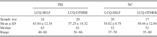
Twenty-four individuals with TBI and 20 normal healthy comparison (NC) participants were recruited for this study. Individuals with TBI were in the chronic stage of their injury (>6 months post injury, as studies have found that most of the recovery occurs in the 6 months following a TBI; Pagulayan, Temkin, Machamer, & Dikmen, Reference Pagulayan, Temkin, Machamer and Dikmen2006) and recruited through the University of Iowa Hospital. For all participants, TBI history was assessed though a combination of medical records and interview with the participant. TBI severity was assigned using the Mayo Classification System (Malec et al., Reference Malec, Brown, Leibson, Flaada, Mandrekar, Diehl and Perkins2007). All participants were classified as moderate to severe, as they all met at least one of the following criteria: (A) Glasgow Coma Scale score lower than 13; (B) positive acute computed tomography findings or focal lesions visible on a chronic MRI; (C) loss of consciousness longer than 30 min; (D) post traumatic amnesia longer that 24 hr. One participant had sustained two TBIs. Cause of injury included motor/non-motor vehicle accidents (9), assaults (1), and falls (15) (see Supplementary Table S1). Participants had no identified aphasia, as determined by screening from a certified speech language pathologist and/or by a score of 93.8 or higher on the Western Aphasia Battery bedside form (Shewan & Kertesz, Reference Shewan and Kertesz1980).
NCs were recruited from the Iowa City community and were included in the study if they had no history of psychiatric, neurological, or learning disorders and if they were compatible with MRI environment. TBI and NC groups did not significantly differ in average age (t(42)=.02; p>.05); sex (χ 2 (1,1)=.3; p>.05), or years of education (t(42)=1.9; p>.05). The study was approved by the Institutional Review Board of the University of Iowa.
Behavioral Data
Participants completed the LCQ (Douglas et al., Reference Douglas, Bracy and Snow2007) as part of a larger battery. Briefly, the LCQ is a two-form questionnaire that measures perceived communication problems in individuals with a history of TBI using two forms: a self-evaluation form administered directly to the participant (LCQ-SELF), and another form administered to a close other (CO) nominated by the participant, who is instructed to evaluate the participant (LCQ-OTHER). LCQ-SELF and LCQ-OTHER are composed of the same 34 items, which include questions about the participant’s ability to initiate and maintain conversational flow, converse effectively, be sensitive to the conversational partner’s needs, and inhibit inappropriate responses (Struchen, Pappadis, et al., Reference Struchen, Pappadis, Mazzei, Clark, Davis and Sander2008).
Items are phrased in the second person for the LCQ-SELF (e.g., When talking to others, do you leave out important details?) and in the third person for the LCQ-OTHER (e.g., When talking to others, does X leave out important details?). Respondents rate each item on a 1–4 Likert scale, where 1 represents “Never or rarely” and 4 represents “Usually or always.” Higher overall scores correspond to lower perceived communication quality. After participants completed the LCQ-SELF, they were asked to indicate a CO to whom experimenters could mail the LCQ-OTHER form. As in previous research (Despins et al., Reference Despins, Turkstra, Struchen and Clark2016), a CO was defined as someone with whom participants frequently communicated, such as a friend or a family member. To characterize relationships closeness, participants were also asked to complete the Inclusion of Other in the Self Scale (IOS) (Aron, Aron, & Smollan, Reference Aron, Aron and Smollan1992), which measures interpersonal closeness on a scale from 1 (very low closeness) to 7 (very high closeness). COs were asked to fill out both the LCQ-OTHER and the IOS in regard to the participant who had named them and their relationship with them. For the TBI group, 20 COs returned the LCQ-OTHER form, and 19 COs returned the IOS form. For the NC group, 17 COs returned both forms.
In addition to the LCQ, to characterize the sample participants, we administered the California Verbal Learning Test (CVLT-Immediate), short-delay verbal recall (CVLT-Short Delay) and long-delay verbal recall (CVLT-Long Delay) (Delis, Freeland, Kramer, & Kaplan, Reference Delis, Freeland, Kramer and Kaplan1988); the Symbol Search and Coding subtests of the Wechsler Adult Intelligence Scale (WAIS), obtaining a composite index of Processing Speed (WAIS-PSI) (Holdnack, Xiaobin, Larrabee, Millis, & Salthouse, Reference Holdnack, Xiaobin, Larrabee, Millis and Salthouse2011); and the Trail Making Test (Trails B) (Gordon, Reference Gordon1972). Finally, participants with TBI were administered the Wide Range Achievement Test – Reading Part (WRAT) to estimate premorbid IQ (Ahles et al., Reference Ahles, Saykin, Noll, Furstenberg, Guerin, Cole and Mott2003; Johnstone, Callahan, Kapila, & Bouman, Reference Johnstone, Callahan, Kapila and Bouman1996; Wilkinson & Robertson, Reference Wilkinson and Robertson2006). Due to study attrition, results from some neuropsychological tests were missing from non-overlapping participants in both groups, resulting in varying sample sizes for each test (CVLT: TBI=22, NC=19; Trails: TBI=22, NC=20; WAIS-PSI: TBI=24, NC=20; WRAT: TBI=23).
Neuroimaging Data Acquisition
Neuroimaging data were acquired at the University of Iowa Magnetic Resonance Facilities, on a 3 Tesla whole-body MRI scanner (Magnetom TIM Trio, Siemens Healthcare, Erlangen, Germany) with a 12-channel radiofrequency head receive coil. High resolution T1-weighted brain images were acquired using a three-dimensional magnetization prepared rapid gradient echo Imaging (MPRAGE) protocol with 208 contiguous coronal slices, echo time (TE)=3.04 ms, repetition time (TR)=2530 ms, field of view (FOV)=256 mm2, voxel size=1 mm3, and flip angle=10º.
DTI images were collected with 70 slices acquired in descending order, TE=86 ms, TR=9000 ms, voxel size=2 mm3, FOV=256 mm2, and flip angle=90º; one T2-weighted image (b-value=0 s/mm2) and one 64-direction diffusion-weighted echo planar imaging scan (b-value=1000 s/mm2) were collected. For one participant in the TBI group, only six directions were recorded due to problems during data acquisition. The participant was excluded from the neuroimaging sample (Final N=43), but included in the behavioral analyses (Final N=44).
Neuroimaging Data Analysis
Briefly, DTI data preprocessing was carried out in FSL 5.0.4 (Smith et al., Reference Smith, Jenkinson, Woolrich, Beckmann, Behrens, Johansen-Berg and Matthews2004) with the FMRIB’s Diffusion Toolbox (FDT) to correct for eddy currents and fit diffusion tensors (Behrens et al., Reference Behrens, Woolrich, Jenkinson, Johansen-Berg, Nunes, Clare and Smith2003) and with the Brain Extraction Tool (Smith, Reference Smith2002) to remove non-brain structures. The measure of interest for the current study was FA, an indicator of compactness and orientation of white matter fibers based on water’s unidirectional diffusion (Hulkower, Poliak, Rosenbaum, Zimmerman, & Lipton, Reference Hulkower, Poliak, Rosenbaum, Zimmerman and Lipton2013; Werring, Clark, Barker, Thompson, & Miller, Reference Werring, Clark, Barker, Thompson and Miller1999). Higher FA values correspond to higher directionality and integrity of white matter pathways (Basser, Reference Basser1995; Beaulieu, Reference Beaulieu2002; Sen & Basser, Reference Sen and Basser2005). FA was chosen over other scalar values (e.g., mean diffusivity, MD), as it has been suggested to be a better marker of white matter abnormalities (Cubon, Putukian, Boyer, & Dettwiler, Reference Cubon, Putukian, Boyer and Dettwiler2011); however, for completeness the same analyses were carried out for FA and MD data, and the results for MD data are reported in the Supplementary Information.
All images were run through a voxel-wise statistical analysis using Tract-Based Spatial Statistics (Smith et al., Reference Smith, Jenkinson, Johansen-Berg, Rueckert, Nichols, Mackay and Behrens2006): the nonlinear registration tool FNIRT (Andersson, Jenkinson, & Smith, Reference Andersson, Jenkinson and Smith2007a, Reference Andersson, Jenkinson and Smith2007b) was used to transform all subjects’ FA and MD data onto the FMRIB58_FA standard-space image and then to project each subject’s FA and MD data onto a mean skeleton.
First, to determine between-group differences in white matter integrity, we computed a whole-brain comparison of FA and MD skeletons using the nonparametric permutation tool randomise (Winkler, Ridgway, Webster, Smith, & Nichols, Reference Winkler, Ridgway, Webster, Smith and Nichols2014). We examined between-group differences in FA and MD values by building a general linear model that included as covariates sex, age, and education. Results were visualized by applying a threshold of p<.05 to the resulting family-wise error corrected FA and MD statistical maps. The JHU-White Matter Atlas and the JHU ICBM-DTI-81 White-Matter Labels Atlas (Hua et al., Reference Hua, Zhang, Wakana, Jiang, Li, Reich and Mori2008; Wakana et al., Reference Wakana, Caprihan, Panzenboeck, Fallon, Perry, Gollub and Mori2007) were used to identify white matter tracts where statistically significant clusters may indicate group differences in local white matter integrity.
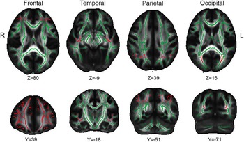
Next, for the primary analysis of interest, correlations between lobar FA and MD and communication abilities were computed using an a priori approach: a total of eight lateralized lobar regions of interest (ROIs) were created in FSLView and used to extract average lateralized lobar FA and MD values (Head et al., Reference Head, Buckner, Shimony, Williams, Akbudak, Conturo and Snyder2004; Voss et al., Reference Voss, Heo, Prakash, Erickson, Alves, Chaddock and Kramer2013). ROIs included right and left frontal, temporal, parietal, and occipital lobe (Figure 1). Frontal and temporal ROIs were used to test our hypothesis, while the other ROIs served as controls. Partial correlations were calculated between LCQ-SELF and OTHER scores and FA and MD values, accounting for sex, age, and education. Bonferroni correction was applied for multiple comparisons (.05/8=.0063; all p-values<.0063 were considered significant).
Lobar ROIs are shown in red, overlaying the white matter skeleton for the sample of the study. Skeleton and ROIs are presented on the FMRIB58_FA template. Left and right lobar ROIs were considered separately during FA analysis.

Lastly, to investigate the presence of specific white matter tracts whose integrity is associated with frequency of communication problems, we performed an exploratory analysis using the tool randomise, and setting as regressors LCQ scores (SELF and OTHER, in separate analyses) and adding age, sex, and education as covariates. These analyses were carried out both within the TBI and the NC group. Results of this analysis are reported in the Supplementary Information.
All T1 (MPRAGE) images were screened by an expert technician blinded to diagnosis for visible trauma-related focal lesions. Eight participants with TBI were identified as clearly displaying visible frontotemporal lesions.
Behavioral Data: Statistical Analysis
Although no attempts were made to match TBI and NC COs for sex or role (e.g., spouse, friend, family member) composition, we investigated whether the two groups were significantly different using a two-tailed chi-squared test. For LCQ data, there was one missing responses to a single item, which was substituted with the participant’s average score for the respective subscale. To examine the relationship between group, communication difficulties, and responder perception, we used a mixed effect analysis of variance (ANOVA) and investigated the presence of effects of Group (TBI vs. NC) and Respondent (LCQ-SELF vs. LCQ-OTHER) and of a Group-by-Respondent interaction. Main effect analyses were carried out using one-tailed Mann-Whitney tests (due to sample size and the non-normal distribution of LCQ-OTHER data) to test our directional hypothesis that individuals with TBI would show higher LCQ-SELF and LCQ-OTHER scores than NCs, and using two-tailed Wilcoxon signed-rank tests to test our non-directional hypothesis that LCQ-SELF scores would significantly differ from LCQ-OTHER in both the TBI and NC group. The same procedure was used to investigate significant group differences between IOS-SELF and IOS-OTHER.
One-tailed independent sample t tests were used to determine group differences for neuropsychological tests, based on the expectation that individuals with TBI would perform worse than NCs. Pearson correlation was used to examine the within-group association between LCQ-OTHER and LCQ-SELF and neuropsychological measures, applying Bonferroni correction and setting a significance level of p<.008. As one participant with TBI was not able to finish Part B of Trails, we re-coded his score with the lowest values from the dataset (Costa, Reference Costa2014).
RESULTS
La Trobe Communication Questionnaire
A chi-squared test to examine the composition of the CO group test revealed that TBI and NC CO groups did not differ for sex (χ 2 (1)=1.15; p>.05). Based on participants’ reports, COs were identified as spouses (including husbands, wives, domestic partners, boyfriends, girlfriends, and so on), non-spouse family members (e.g., parents, siblings), or friends. A chi-squared test showed no significant difference in CO compositions between the TBI and NC group (χ 2 (2)=.08; p>.05) (Table 1).
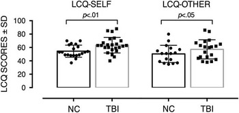
LCQ scores across groups

LCQ=LaTrobe Communication Questionnaire.
A mixed-effect ANOVA on LCQ data revealed significant main effects of Group (F 1,35=5.19; p<.05; ηp 2 =.15) and Respondent (F 1,35=4.47; p<.05; ηp 2 =.13), but no significant Group-by-Respondent interaction (F 1,35=.06; p>.05; ηp 2 =.002). Individuals with TBI had significantly higher LCQ-SELF scores (U=135; p<.01; r=.26) and LCQ-OTHER scores (U=117; p<.05; r=.38) when compared with NCs. This indicates that participants with TBI self-reported more communication problems and were also rated by their COs as having more communication difficulties than NCs. In addition, there was no significant difference between LCQ-SELF and LCQ-OTHER scores within the TBI group (Z=1.75; p>.05; r=.39) or the NC group (Z=1.58; p>.05; r=.38), meaning that on average the self-ratings of communication skills of TBIs or NCs were not significantly different from their COs ratings (Figure 2).
Individuals with TBI reported significantly more communication difficulties than NCs. In addition, COs reported that individuals with TBI have more communication difficulties than NCs. There were no significant differences between LCQ-SELF and LCQ-OTHER scores within the TBI or NC groups.

Inclusion of Other in the Self Scale
A mixed-effect ANOVA showed no significant main effect of Group (F 1,34=.66; p>.05; ηp 2 =.02), main effect of Respondent (F 1,34=.6; p>.05; ηp 2 =.02), nor a Group-by-Respondent interaction (F 1,34=3.3; p>.05; ηp 2 =.07). This reveals that participants in the TBI and NC groups did not differ in ratings of interpersonal closeness with their COs. Similarly, COs did not rate individuals with TBI as significantly more or less close than NCs. There was no significant difference in participants’ and COs’ ratings of closeness within either groups.
Correlations between LCQ and Neuropsychological Indexes
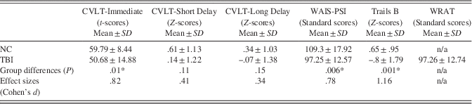
We first used one-tailed t tests to examine group differences on neuropsychological tests. When correcting for multiple comparisons, participants with TBI performed significantly worse than NCs on the CVLT-Immediate (t(39)=2.4; p=.01; d=.82), on the Trails B (t(40)=3.32; p<.01; d=1.16), and on the WAIS-PSI (t(42)=−2.6; p<.01; d=.78) (Table 2).
Comparison of performance on neuropsychological tasks across groups

* Results statistically significant following Bonferroni correction for multiple comparisons are marked with an asterisk.
CVLT=California Verbal Learning Test; WAIS-PSI=Wechsler Adult Intelligence Scale-Processing Speed Index; WRAT=Wide Range of Achievement Test.
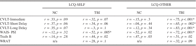
Correlations between LCQ and neuropsychological scores were carried out to examine the relationship between self- and other-reported communication abilities and cognitive skills. Within the TBI group, the only test showing significant negative correlation with the LCQ-SELF was the WAIS-PSI (r=−.58, p=.001; all other rs>−.44, ps>.02): participants with more self-reported communication problems had slower processing speed. Within the NC group, there were no significant correlations between LCQ-SELF and any of the neuropsychological measures (−.15<all rs<.37, ps>.05). In the TBI group, LCQ-OTHER scores significantly correlated with CVLT–Immediate (r=−.75; p<.001), CVLT-Short Delay(r=−.65; p<.01), CVLT-Long Delay (r=−.83; p<.001), and WAIS-PSI (r=−.54; p<.01). In the NC group, there were no significant correlations between LCQ-OTHER and neuropsychological tests (all rs<−.53; ps>.02) (Table 3).
Correlations between LCQ scores and performance on neuropsychological tests

Note. Pearson correlations between performance on neuropsychological tests and LCQ-SELF and OTHER within the TBI and NC group.
* Correlations statistically significant following Bonferroni correction for multiple comparisons are marked with an asterisk.
LCQ=LaTrobe Communication Questionnaire; CVLT=California Verbal Learning Test; WAIS-PSI=Wechsler Adult Intelligence Scale-Processing Speed Index; WRAT=Wide Range of Achievement Test.
White Matter Integrity: Between-Group Comparisons
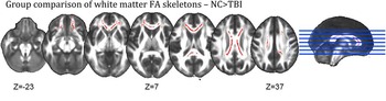
Individuals with TBI had significantly lower FA than NCs in bilateral frontal and occipital lobes and the corpus callosum. In particular, the TBI group had lower FA in the forceps minor; anterior thalamic radiation; frontal parts of the bilateral uncinate fasciculi; bilateral inferior fronto-occipital fasciculi; genu, body, and splenium of the corpus callosum; forceps major; and posterior part of the cingulum (Figure 3).
A whole-brain comparison of FA skeletons using the nonparametric permutation tool randomise with 5000 permutations and adding sex, age, and adding sex and age as covariates. The resulting FA family-wise error-corrected statistical map is shown in red thresholded at .95 (5% confidence interval) and overlaid on the FMRIB58 1 mm template. Individuals with TBI had significantly lower FA than NCs in bilateral frontal (uncinate fasciculi, fronto-occipital fasciculi) and occipital lobes (forceps mayor) and the corpus callosum (genu, body, and splenium).

White Matter Integrity: Correlations with LCQ
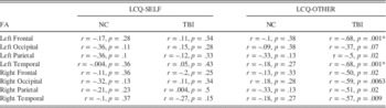
LCQ-SELF scores did not significantly correlate with lobar FA in the TBI group (−.27> all rs<.15 and; all ps>.05 ) or the NC group (all rs<−.36; all ps>.05) (Table 4). In the TBI group, LCQ-OTHER scores significantly and negatively correlated with left frontal lobe FA (r=−.68; p<.0063), left temporal lobe FA (r=−.68; p<.0063), with a marginal correlation with right occipital lobe FA (r=−.59; p=.0063). This indicates that participants with lower left frontal and temporal white matter integrity were perceived by COs to have more communication problems. There were no significant correlations between LCQ–OTHER scores and other lobar ROIs FA within the TBI group (all rs>−.57; all ps>.0063) or within the NC group (−.33> all rs<.18; all ps>.05).
Correlations between LCQ scores and lobar white matter FA

Note. Partial correlations, using sex, age, and education as covariates, between LCQ-SELF and OTHER and lobar FA within the TBI and the NC group.
* Correlations statistically significant following Bonferroni correction for multiple comparisons are marked with an asterisk.
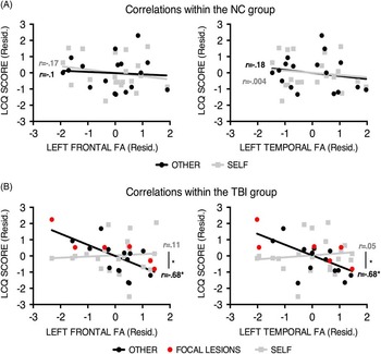
A Fisher r-to-z transformation revealed that within the TBI group the correlation coefficients between LCQ-OTHER and left frontal FA and the correlation between LCQ-SELF and left frontal FA were significantly different (z=2.11; p<.05). Similarly, within the TBI group the correlation coefficients between LCQ-OTHER and left Temporal FA and LCQ-SELF and left Temporal FA were significantly different (z=2.56; p<.05). When accounting for factors such as presence of focal lesions, chronicity, or cause of injury, the correlations between frontal and temporal FA and LCQ-OTHER remained significant (see Supplementary Information) (Figure 4).
(A) No significant correlations between frontal or temporal white matter FA and LCQ-SELF or LCQ-OTHER scores within the NC group. (B) Significant and negative correlations between both frontal and temporal white matter FA and LCQ-OTHER scores, but not with LCQ-SELF scores. For both frontal and temporal ROIs, the correlation coefficients between FA and LCQ-OTHER were significantly higher than the correlation coefficients between FA and LCQ-SELF. Participants with frontotemporal lesions are marked in red, showing that individuals with frontotemporal lesions did not necessarily have lower left frontal or temporal FA and that FA analysis provided more detailed information on lobar integrity than lesion identification.

Given the results, we also explored potential effects of lateralization. Within the TBI group, the difference between the correlation of LCQ-SELF with left Frontal FA and the correlation of LCQ-SELF with right Frontal FA was not significant (z=.82; p>.05). Similarly, the difference between the correlation of LCQ-SELF with left temporal FA and the correlation of LCQ-SELF with right temporal FA was not significant (z=.53; p>.05).
Follow-up analyses revealed no significant correlations between frontotemporal white-matter and scores on other neuropsychological tests (see Supplementary Information).
DISCUSSION
This study examined whether communication problems were associated with specific patterns of axonal injury in adults with TBI. Replicating previous studies, our results revealed more self- and other-reported communication problems in adults with TBI than in typical adults, as well as links between impairments and communication problems (Despins et al., Reference Despins, Turkstra, Struchen and Clark2016; Douglas, Reference Douglas2010; Douglas et al., Reference Douglas, Bracy and Snow2007; Struchen, Pappadis, et al., Reference Struchen, Pappadis, Mazzei, Clark, Davis and Sander2008). Novel findings in this study were that other-reports of communication problems were linked to lower white matter integrity in the left frontal and temporal lobes in participants with TBI. These findings are discussed in detail next.
Analysis of LCQ indicated that participants with TBI report more communication problems and are considered by COs as having more communication problems than healthy controls. The strong correlations between both DTI and scores on neuropsychological tests and LCQ-OTHER, compared to the lack of correlations with LCQ-SELF, support that self-perception of communicative problems might be altered in individuals with TBI and that LCQ-OTHER might be a more accurate measure of communication difficulties after TBI. Of interest, premorbid IQ was the only neuropsychological variable that did not correlate with CO-perceived communication problems, supporting the notion that the LCQ measures post injury communication issues.
In the NC group, there were no significant correlations between neuropsychological variables and LCQ scores. This finding suggests a strong relationship between communication problems and factors such as verbal memory and processing speed among individuals whose cognitive functions are suddenly and chronically disrupted due to brain injury but not in a healthy population. Another possibility is that communication problems only become apparent to close others when one or more cognitive skills are effectively impaired. This explanation is supported by the fact that in our sample individuals with TBI performed worse than NCs on several neuropsychological tests.
The numerous significant correlations between verbal memory, verbal learning, processing speed, and other-perceived communication problems support the idea that communication is a complex cognitive ability, requiring the orchestration and integrity of several different cognitive domains. Moreover, the fact that all participants were free of aphasia supports the notion of dissociations between basic linguistic and overall communication abilities. In the context of diffuse damage and a constellation of cognitive disruptions, communication deficits in TBI are more likely to be due to a failure in the successful and timely coordination of different skills and resources during interpersonal interaction.
The present study aimed to investigate whether participants with TBI with higher communication abilities show more white matter integrity in the temporal and frontal lobe and found that left frontotemporal FA was significantly associated with communicative difficulties reported by COs. However, follow-up analyses revealed that processing speed and verbal memory and learning were not significantly associated with frontotemporal white matter integrity; thus, the strong association between left frontal and temporal FA and communication difficulties could not be reduced to be completely explained by the relationship between white matter integrity and processing speed or verbal memory and learning.
We hypothesized that frontal lobe damage would be significantly associated with communication skills because of its importance for working memory, planning, response inhibition, and linguistic production (Bernicot & Dardier, Reference Bernicot and Dardier2001; Duncan & Owen, Reference Duncan and Owen2000), all indexes crucial to achieve successful communication and measured by the LCQ. Similarly, the temporal lobe has been found to be important for both language and cognitive functions highly related to memory (Duff & Brown-Schmidt, Reference Duff and Brown-Schmidt2012) and social processing functions including theory of mind, biological motion perception (Beauchamp, Reference Beauchamp2015), emotion recognition, and pragmatic language use (Saxe, Reference Saxe2006). Indeed, interpersonal and communication impairments are the hallmarks of frontotemporal dementia, a neurodegenerative condition characterized by progressive atrophy of frontal and temporal lobes (Edwards-Lee et al., Reference Edwards-Lee, Miller, Benson, Cummings, Russell, Boone and Mena1997).
Our results indicate that participants who have identifiable frontotemporal lesions based on their T1 images are not necessarily worse communicators. However, when focusing on lobar axonal injury instead of lobar atrophy, the association between frontotemporal damage and communication deficits becomes clearer, supporting the involvement of these brain regions in communicative abilities and providing a possible explanation for the individual differences in social functioning found in TBI populations. Of interest, although in our TBI sample disruptions in white matter could be found throughout the brain, only changes in frontal and temporal FA correlated with communication ability.
Furthermore, our results suggest a role of the left, but not of the right, temporal and frontal lobes in communication after TBI. This finding was somewhat surprising, because although previous research has found that the left frontal and temporal lobes play a more critical role in language production (Vigneau et al., Reference Vigneau, Beaucousin, Herve, Duffau, Crivello, Houde and Tzourio-Mazoyer2006), the right hemisphere is involved in the more pragmatic aspects of language (Champagne-Lavau, Stip, & Joanette, Reference Champagne-Lavau, Stip and Joanette2007; Tompkins, Lehman, Wyatt, & Schulz, Reference Tompkins, Lehman, Wyatt and Schulz1998), which are evaluated by the LCQ. However, the correlations between LCQ-OTHER and left-hemisphere ROIs were not significantly different from the correlations with right-hemisphere ROIs, indicating no statistically significant lateralization of findings, and that LCQ-OTHER correlations with right-hemisphere ROIs might be significant in a larger sample.
Furthermore, correlations between frontotemporal FA and communication were present in the TBI sample, but not in the NC group. This finding suggests that lobar FA does not play a role in communication abilities pre-injury and that its negative association with communicative difficulties is limited to TBI populations. One possible interpretation is that communication disruption occurs not as a consequence of frontotemporal damage, but following widespread brain damage, and that injured individuals with higher frontotemporal WM integrity are better able to recruit the cognitive abilities necessary to achieve successful interpersonal interaction and navigate the social world. Although the use of a single neuroimaging modality prevents us from reaching further conclusions, the addition of other structural (e.g., voxel based morphometry) and functional (e.g., resting state fMRI) modalities might further clarify the role of different brain regions in communication skills by allowing the adoption of a system perspective and elucidating the role of lobar DAI in large-scale functional organization. Moreover, it should be mentioned that other white-matter imaging methods, such as susceptibility-weighted imaging (SWI) or fluid attenuation inversion recovery (FLAIR) could also provide converging methods to evaluate white matter damage, which could further improve precision in determining the relationship between white matter damage and communication abilities for both scientists and clinicians.
Lastly, collecting both neuroimaging data and communication information longitudinally following a TBI would aid in determining whether the association between frontotemporal white matter integrity and communicative skills is also evident in acute and sub-acute settings, or whether this association results from changes and reorganizations that occur over time. This knowledge will be critical to advance our ability to make timely and accurate diagnostic statements and to more efficiently deploy clinical services.
Limitations
Our results should be viewed within the context of several limitations. First, our dataset included several missing data-points due to both the fact that LCQ-OTHER forms were mailed to COs and to the still ongoing data collection. In addition, a limitation of the study is the relative small sample size, especially considering the great inter-individual variability that has been reported in moderate to severe TBI populations. It should also be noted that, although every effort was made to match the TBI and comparison group for age, sex composition, and education, we could not account for possible risk factors that might predispose certain individuals to TBI, and which might affect DTI and LCQ results.
As mentioned above, another limitation lies in the lack of imaging information necessary to examine the presence of visible white matter or gray matter damage. Although by using the MP-RAGEs we were able to ascertain that no patients had obvious overlaps between focal lesions and DTI ROIs, it should be kept in mind that ours was a relatively coarse analysis, as the images available to us were not ideal to identify small contusions or to quantify atrophy. Future studies should examine more closely the relationship between white matter damage and brain atrophy and lesions, to rule out definitively the possibility that gray matter damage can account for the association between white matter integrity and communication difficulties.
Finally, as this was the first study investigating neural correlates of communication in TBI, we decided to use as dependent variables the total LCQ scores, without exploring the different dimensions of communication, such as conversational flow or partner sensitivity. Future work should examine whether different aspects of communication impairment are related to different neuropsychological variables or patterns of white matter damage.
CONCLUSIONS
This study aimed to identify links between diffuse axonal injury and communication problems in adults with TBI. Results showed that white matter integrity related to frequency of communication problems reported by communication partners of adults with TBI. This knowledge has the potential to guide the development of diagnostic tools that differentiate individuals with TBI at high risk of communication impairment during the acute phase of their brain injury as well as shed light on the mechanisms underlying communication impairment in adults with TBI.
Acknowledgments
This work was supported by NICHD/NCMRR grant R01 HD071089, the University of Iowa Magnetic Resonance Research Facilities and a research grant from the University of Iowa Graduate and Professional Student Government. The authors thank Joel Bruss, Ruth Hansen, and Kristina Warndahl for their help. The authors have no conflicts of interest to disclose.
Supplementary material
To view supplementary material for this article, please visit http://dx.doi.org/10.1017/S1355617716000539