Pulmonary agenesis is a rare congenital abnormality, defined as the complete absence of bronchus, parenchyma, and associated blood vessels.Reference Furia, Biban, Benedetti, Terzi, Soffiati and Calabro 1 In cases of right lung agenesis, the heart can be rotated into the right pleural cavity. As a result, the airway tends to be compressed between the aorta and the vertebral body. Here, we discuss a case of right lung agenesis in a 1-month-old baby.
Case report
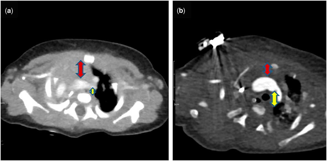
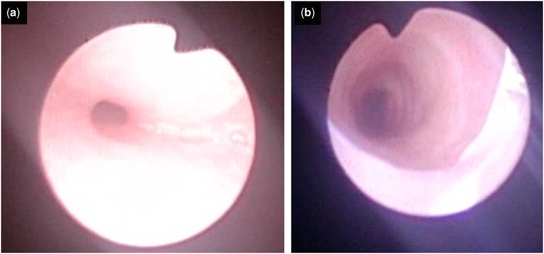
The patient was born in our hospital at a gestational age of 37 weeks, weighing 2.05 kg. His initial Apgar score was 4 at 1 minute and 5 at 5 minutes. He was admitted to the neonatal ICU where CT revealed right lung and bronchus agenesis (Fig 1a). No airway problems were evident at the time. After neonatal management, the patient was discharged. However, he was subsequently readmitted to the hospital because of respiratory distress, which began 1 week after initial discharge. Airway status was re-evaluated by CT scan and bronchoscopy, confirming almost complete compression of the trachea between the aorta and the vertebral body (Figs 1a and 2a). The severely compressed airway was considered to be the cause of the repeated respiratory distress. Therefore, anterior aortopexy was planned to pull the ascending aorta anterior up to the sternum.
Chest CT before and after aortopexy with thymectomy. ( a ) Before aortopexy, ( b ) after aortopexy.

Bronchoscopic findings before and after aortopexy with thymectomy. ( a ) Before aortopexy, ( b ) after aortopexy.

Partial sternotomy was performed from xiphoid to just below the level of the manubrium. The manubrium remained intact and was used to anchor the ascending aorta. The majority of the thymic tissue was removed, providing sufficient space in the anterior mediastinum. The pericardium was opened. Without further dissection or mobilisation of great arteries, two 4-0 Ethibond pledgeted sutures were placed at both lateral sides of the distal ascending aorta where the trachea was mostly compressed on CT findings. The two sutures penetrated the second intercostal spaces of both sides of the manubrium; the left- and right-side sutures penetrated each side of the manubrium. As a result, these sutures pulled the ascending aorta to the sternum and leftwards with adjacent structures which compressed the trachea. We pulled these sutures until the anchored ascending aorta almost reaches the anterior chest wall. After bronchoscopic confirmation of an improvement in the extrinsic compression of the tracheal lumen, the sutures were tied and fixed outside the anterior chest wall (Fig 2b). The patient was transferred to the paediatric ICU with an endotracheal tube for mechanical ventilation because of an airway secretion. Extubation was possible at post-operative day 3 without respiratory difficulty. Follow-up CT showed a widened space between the aorta and the vertebral body and improved extrinsic compression (Fig 1b). The patient was discharged at 24 days post-surgery following management of the urinary tract infection and received follow-up in the outpatient clinic. To date, he has progressed well and has shown acceptable weight gain and no respiratory difficulties.
Discussion
Most patients with right lung agenesis suffer from airway problems as a result of tracheal or bronchial compression.Reference Agrawal, Patri and Kalavakunta 2 Of the available surgical interventions (such as diaphragm translocation or the application of tissue expander in the empty pleural cavity), we selected aortopexy and near total thymectomy to relieve tracheal compression, as suggested by Dohlemann et al.Reference Furia, Biban, Benedetti, Terzi, Soffiati and Calabro 1 – Reference Dohlemann, Mantel, Schneider, Guntner, Kreuzer and Hecker 3 As the use of tissue expanders requires some time to fill the pleural cavity, we would not have achieved a rapid response for our patient. We did not consider the use of rapid expansion of a tissue expander for fast weaning from the ventilator to be appropriate because of the risk of haemodynamical instability. Regarding diaphragm translocation, this approach is challenging in such a young patient in the peri-neonatal period because the tissue is fragile and not mature enough to tolerate the required level of dissection and suture.
Our approach differed to that reported in the previous trial. First, we did a lower partial sternotomy with preservation of the manubrium of the anterior chest wall, which served as the anchoring area. In our experience, we have seen cases where funnel chest deformity after anterior aortopexy developed over time in young patients whose sternum had not completely matured, probably owing to the force exerted by the anchoring aortic sutures. We therefore consider that preservation of the manubrium minimises the risk of chest wall deformity after aortopexy. Second, we removed the majority of the thymus. This helps not only to obtain a space in which to move the ascending aorta to the anterior chest wall but also to relieve compression by the thymus itself.
In conclusion, the surgical approach described here is less invasive than other surgical options and may be particularly effective in the peri-natal patients with airway compression accompanying right lung agenesis.
Acknowledgements
None.
Conflicts of Interest
None.