Introduction
Tuberculosis is an infectious disease which has destructive effects unless treated. It most commonly affects the lungs, but can also affect the central nervous system, lymphatic system, circulatory system, genitourinary system, bones, joints and even the skin. The prevalence of the condition declined until the mid-1980s but since that time has begun to rise, due to acquired immunodeficiency syndrome and to emerging Mycobacterium tuberculosis drug resistance.Reference Harisinghani, McLoud, Shepard, Ko, Shroff and Mueller1
Tuberculosis otomastoiditis and tuberculosis meningitis are rare forms of extrapulmonary tuberculosis. Therefore, infections in these areas may be overlooked in the differential diagnosis, even in the absence of pulmonary tuberculosis findings.
Early diagnosis and treatment of tuberculosis otomastoiditis is important in order to prevent progression of the disease and development of neurological complications.Reference Cavallin and Muren2 Tuberculosis meningitis may be a complication of tuberculosis otomastoiditis. Miliary tuberculosis may occur concomitantly with tuberculosis otomastoiditis. Since the clinical findings of tuberculosis otomastoiditis are non-specific, the diagnosis may be difficult, especially when the condition occurs together with tuberculosis meningitis, which presents a severe clinical picture.Reference Vaamonde, Castro, Garcia-Soto, Labella and Lozano3
Materials and methods
Patient population
A retrospective chart review of hospital medical records identified 32 patients (21 male, 11 female) who had been diagnosed with tuberculous meningitis between 1999 and 2006. The patients' average age was 24.2 years (standard deviation 1.4 years).
Magnetic resonance imaging
A 1.5 T magnetic resonance imaging (MRI) scanner (Siemens Magnet Vision, Erlangen, Germany) was used. We obtained the following images: conventional T1-weighted (640/14 (Time of Repetition (TR)/Time of Echo (TE)) matrix, 192 × 256); T2-weighted (3900/99 (TR/TE) 264 × 512); fluid-attenuated inversion recovery (inversion time 2500 ms; 9000/110 (Gulhane Military Medical Academy Haydarpasa Teaching hospital) matrix, 132 × 256); and contrast-enhanced T1-weighted.
The diagnosis of tuberculous meningitis was based on the clinical picture (including the patient's response to antibiotic therapy) and on laboratory and clinical findings. In cases diagnosed radiologically with otomastoiditis, we reviewed the patient's previous history of tuberculous meningitis, audiological examination findings and response to anti-tuberculous treatment. Patients with otomastoiditis who had no response to long-term, non-tuberculous antibiotic treatment and who demonstrated a dramatic radiological improvement following anti-tuberculous treatment were regarded as positive for tuberculous otomastoiditis. Patients without otomastoiditis, and those with otomastoiditis who did not respond to anti-tuberculous treatment and did not have clinical or laboratory findings supporting tuberculosis, were regarded as negative for tuberculous otomastoiditis.
Each case was separately reviewed by two radiologists who were blinded to all patients' medical records. Two reviewers independently rated each patient as positive or negative for tuberculous otomastoiditis. Any discrepancies between the two reviewers were resolved by consensus.
Results
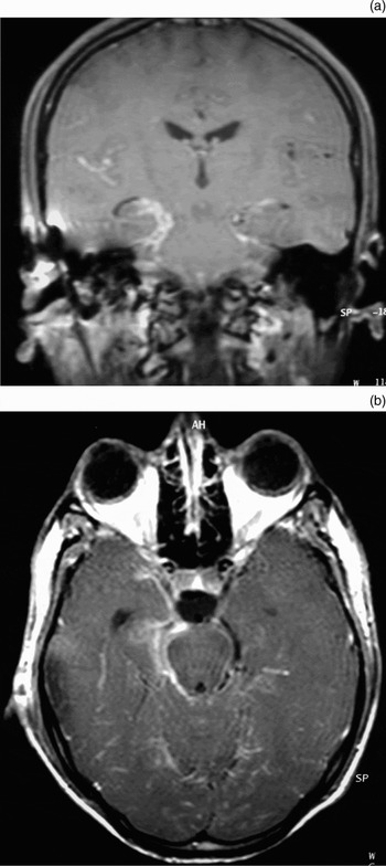
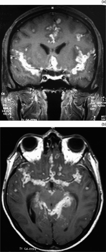
The meningeal involvement site was unilateral (sylvian fissure and perimesencephalic cistern) in 28 patients (87.5 per cent), and bilateral and widespread in four patients (12.5 per cent) (Figure 1). Two patients (6.25 per cent) had parenchymal involvement (in the form of multiple tuberculomas), in addition to meningeal involvement (Figure 2).
(a) Coronal and (b) axial, contrast-enhanced, T1-weighted magnetic resonance images showing meningeal enhancement in the right perimesencephalic cistern and sylvian fissure. Note the ipsilateral otomastoiditis.

(a) Coronal and (b) axial, contrast-enhanced, T1-weighted magnetic resonance images showing extensive meningeal enhancement and multiple parenchymal tuberculomas.

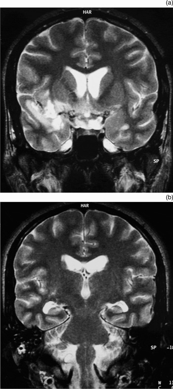
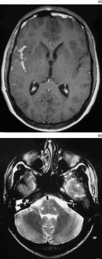
Tuberculous otomastoiditis was determined to be present in 11 of the tuberculous meningitis patients (34.3 per cent). Four of these were women and seven were men. The otomastoiditis was on the same side as the meningeal involvement in nine of these 11 patients (Figures 3 and 4). Bilateral otomastoiditis was seen in two patients with widespread meningeal involvement.
(a) Coronal, T2-weighted magnetic resonance imaging (MRI) scan showing signal intensity increase in the grey matter adjacent to the right sylvian fissure. (b) Coronal, T2-weighted MRI scan through a different level, showing high signal intensity in the right mastoid cells.

Another patient with ipsilateral meningeal enhancement and otomastoiditis. (a) Axial, contrast-enhanced, T1-weighted magnetic resonance imaging (MRI) scan showing meningeal enhancement in the right sylvian fissure. (b) Axial, T2-weighted MRI scan of the same patient, showing high signal intensity in the ipsilateal middle-ear cavity and mastoid cells.

Discussion
Tuberculous infection in the central nervous system generally arises as the result of haematogenous spread from a primary focus. It exists in two forms: tuberculous meningitis and intracranial tuberculoma. Tuberculous meningitis is the most common manifestation of neurotuberculosis. It is frequently seen in children, but may be observed in all age groups.Reference Harisinghani, McLoud, Shepard, Ko, Shroff and Mueller1, Reference Mutlu, Silit, Pekkafali, Basekim, Kantarcı and Kizilkaya4, Reference Ozates, Kemaloglu, Gurkan, Ozkan, Hosoglu and Simsek5 The most frequent sites of tuberculous meningitis are the sylvian fissure, the chiasmatic cistern and the perimesencephalic cistern (92 per cent).Reference Ozates, Kemaloglu, Gurkan, Ozkan, Hosoglu and Simsek5 The MRI findings vary depending on the stage of the disease. In the early stages, unenhanced spin echo images may be normal. In the later stages, T1 and T2 relaxation times become shorter than the cerebrospinal fluid. Increased meningeal enhancement compared to the normal meninges could be seen in contrast-enhanced T1-weighted images. The sylvian fissure and perimesencephalic cistern were involved in 87.5 per cent of our cases; post-contrast images showed strong enhancement of these areas.
Tuberculous infection of the middle-ear cavity and mastoid cavity comprises approximately 0.04 to 0.06 per cent of all cases of suppurative otitis media.Reference Weiner, O'Connell and Pahor6, Reference Mongkolrattanothai, Oram, Redleaf, Bova and Englund7 Infection occurs due to haematogenous or lymphogenic spread from a previously existing tuberculous focus, by means of the eustachian tube, or by direct implantation from the perforated tympanic membrane.Reference Awan and Salahuddin8–Reference Cho, Lee, Kim, Chung, Lee and Koh10 There are no other tuberculous findings elsewhere in the body in 40 to 50 per cent of patients.Reference Awan and Salahuddin8
Diagnosis of tuberculous otomastoiditis is made difficult by the rarity of the condition, the variability of clinical findings, the fastidious nature of M tuberculosis, the frequency of false negative cultures, and superinfection by other bacterial infections.Reference Vaamonde, Castro, Garcia-Soto, Labella and Lozano3, Reference Awan and Salahuddin8, Reference Cho, Lee, Kim, Chung, Lee and Koh10 Since tuberculous otomastoiditis leads to serious complications, such as hearing loss, facial nerve palsy, meningitis, multiple cranial nerve palsies and sigmoid sinus thrombosis, early diagnosis is of great importance.Reference Saunders and Albert11–Reference Chiu, Pang, Juh and Huang13 Early diagnosis of tuberculous otomastoiditis depends on a high index of clinical suspicion, especially in regions where tuberculosis is common. In developed countries, tuberculous mastoiditis is generally overlooked.
The isolation of M tuberculosis, particularly in specimens taken pre- or intra-operatively, confirms the diagnosis of tuberculous otomastoiditis. In addition, since isolation of the tuberculous bacillus is not always possible, analysis of clinical, radiological and histopathological findings and response to anti-tuberculous treatment are of great importance in establishing an early and accurate diagnosis.Reference Saunders and Albert11, Reference Pitcher and Thandar12, Reference Thandar, Fagan and Garb14, Reference Unuvar, Oguz, Sadikoglu, Sidal, Ones and Tetikkurt15 In our study, individuals' previous clinical findings, radiological findings and response to anti-tuberculous treatment were adopted as diagnostic criteria. Since ours was a retrospective study, we were unable to obtain material from the infection area or to isolate the tuberculous bacillus. This was a limitation of our study. However, it has been reported that there may be a high level of false negative results for histopathological investigations of tuberculous mastoiditis.Reference Weiner, O'Connell and Pahor6, Reference Saunders and Albert11 For that reason, we consider the clinical response to anti-tuberculous treatment to be one of the best diagnostic markers. Our diagnosis was further supported by the fact that meningeal involvement was on the same side in 81 per cent of our patients determined as having otomastoiditis.
• Otomastoiditis and meningitis are rare forms of extrapulmonary tuberculosis
• Early diagnosis and treatment of tuberculous otomastoiditis is important in preventing progression of the disease and neurological complications
• The aim of this study was to determine the correlation between tuberculous meningitis and tuberculous otomastoiditis
• Tuberculous meningitis is frequently accompanied by otomastoiditis, although the exact causal relationship between the two conditions is unclear
Tuberculous meningitis is a serious complication of tuberculous otomastoiditis. However, cases of tuberculous meningitis together with tuberculous otomastoiditis have rarely been reported.Reference Chiu, Pang, Juh and Huang13, Reference Bitsori, Galanakis, Kokori, Amanakis and Sbyrakis16, Reference Samuel and Fernandes17 Haematogenous and lymphogenous spread have been implicated more often than direct extension from the mastoid.Reference Harisinghani, McLoud, Shepard, Ko, Shroff and Mueller1, Reference Mongkolrattanothai, Oram, Redleaf, Bova and Englund7, Reference Lee, Chae, Lim, Hwang and Suh18
Awan and Salahuddin stated that tuberculous meningitis could be an initial indication of tuberculous otomastoiditis and otitis.Reference Awan and Salahuddin8 Samuel and Fernandes found tuberculous meningitis in two of 23 patients with tuberculous otomastoiditis.Reference Samuel and Fernandes17 We investigated mastoid involvement in patients with tuberculous meningitis and found tuberculous otomastoiditis in 11 of 32 patients. To our knowledge, no previous reports have indicated such a high level of correlation between tuberculous meningitis and tuberculous otomastoiditis. In our 11 patients with tuberculous otomastoiditis, the lesion sites, patient histories and laboratory findings suggested that tuberculous meningitis had developed as a complication of tuberculous otomastoiditis. Unfortunately, no radiological images were obtained during the onset of symptoms, before diagnosis of meningitis; this was another limitation of our study.
Conclusion
Tuberculous otomastoiditis frequently accompanies tuberculous meningitis. However, it is uncertain which condition causes the other. Since the clinical picture of meningitis is serious, accompanying otomastoiditis is generally overlooked. Possible tuberculous otomastoiditis must therefore be carefully investigated in patients with tuberculous meningitis. More importantly, the initial clinical picture may be tuberculous otomastoiditis or tuberculosis-related chronic suppurative otitis media. This pathology may represent a focus of tuberculous meningitis. In addition, tuberculosis must also be suspected in cases of suppurative otitis media or mastoiditis which do not respond to empirical antibiotic therapy, or when general bacteriological cultures of ear discharge are negative.