Introduction
Carcinoma breast is the most common cancer among Indian females Reference Malvia, Bagadi, Dubey and Saxena1 usually has a long disease-free survival (DFS) after adequate treatment. Breast cancer metastasize most commonly to bone, liver, brain and lung, while oesophagus is a rare site of metastasis. Reference Rampado, Ruol and Guido2 Autopsy studies reveal that most oesophageal metastases are subclinical and dysphagia due to stricture is commonly attributed to post-radiotherapy changes or other benign conditions. Reference Abrams, Spiro and Goldstein3,Reference Wada, Harada and Ohara4 Oesophageal metastases are usually submucosal depositions posing a challenge during diagnosis as they are usually missed in simple mucosal biopsies. Failure to detect secondary oesophageal cancers can eventually lead to decreased nutritional intake by the patient resulting in cachexia, increased morbidity and decreased survival of the patient. As a considerable percentage of oesophageal metastases go undetected during patients’ lifetime, there is a lack of consensus in management guidelines in this condition. Herein, we report a case of a 65-year-old female diagnosed with oesophageal metastases 14 years post-surgery for breast cancer. We also performed a comprehensive review of literature on breast cancer with oesophageal metastases, aiming to improve the management of this rare presentation.
Case Presentation
A 65-year-old female presented with progressive dysphagia for 1 year. 12 years back, she was diagnosed with carcinoma of left breast, pathological stage T2N1. Tumour was histologically confirmed as invasive ductal carcinoma and immunohistochemistry (IHC) studies were positive for estrogen (ER) and progesterone (PR) receptors and negative for Her2neu receptors. Patient underwent left modified radical mastectomy with axillary lymph node dissection, followed by seven cycles of CMF (Cyclophosphamide, Methotrexate, 5-fluorouracil). She then completed adjuvant radiotherapy of dose 45 Gray (Gy) in 20 fractions by conventional technique. She was later started on hormonal therapy and was asymptomatic for next the 11 years, when she was diagnosed with lung and bone metastases. Patient underwent four cycles of trastuzumab, letrozole and zoledronic acid. This was followed by external radiotherapy of dose 30 Gy in ten fractions to tender sites to palliate pain. Patient was further administered three cycles docetaxel and six cycles gemcitabine, but the disease progressed. Also, patient started presenting with mild dysphagia which was attributed to benign oesophageal stricture due to radiation-associated fibrosis as the initial biopsy from the stricture was negative for malignancy.
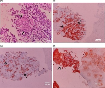
In the next year, complaints of dysphagia exacerbated. Due to the persistence of symptoms, upper gastrointestinal (GI) endoscopy was repeated which revealed submucosal mass with stricture at 23 cm from incisor. Also endoscopic-guided Ryle’s tube insertion was done to maintain adequate nutrition. Endoscopic ultrasound (EUS)-guided fine-needle biopsy (FNB) was performed and revealed an adenocarcinomatous lesion (refer Fig. 1A – focus of adenocarcinomatous changes in the oesophageal wall), and IHC was positive for transcription factor GATA3 (GATA Binding Protein 3) and Cytokeratin 7 (CK7) confirming a metastatic lesion and positive for ER and PR receptor indicating breast primary (refer Fig. 1B – oesophageal lesion showing ER positivity, Fig. 1C – oesophageal lesion showing CK7 positivity and Fig. 1D – oesophageal lesion showing GATA3 positivity). Positron emission tomography–computed tomography (PET CT) imaging revealed an asymmetrical lesion involving upper to mid-thoracic oesophagus of 4 cm length with standardised uptake value maximum of 4.8, along with residual bone and adrenal metastases. Patient was administered nine cycles of gemcitabine but yielded poor subjective and clinical response with regard to relieving dysphagia and patient symptoms eventually progressed to absolute dysphagia. CT thorax images presented a 5.4-cm lesion involving mid-thoracic oesophagus with maximum thickness of 6 mm. Gastroesophageal junction was not involved by the disease.
(A) Histopathological findings of oesophageal metastases by light microscopy. Highlighted by the arrows is the focus of poorly differentiated adenocarcinomatous. (B) IHC for ER shows strong (3+) positivity. Arrows indicate the strong presence of estrogen receptor in the metastatic cells. Scale bar – 100 μm. (C) IHC for CK7. Arrows highlight strong diffuse CK7 immunostaining. Scale bar – 100 μm. (D) IHC for GATA3. Arrows highlight strong diffuse GATA3 immunostaining. Scale bar – 200 μm.

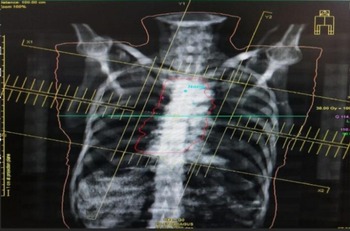
Patient general condition was good with Karnofsky performance score of 90 and weight of 43 kg. Patient was thus planned for short-course external beam radiotherapy (EBRT) followed by brachytherapy. Mid-oesophageal location of the lesion with the length of 5.4 cm made the patient a suitable candidate for intraluminal radiotherapy (ILRT). Oesophageal metastases were irradiated to 30 Gy in ten fractions, 3 Gy per fraction, five fractions per week by conformal EBRT technique (refer Fig. 2 – EBRT plan by conformal technique) followed by 13 Gy in two fractions by ILRT, achieving a total biological equivalent dose of 60.5 Gy. Patient responded well to radiotherapy, and dysphagia was relieved substantially. Patient was able to consume liquid and semisolid diet by the end of the treatment and gained 5 kg by the end of 3 months follow-up. Patient is currently on hormonal therapy and on regular 2-month follow-up.
External beam radiotherapy plan of the patient by conformal technique.

Patient has given written informed consent to publish the case, including accompanying images. Since this is case study, ethical approval is not needed.
Discussion
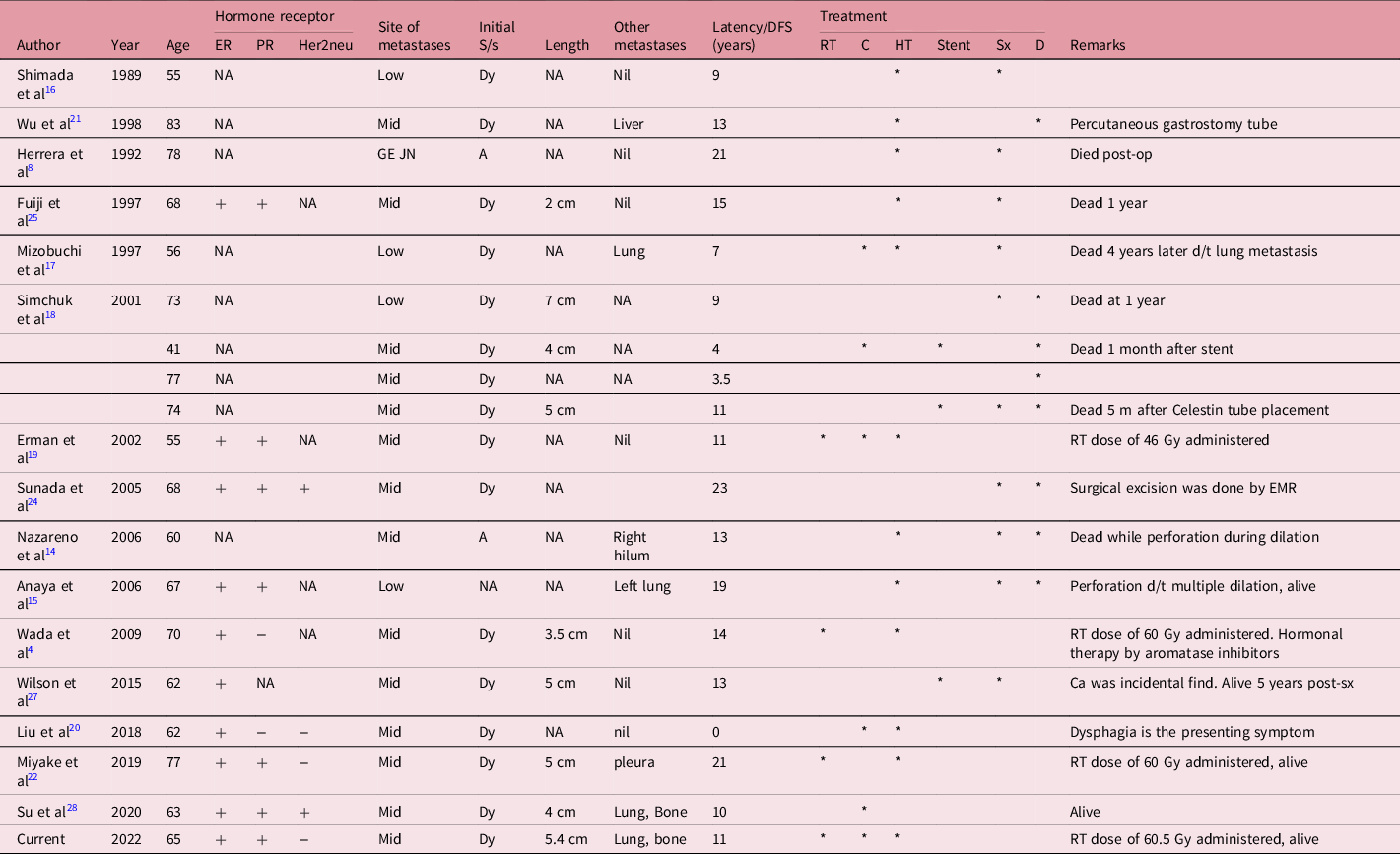
Carcinoma breast with oesophageal metastasis is a rare presentation comprising only 0.4%. Reference Borst and Ingold7 Most patients present initially with dysphagia, while other less common symptoms include achalasia Reference Herrera8 and other motility disorders. Reference Isaacs, MacGillivray and Springett9 Lesions most commonly arise in the middle and distal third of thoracic oesophagus, usually seen in post-menopausal women. Reference Rampado, Ruol and Guido2,Reference Wada, Harada and Ohara4 The mean time from mastectomy to the onset of dysphagia was calculated to be 7.1 ± 4.2 years by Anderson and Harell. Reference Anderson and Harell13 In this review of literature, we observed 83% patients presented with complaints of dysphagia after a median of 13 years post-breast surgery, with 72% metastases involving mid-thoracic oesophagus (Table 1). Mechanism of metastases is usually lymphatic, but intramural spread from breast primary is not uncommon. Reference Rampado, Ruol and Guido2
Oesophageal metastases from breast carcinoma in medical literature reviewed for our paper

Mid, middle 1/3rd oesophagus; Low, lower 1/3rd oesophagus; GE JN, gastroesophageal junction; NA, not available; Dy, dysphagia; A, achalasia; CA, carcinoma; RT, radiotherapy; C, chemotherapy; sx, surgery; HT, hormonal therapy; D, dilation.
Diagnosis of oesophageal metastases pose a considerable challenge as the metastatic deposition is submucosal with normal mucosal covering. Thus, most cases go undetected during patients’ lifetime, and this explains the aberration seen in the prevalence of 6% in autopsy studies Reference Graham and Goldman10 compared to clinically significant lesions which is less than 1%. While explorative surgeries like thoracoscopy and mediastinal exploration are diagnostic, EUS-FNB and endoscopic mucosal resection (EMR) are safe and effective alternatives. Reference Rampado, Ruol and Guido2,Reference Wada, Harada and Ohara4 In our case, initial biopsy from the oesophageal stricture site was negative for malignancy and EUS-FNB was needed to confirm diagnosis. Histopathological examination (HPE) and IHC studies are necessary to differentiate primary and secondary oesophageal lesions. Oesophageal metastases are usually hormonally receptive owing to their breast origin. In our patient, while HPE revealed adenocarcinomatous lesion, IHC was required to confirm metastatic lesion from breast.
Oesophageal metastases from breast primary usually present after long DFS. Reference Atkins11,Reference Polk, Camp and Walker12 In our case, the interval was 11 years, while the review of literature revealed a median of 13 years and mean of 12 years (Table 1). Breast cancer recurrence as isolated oesophageal lesion is uncommon, and by the time a patient becomes symptomatic for oesophageal lesion, they are already diagnosed with other sites of distant metastases. In our case, the patient presented with breast cancer recurrence involving bone, lung and oesophageal metastases. Thus, a curative treatment is not a legitimate approach to this presentation, and the goal is usually palliation of symptoms. Reference Rampado, Ruol and Guido2,Reference De Palma, di Matteo, Romano, Fimmano, Rondinone and Catanzano6
Management of secondary oesophageal cancers have undergone considerable change over the years. We reviewed a wide range of publications belonging to even late 20th century, to get a more holistic picture of evolution of treatment strategies used in his rare condition. Initially treatment included surgery, dilation and stent. Endoscopic dilation has been attempted to relieve symptoms, but evidence suggests that repeated dilation is associated with perforation. Reference Holyoke, Nemoto and Dao5,Reference Nazareno, Taves and Preiksaitis14,Reference Anaya, Yu and Karmy-Jones15 While expandable metal stents are effective in inoperable Reference Borst and Ingold7 and chemoresistant cases, they provide only a temporary palliation of symptoms and associated with a high incidence of oesophageal perforation. Reference Rampado, Ruol and Guido2 Surgical management in oesophageal metastases is ideally not justified due to palliative intent of treatment, but various surgical procedures have been practiced over the years to different outcomes. Adrenalectomy was shown to improve symptoms of dysphagia, especially in post-menopausal women. Reference Holyoke, Nemoto and Dao5 Explorative laparotomy was attempted by Herrera et al, Reference Herrera8 but the patient eventually died due to aspiration pneumonia and sepsis. Some studies have supported an aggressive surgical approach like oesophageal resection as a safe and effective palliation for oesophageal obstruction due to metastatic breast cancer, Reference Shimada, Imamura and Tobe16,Reference Mizobuchi, Tachimori, Kato, Watanabe, Nakanishi and Ochiai17 but preoperative assessment of disease status is necessary for achieving adequate resection. Reference Anaya, Yu and Karmy-Jones15 Surgical placement of Celestin tube was attempted by Simchuk et al, Reference Simchuk and Low18 but patient died due to massive upper gastrointestinal bleeding just 5 months post-surgery.
Recurrent metastatic disease requires systemic treatment, and thus chemotherapy is an effective treatment. Our patient was administered multiple cycles of docetaxel and gemcitabine, while other studies have supported the use of adriamycin, epirubicin and cyclophosphamide. Reference Erman, Karaoğlu, Öksüzoğlu, Aydıngöz, Ayhan and Güler19,Reference Liu, Feng, Chen, Li, Wu, Qian and Yang20 Oesophageal metastases are commonly seen in ER- and PR-positive patients, and thus hormonal therapy have been part of the treatment and are also effective in improving survival. Reference Wada, Harada and Ohara4,Reference Erman, Karaoğlu, Öksüzoğlu, Aydıngöz, Ayhan and Güler19 But some studies have reported exacerbation of dysphagia after years of hormonal therapy, thus warranting a local therapy. Reference Wada, Harada and Ohara4,Reference Miyake, Yamada, Miyake and Endo22 Radiotherapy was actively adopted for treatment only in the 21st century and practiced only by a handful of clinicians. Importance of local control on survival was emphasised by Miyake et al. Reference Miyake, Yamada, Miyake and Endo22 In our patient, radiotherapy was highly effective in the palliation of symptoms. Improvement of dysphagia after administration of EBRT has also been reported in recent studies. Reference Wada, Harada and Ohara4,Reference Miyake, Yamada, Miyake and Endo22 We also performed ILRT in this patient in addition to the short-course EBRT. The American Brachytherapy Society approves brachytherapy as an effective treatment modality for palliation of dysphagia. Short-course EBRT followed by ILRT, especially in patients with life expectancy >6 months, achieve superior palliation in a shorter duration than the conventional 5-week course EBRT. Reference Gaspar, Nag, Herskovic, Mantravadi and Speiser26
Therefore, it is principal that treatment should include both systemic therapy and local therapy for effective palliation of symptoms and disease control. At present, systemic chemotherapy with hormonal therapy and local radiotherapy for palliation of symptoms is the best approach for management of secondary oesophageal lesion from breast primary. Also, as oesophageal obstruction is associated with weight loss and cachexia due to decreased oral intake, it is pertinent to ensure adequate nourishment for patients. Ryle’s tube insertion, percutaneous gastrostomy tube and feeding jejunostomy tube have been attempted to aid patient’s nutrition. Reference Wu, Hruban and Fishman21
Breast cancer with oesophageal obstruction is usually associated with multiple distant metastases, and thus it is difficult to assess the effect of oesophageal metastases on survival. Goldberg et al Reference Goldberg, Rams, Stone and Barkin23 claimed that patients with oesophageal metastases survived only 5 years after dysphagia, but at the same time, oesophageal metastases are not an established prognostic factor. Thus, treatment should be decided by the severity of other metastases and on the degree of stenosis. Reference Miyake, Yamada, Miyake and Endo22 Even though oesophageal cancers are associated with terminal stage of disease, we have observed better survival than expected, which can be attributed to the effectiveness of multimodal treatment. Reference Wada, Harada and Ohara4,Reference Herrera8,Reference Erman, Karaoğlu, Öksüzoğlu, Aydıngöz, Ayhan and Güler19
Conclusion
Breast cancer recurrence involving oesophagus is an infrequent presentation. It is pertinent to rule out oesophageal metastases when a patient of carcinoma of breast present with dysphagia after a long DFS. As malignancy is usually missed by simple endoscopic biopsy, clinicians should prefer EUS-FNB or EMR before ruling out malignancy. Most effective treatment is systemic management by chemotherapy, hormonal therapy and local control by radiotherapy, but more extensive studies using current advancements are warranted. Even though prognosis is difficult to predict in these patients, this combined modality of treatment seems to achieve a better overall survival and an effective palliation of symptoms. As clinicians, it is our prime responsibility to assess this rare presentation early and provide an effective intervention so that patients can enjoy maximum palliation from the distressing symptoms of this condition.
Data availability
Patient’s data are available in GCRI official patient information website but cannot be publicly accessed to protect patients’ privacy.
Acknowledgement
This case was studied, diagnosed and treated in Gujarat Cancer and Research Institute, Ahmedabad, India.
Authors contribution
JB – conceptualisation, methodology, software, data curation, writing – original draft preparation and investigation; AP – writing – reviewing and editing, data curation, and conceptualisation; MM – visualisation, reviewing and editing, and validation; US – supervision and validation.
Funding source
NA.
Competing interests
The authors have no conflicts of interest to declare.
Presentation
This paper was presented as poster in ESMO Asia conference conducted in Singapore.
Ethics statement
Informed written consent has been procured from patient to publish this case report and accompanying images. Due approval has been obtained from Institute Review Committee of GCRI, Ahmedabad.
Consent to publish
Patient has given written informed consent to publish the case including images.