The high possibility of morbidities and mortality associated with the modified Blalock–Taussig shunt is well established.Reference Petrucci, O’Brien and Jacobs1,Reference Dorobantu, Pandey and Sharabiani2 Therefore, the patent ductus arteriosus stenting for duct-dependent cardiac anomalies is considered a reasonable less-invasive alternative. The ductal stenting was first described by Gibbs et al in 1992.Reference Gibbs, Rothman, Rees, Parsons, Blackburn and Ruiz3
Over the last two decades, ductal stenting has undergone a massive evolution. However, the morphology of the ductus arteriosus is still a predictor for the technical difficulty of stenting, the risk of restenosis, and the necessity for reintervention.Reference Qureshi, Goldstein and Glatz4,Reference Bauser-Heaton, Qureshi and Goldstein5 On the one hand, in the normal heart with a left-sided aortic arch, the ductus arteriosus connects the left pulmonary artery to the descending aorta just distal to the origin of the left subclavian artery and has a short, straight course. On the other hand, the ductus arteriosus in the neonates with severe right-sided cardiac obstructive lesions has variable morphologies and anatomies. Ductus arteriosus in these patients is longer, tortuous in different planes, and mostly has a vertical origin from the aortic arch.Reference Hussain, Al-Zharani, Muhammed, Al-Ata and Galal6 This type of vertical patent ductus arteriosus generally is challenging to stent via the retrograde femoral artery route because of the difficulty to engage the ampulla or securing a stable guidewire position for tracking the balloon-stent ensemble. Moreover, this route makes catheter control more difficult and can cause hemodynamic instability especially in small neonates.Reference Alwi7
Femoral vascular access is the routinely obtained percutaneous access for ductal stenting due to the familiarity with this approach and technical ease with the positioning of personnel and equipment.Reference Choudhry, Balzer, Murphy, Nicolas and Shahanavaz8 Regarding the transcarotid catheterisation, it was initially approached through surgical carotid cut-down involved placement of a purse-string suture, followed by the creation of a longitudinal arteriotomy through which catheters were placed.Reference Azzolina, Eufrate and Allella9 But it is invasive, time-consuming, requires surgeon availability, and scarring as well as significant carotid stenosis in up to 31% of cases.Reference Robinson, Brzezinska-Rajszys and Weber10,Reference Weber, Mart, Kupferschmid, Myers and Cyran11
In preliminary studies, the percutaneous carotid access in neonates with congenital heart interventions seems to be feasible and safe with limited adverse events.Reference Choudhry, Balzer, Murphy, Nicolas and Shahanavaz8,Reference Justino and Petit12 Nevertheless, comparisons between conventional femoral access and carotid access in terms of procedure duration, fluoroscopy duration, success, and complications are limited. We tried to evaluate the use of carotid access for ductal stenting in relation to the traditional femoral access in neonates.
Subject and methods
Forty neonates who underwent percutaneous ductus arteriosus stenting were retrospectively reviewed, 20 cases via carotid access, and 20 via femoral access. The included cases were performed between November 2016 and May 2020. The study was conducted in Mansoura University Children Hospital after approval of the institutional review board of Mansoura faculty of medicine.
We included neonates with cyanotic congenital heart diseases dependent on the ductus as a sole source of blood flow or those who require additional blood flow for some time as recommended in the American Heart Association 2011 guidelines.Reference Feltes, Bacha and Beekman13
Demographic, procedural, and post-procedural variables were assessed with particular emphasis on vascular access, post-procedural vessel patency, and neurologic complications.
Detailed diagnoses of congenital heart disease were established using transthoracic two-dimensional and Doppler echocardiography. Ductal size and morphology were determined before the stenting procedure using a multi-slice computed tomography (CT) angiogram. Prostaglandin E1 (PGE-1) was started and maintained until the stent was deployed to avoid ductal spasm.
Procedure
All procedures were performed in the cardiac catheterisation laboratory under general anaesthesia with endotracheal intubation under biplane fluoroscopy. The intravenous prostaglandin administration was continued till the end of the procedure at the lowest dose maintaining ductal patency. Patients received cefazolin (25 mg/kg, intravenously) during the procedure, followed by two more doses post-procedure. All patients received intravenous heparin sodium (50–100 U/kg) to maintain activated clotting time of >200 seconds. Heart rate, respiratory rate, oxygen saturation, and blood pressure were monitored during the procedure.
For carotid access, we put a rolled small towel below the shoulders with a slight neck tilting to the opposite side of the intended access. Percutaneous access was obtained via a single puncture using a 21-gauge needle through the anterior wall of the carotid artery in the anterior triangle of the neck on palpating its pulsation approximately midway between the angle of the mandible and clavicle without the need of ultrasound guidance. A 0.014″ or 0.018″ floppy coronary wire was advanced into the carotid artery till the aorta, and then the position was confirmed fluoroscopically before insertion of a 4-Fr dilator. Fluoroscopy was then used to advance the wire into the ascending aorta and then across the patent ductus arteriosus. Next, the wire was removed and replaced with the sheath. Right carotid was chosen unless if one carotid seemed more directly aligned with the duct.
For femoral access, the right or left femoral artery was approached via the modified Seldinger technique using a cut butterfly needle attached to a syringe at a 30-to 60-degree angle over the point of maximal impulse.
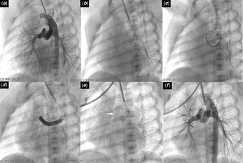
In all the cases, after vascular access was obtained, the appropriate (4F or 5F) sheath and catheter were introduced. An aortic arch angiography was performed in the antero-posterior and lateral views, but angled projections might be obtained especially with tortuous duct to best profile the patent ductus arteriosus to evaluate the ductal size, length, and morphology. Thereafter, a 0.014-inch floppy guidewire was passed through the patent ductus arteriosus and placed in the left or right branch pulmonary arteries. Then, a coronary stent was delivered over the wire and implanted into the patent ductus arteriosus. The stent was chosen according to neonatal weight for neonates >3.0 kg, a 3.5–4 mm diameter stent is chosen, for those weighing 2.0–3.0 kg, a 3.5 mm diameter. The length of the chosen stent was 2–3 mm longer than the ductal length between the aortic and pulmonary ends. Once the stent is deployed, the patent ductus arteriosus stent anatomy is reassessed, and another angiogram with the wire still in place to ascertain whether another stent is required. (see Fig 1)
Transcarotid ductal stent of a neonate with pulmonary atresia intact septum through right carotid artery access. (a) Aortic arch angiography was performed to evaluate the ductal morphology in modified oblique fluoroscopic view. (b) A 0.014-inch floppy guidewire is seen passing from right carotid artery across PDA. (c) Coronary stent was positioned across PDA. (d) Inflation of the balloon. (e) PDA stent in place. (f) Angiogram post stent placement showing good filling through the stent to both branch pulmonary arteries.

Hemostasis was obtained by manual compression in all the cases. A small gauze or non-occlusive bandage was applied to the access site after hemostasis was attained. Following the procedure, the neonate was monitored in the hospital for at least 24 hours after the procedures and was kept on intravenous heparin (20 units/kg/hour), then changed to acetylsalicylic acid (3–5 mg/kg/day) with follow-up chest radiographs and echocardiograms the day after the procedure.
Post-procedural artery patency was assessed using a Doppler ultrasound. The artery was classified as normal, mildly stenotic (lumen >50% of the normal adjacent vessel), moderately stenotic (lumen reduced to <50% of normal), or completely occluded.Reference Justino and Petit12 In symptomatic cases, it was performed on the day of the procedure or as required on admission; otherwise, it was performed on an outpatient basis during a follow-up visit.
Studied variables
The retrospective comparison was performed between the cases who had patent ductus arteriosus stenting through carotid access and those through femoral access. The comparison included demographic data such as age, the weight of the neonate at the procedure, cardiac diagnosis, and ventricular physiology. Procedure details such as the time required to achieve the access, procedure duration, duration to hemostasis, sheathes used, catheter types needed. The complications and outcomes compared were mortality, stroke, bleeding, conduction disturbances including arrhythmias, and vascular access-related local complications either immediate as hematomas, thrombosis, stenosis, pseudoaneurysm formation or late as stenosis or occlusion.
Retrospective analysis of angiographic studies was performed to classify the patent ductus arteriosus according to the novel patent ductus arteriosus morphology classification described by Qureshi et al It is based on the tortuosity index on angiographic measurements dividing the patent ductus arteriosus into three types: Type I (straight), Type II (one turn), and Type III (multiple turns).Reference Qureshi, Goldstein and Glatz4
Statistics
All the statistical analyses were carried out using SPSS software (version 25, IBM Corp., Armonk, NY, USA). The results for the numeric variables are presented as median (interquartile range) while the categorical variables as frequencies and percentages. Comparisons between variables related to carotid access and femoral access patent ductus arteriosus stenting procedures were assessed using unpaired t-test or Mann–Whitney or Chi-square test or Fisher’s exact test according to the variable type. A p value < 0.05 was considered statistically significant.
Results
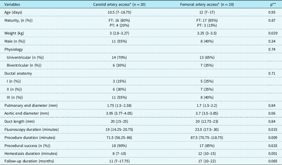
Forty neonates with median age (25th–75th percentile) of 10.5 (3–28) days who underwent patent ductus arteriosus stenting, were retrospectively analysed. Twenty patients via percutaneous carotid access and 20 via femoral artery access. Table 1 demonstrates the patients’ characteristics, angiographic-derived ductus details, and some procedure-related information. Although both groups matched in age, sex, and maturity, patients’ weight was significantly lesser in the carotid access group with a median (25th–75th percentile) of 3 (2.8–3.27); p: 0.029. Different duct anatomies were approached using both accesses, but carotid access was used more frequently in neonates with complex duct anatomy (class III). The median of procedure duration, fluoroscopy time, and hemostasis time were significantly lower in the group of carotid artery access in correlation with femoral access with p-values of 0.015, 0.009, and 0.001, respectively.
Comparison of ductal stenting via the carotid artery and femoral artery including patients’ characteristics, angiographic PDA size and anatomy, and procedure details

FT: full term, min: minutes, mm: millimetre, n (%): number (percentage), PT: premature
* Values are expressed as median (interquartile range) or frequencies (percentage)
** p-value is significant if < 0.05
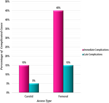
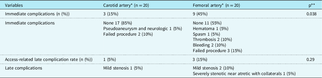
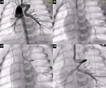
There was no procedure-related mortality. Nevertheless, immediate and late access-related complications were more frequent in the femoral artery group in correlation with the carotid artery group (Fig 2). Details of morbidities are presented in Table 2. A total of nine immediate procedure-related complications in the femoral group, including hematoma at the puncture site without evidence of flow disturbance in 1 patient, bleeding in 1 patient, arterial spasm in a patient, non-occlusive thrombus in another one patient, and occlusive thrombus in another neonate resolved on low-molecular-weight heparin in addition to three cases with failure in obtaining access or failure in introducing the stent in complex anatomies. On the other hand, on the carotid artery access group, complications were limited to one case of criss-cross heart with critical pulmonary stenosis suffered from a small pseudoaneurysm in the carotid artery that required no intervention, but the patient had mild weakness on one side, 3 days after the stenting. The patient was maintained on heparin infusion followed by warfarin and fortunately, the patient regained motor power within 2 weeks. Additionally, two cases with failure to complete the procedure due to failure to advance the stent through the complex ductal anatomy type III and another case with type II but with sharp-angled single turn simulating V shape (see Fig 3).
Two-dimensional bar chart showing the percentage of immediate and late access-related complications in femoral artery versus carotid artery access in neonatal PDA stenting.

Complications of ductal stenting via the carotid artery and femoral artery

* Values are expressed as frequencies (percentage)
** p-value is significant if < 0.05
Transcarotid ductal stent of a neonate with double outlet right ventricle and pulmonary atresia through left carotid artery access (a) Aortic arch angiography was performed to evaluate the ductal morphology in antroposterior fluoroscopic view. (b) Floppy guidewire is seen passing across the PDA. (c) Coronary stent was advanced till the level of the middle angle of the V-shaped duct. (d) Angiogram (via hand injection) showing the PDA and both branch pulmonary arteries with stent failure to progress.

Doppler ultrasound examinations on follow-up were performed at a median of 11 (7–17.75) months after the intervention for the carotid artery access cases and a mean of 17 (10–22) months following patent ductus arteriosus stenting for the femoral artery group. In the femoral artery access, two had mild luminal narrowing and one case had severe stenosis at the access site while with the carotid artery group, only one case had mild stenosis of the access artery.
At the last follow-up, seven patients completed biventricular repair, and 11 received bidirectional Glenn’s shunt. The remaining were waiting for their surgical intervention.
Discussion
A sole supply of pulmonary blood flow is considered a Class II b indication for patent ductus arteriosus stenting (level of evidence: C), according to the guidelines of American Heart Association in 2011. Moreover, in the presence of branch pulmonary artery stenosis, it is a Class III indication (level of evidence: C).Reference Feltes, Bacha and Beekman13 Patent ductus arteriosus stenting in neonates can be a technically challenging cardiac intervention with possible variable approaches but without established preferences or differences among them.
Although the femoral artery approach is the traditional access, complications following femoral artery access have been explored by various studies and have varied from 2.9to 39%.Reference Vitiello, McCrindle, Nykanen, Freedom and Benson14–Reference Bergersen, Marshall and Gauvreau16 Vermilion et al reported that children under 1 year of age have a higher risk of developing arterial obstructions after balloon dilation of left obstructive lesions using the femoral approach.Reference Vermilion, Snider, Bengur and Beekman15 Cohn et al reported 10-fold higher complications risk for children less than 4 months old.Reference Cohn, Freed and Hellenbrand17 On using ultrasound assessment for 120 arterial catheterisation procedures, arterial thrombosis was detected in 16% of patients less than 10 kg.Reference Kulkarni and Naidu18 In the current work, we had comparable rates of femoral artery thrombosis (10%).
Carotid artery access may play an important role in preventing the complications encountered with the femoral artery. Given that the neonatal carotid artery is larger in diameter than the femoral artery.Reference Steinberg, Weinstock and Gold19,Reference Gasparella, Milanesi and Biffanti20 The potential benefit of the carotid approach is the ability to manoeuver the catheter directly to left heart structures with potentially less technical difficulty. The angle of approach from the carotid artery is straighter than with the femoral approach, thus it facilitates wire and stent passage across tortuous anatomy and provides more support.Reference Choudhry, Balzer, Murphy, Nicolas and Shahanavaz8
The patent ductus arteriosus morphology classification described by Qureshi et al may help in anticipating outcomes in patients undergoing patent ductus arteriosus stenting as higher tortuosity indicated more possibility of carotid artery access as well as increased reintervention rates.Reference Qureshi, Goldstein and Glatz4 Nevertheless, we had one patient with a V-shaped sharp-angled patent ductus arteriosus with failure of stent progression although the wire was in a good position. Thus, we suggest that type 2 can be subdivided into a: patent ductus arteriosus with a single smooth turn and b: ductus with a sharp-angled single turn as difficulty in stenting is more expected with the acute-angled patent ductus arteriosus turns.
Carotid access safety was established even in small infants, and hemostasis can be achieved without surgical repair, with a patency rate superior to surgical cut-down.Reference Choudhry, Balzer, Murphy, Nicolas and Shahanavaz8,Reference Justino and Petit12,Reference Polat21 Justino et al described 16 successful cases of carotid access patent ductus arteriosus stenting with four failed cases. Only one patient had occlusive thrombosis followed by mild residual narrowing and another with non-occlusive thrombus; both were treated with low-molecular-weight heparin but no neurologic complications.Reference Justino and Petit12 Choudhry et al proved that percutaneous carotid artery access in infants <3 months of age is feasible with preserved vascular patency and no neurological adverse events. They described eight patients with PDA stenting through carotid access with one case of a ductal spasm and another with a carotid pseudoaneurysm which was repaired surgically.Reference Choudhry, Balzer, Murphy, Nicolas and Shahanavaz8 Similarly, Polat et al reported 23 stenting or restenting procedures of PDA with two failed cases, a single case of mild luminal narrowing, one with non-occlusive thrombosis, another with occlusive thrombosis, and one case with a migrated stent but with no documented neurologic adverse events.Reference Polat21 However, another study included 43 cases of carotid access and six axillary access for patent ductus arteriosus stenting, complications rate was comparable with the femoral access with a non-significant higher rate of access site clotting or bleeding as well as arrhythmia in the femoral group. They reported a case of aortic dissection on a carotid approach for patent ductus arteriosus stent placement. On ultrasound surveillance, they had one case of carotid artery occlusion, one with carotid pseudoaneurysm, and another case with carotid stenosis but no documented neurologic sequel or limb ischemia.Reference Bauser-Heaton, Qureshi and Goldstein5 In a study by Ligon et al on 55 patients with carotid access procedures, 32 of them had patent ductus arteriosusstenting,, 2 cases had non-occlusive thrombi. Moreover, angiography in subsequent catheterisation at a median of 98 days revealed only mild stenosis in 35% of cases.Reference Ligon, Kim and Vincent22 In our cohort, we had one case with carotid artery small pseudoaneurysm and hemiparesis 3 days after the intervention with an improvement of neurologic signs after 2 weeks on therapy.
Carotid puncture site manual compression in our cases was successful at achieving hemostasis without reversal of heparin. No patient in our series experienced significant carotid bleeding or airway compression. Similar findings were reported by Justino and Petit.Reference Justino and Petit12 These data are against the reports suggesting the need for carotid repair after a large-bore catheter or sheath placed in the carotid to avoid hematoma, external bleeding, airway compression, or neurological injury.Reference Golden23,Reference Guilbert, Elkouri and Bracco24 Polat found that the median duration of hemostasis in carotid access interventions is 9 minutes (5–22).Reference Polat21 These are consistent with our carotid access cohort.
It was suggested by Justino and Petit that the operator should be at the head of the patient in case of using the carotid artery during catheter manipulations to avoid accidental displacement of the sheath resulting in significant hemorrhage.Reference Justino and Petit12 A flip technique was described recently by Bauser-Heaton et al for easier carotid or axillary approach by orienting the head of the patient on the table toward the foot of the bed, and they found that it decreased procedure time.Reference Bauser-Heaton, Qureshi and Goldstein5
Another access for ductal stenting is the axillary artery, but there are no other anatomical landmarks available for a blind puncture if the axillary artery is not well palpable. But it is safe and feasible for vertical duct especially on using the wire-target technique.Reference Polat25 Although the retrograde approach to patent ductus arteriosusfrom the aorta is the most widely used, antegrade approach through femoral vein was described for deploying the stent but with a high complication rate in 55% of cases.Reference Polat25
Ductal stenting as well as any other complex intervention in a neonate requires a highly skilled interventionist, expert paediatric cardiac anesthesiologist, and ready access to the surgical backup. The limitations of this study include the retrospective single-centre nature of the study and the small sample of patients that could explain some of the insignificant data such as on comparing morphologic classification.
Conclusion
Percutaneous carotid artery access in neonates is a more convenient approach for patent ductus arteriosus stenting in correlation to the traditional trans-femoral access with more preserved vascular patency and limited local complications, but potential neurological adverse events should not be overlooked.
Acknowledgements
None.
Financial support
This research did not receive any specific grant from funding agencies in the public, commercial, or not-for-profit sectors.
Conflict of interest
The authors have no conflicts of interest to declare.
Ethical standards
The authors assert that all procedures contributing to this work comply with the ethical standards of the relevant national guidelines on human experimentation and with the Helsinki Declaration of 1975, as revised in 2008, and has been approved by the institutional research board (IRB) of Faculty of Medicine, Mansoura University.