Introduction
The dosimetry in radiotherapy has been challenged when the beam passes through prostheses and implants components with high density and atomic number. This situation happens more for patients with the hip prosthesis undergoing pelvic radiotherapy than other patients. The heterogeneous materials on the beam path are not taken into account in the routine treatment planning, and it results in the dose distribution would be different from the prescribed dose to the target and surrounded tissues. This conception has created enough attention to justify the formation of an American Association of Physicists in Medicine Task Group to investigate. Reference Reft, Alecu and Das1 Several published literatures reported the effects of the prosthesis on dose distributions. Some authors calculated the effect of various prostheses on the radiation dose for 6 MV, Reference Lin, Chu, Lin and liu2–Reference Keall, Siebers, Jeraj and Mohan4 9 MV photon beam Reference Mesbahi and Nejad5 and Co60 source. Reference Buffard, Gdchwind, Makovicka, Martin, Meunier and David6 Some researchers evaluated the dose including metal objects during the intensity-modulated radiation therapy. Reference Inal and Sarpün7 Buffard et al studied the impact of the hip prosthesis for 25 MV photons. Reference Buffard, Gschwind, Makovicka and David8 Bhushan et al investigated the effect of the hip prosthesis on 6 and 15 MV photon beam energies for stainless steel and Ti prostheses. Reference Bhushan, Tripathi, Yadav, Kumar, Dewan and Kumar9 Electron contamination was also investigated by Bahreyni et al from 15 MV photons for one thickness of the prosthesis. Reference Bahreyni Toossi, Behmadi, Ghorbani and Gholamhosseinian10 The effect of these two prostheses was measured for 6 and 18 MV photons for the treatment planning system. Reference Çatlı and Tanır11,Reference Wieslander and Knöös12 The published results indicated that the dose perturbation depended on the photon energy, density and location of the prosthesis. Also, the performed calculations showed that the Monte Carlo simulation can accurately predict dose distributions inside a phantom where experimental measurements are difficult. Reference Ding and Christine13
More carried-out studies were for the limited number of the prosthesis in a specific location. This work aimed to investigate the dose perturbations in the vicinity of the complete set of prosthesis materials at different positions, which are used in the patient’s body during the external radiotherapy of 15 MV photons. The MCNP Monte Carlo code was applied for the dose calculations, and the effects of locations and prosthesis thicknesses were evaluated to make better clinical decisions for treatment corrections. Finally, a novel polymer prosthesis was introduced which cause to slightly modify the dose distribution and be replaced with other common prostheses.
Materials and Methods
In the present study, the MCNP-4C radiation transport Monte Carlo code was used to build a virtual medical linear accelerator and a 3D dose distribution. Reference Briesmeister14 The 15 MV photon mode of a Siemens Primus medical linear accelerator (Siemens AG, Erlangen, Germany) was simulated based on the manufacturer’s geometry data. The main components of the linear accelerator’s head consist of the target, primary collimator, absorber, flattening filter, photon dose chamber as well as jaws. The calculated percentage depth dose and beam profile curves were compared with measured ones to validate the model. The validation results were reported in the previous study in detail. Reference Mohammadi, Miri-Hakmiabad, Rafat-Motavalli, Akbari and Abdollahi15
Five types of common frequent implants including Co-Cr-Mo, stainless steel, Ti-alloy, Ti and Ti 6A14V were selected to evaluate the metal inhomogeneous effects. These prostheses were modelled as the simplest cylindrical shape and placed in a water phantom to calculate the dose distribution. The elemental compositions of the five hip prostheses are shown in Table 1.
The main characteristics and their percentage by weight (%) of the used prosthesis.

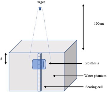
The water phantom was modelled as a cube with 50 × 50 × 50 cm3 dimensions and was placed at 100 cm of the linac head target to irradiate with 10 × 10 cm2 field size. Figure 1 illustrated the schematic view of the MCNP simulation. The total 2e9 initial electrons were transported, and the dose distribution calculations were carried out with mesh tally (type 3). The depth dose was calculated in the cubic scoring cells of 1 cm × 1 cm × 1 mm, and electron and photon energy cutoffs were set to 0·5 and 0·01 MeV, respectively. It should be noted that the relative error of calculations was nearly 3%.
The schematic of MCNP simulation (not to scale).

Results and Discussion
Prosthesis at different depths
The different cylindrical prostheses with diameter of 4 cm and height of 3 cm were placed at the water phantom to study the effects of metal implant on the dose distribution. To simulate a standard irradiation with the presence of pelvic implant, the prosthesis was placed at a 5 cm depth, which reported as the mean value measured on scanner images of some patients. Reference Buffard, Gdchwind, Makovicka, Martin, Meunier and David6
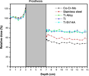
For data analysis, the relative dose was introduced as the ratio of the dose in the presence of prostheses to the dose without prostheses which was informed in percentage terms. The relative dose at different depths of the water phantom is shown in Figure 2. It was predicted that the backscattered electrons of the prosthesis surface result in the dose intensification at the interface of water and implant. It is noted that dose increasing was remarkable at the range of 1–3 mm away from themetal prosthesis. When the implants were set at 5 cm depth, it was seen that the relative dose at the edge of the prosthesis was 121, 117·6, 116·8, 113·4 and 115·75 for Co-Cr-Mo, stainless steel, Ti alloy, Ti and Ti 6V14A. Therefore, it could be concluded the increment magnitude of the relative dose depends on the implant density. The results were in accordance with the results of Mohammadi et al, in which comparable results for titanium and stainless steel were found. Reference Mohammadi, Hassani, Ghorbani, Farhood and Knaup16 They observed an increased dose of about 114·3 and 111·7% due to the backscattering of electrons from a stainless steel and Ti implant, respectively. This increment may lead to localised hotspots around the prosthesis that will not be calculated or considered by the treatment planning system. Reference Carolan and Dao17
Relative dose (%) for different prosthesis.

The backscattered electrons effect was reduced beyond the prosthesis as a result of electrons absorption in the metal prosthesis. The metal components could be able to provide a significant reduction in the absorbed dose at the points located after the prosthesis, and it started to fall rapidly as the distance from the interface was increased. The evaluation of five types of hip prosthesis materials indicated that high-density materials could attenuate significantly the photon beam compared to water. This study’s findings showed that the reduction dose was 56, 62·39, 72·59, 73·76 and 76·09% for Co-Cr-Mo, stainless steel, Ti alloy, Ti and Ti 6V14A, respectively.
It was seen that as the density of the material transversed by the 15 MV photon beams increases, there was an increase in attenuation of the beam and perturbation of the dose. Therefore, there was the maximum dose-difference for the densest (Co-Cr-Mo) prosthesis.
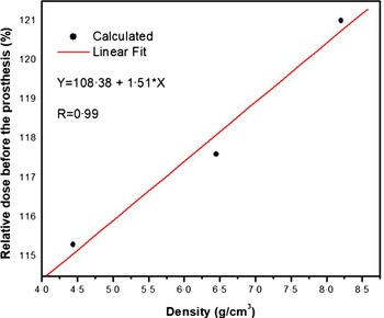
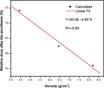
As can be seen, the amount of relative dose before and after the implant depended on its density. Also, the calculations showed that the dose distribution from implants constructed from Ti (Ti alloy, Ti and Ti 6V14A) was close together. Thus, the mean value of density and its relative dose was considered to take the plot of the relative dose by the implant density. These curves are illustrated in Figures 3 and 4. It can be seen that there was a linear relationship between the relative dose and the implant density. Therefore, the calculated points were fitted by linear curves which linear equations were calculated by the Origin software. The obtained equations of linear fit can be beneficial for any treatment planner to increase the knowledge of the beam characteristics in the presence of such metallic objects. It helps to have a better assessment of the treatment plans.
Relative dose (%) before the prosthesis versus implant density.

Relative dose (%) after the prosthesis versus implant density.

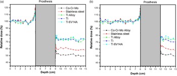
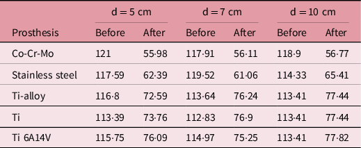
It should be considered that the prosthesis may be located at different depths of the skin; thus, the influence of the prosthesis at various depths was examined. For this purpose, prostheses were placed at the depth of 7 and 10 cm in the simulation. The result of these situations is shown in Figure 5. As was presented in the figures, the dose intensification did not experience a significant change at the entrance of the prosthesis compared to 5 cm depth. It means that the location of the prosthesis did not have a noticeable influence on dose variation at the interface of metal tissue. The magnitude of relative dose at the interface of water and prosthesis is given in Table 2 for 1 mm distance before and after passing the metal.
Relative dose for the prosthesis at depths of (a) 7 cm and (b) 10 cm.

Relative dose (%) before and after the prosthesis at different positions.

Prostheses with different thicknesses
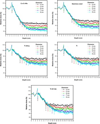
The different thicknesses of the prosthesis (cylinder diameter: 4, 5, 6, 7 and 8 cm) were simulated and placed in the water phantom to determine the effect of prosthesis thicknesses used for various patients. The diagrams of relative dose versus depth in the water phantom are presented in Figure 6. The results showed that the increased dose due to backscattered electrons was not changed considerably, while the dose passed of the prosthesis was strongly depended on the prosthesis thickness and decreased because of photons attenuation by the metal. In other words, the size of the prosthesis could affect the transmission but would not affect the dose due to the backscattering. It can be seen that the dose at the end of the prosthesis decreased as the thickness was increased. The dose value dropped to around 25% in the diameter of 8 cm of the Co-Cr-Mo prosthesis. The amount of reduction for this diameter was 35% for stainless steel implant, and it went down about 50% in other prostheses composed from Ti.
Relative dose (%) for different diameter of the prosthesis.

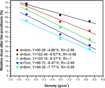
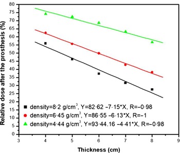
The relative dose after the prosthesis for each diameter of the implant is plotted by its density in Figure 7. The results indicated a linear correlation which was shown with the linear fit line. Moreover, the relative dose of each implant for different thickness and their linear fit is displayed in Figure 8. As can be seen and expected, the slope of the linear fit is greater for high implant density because of intensive beam attenuation. The present findings and obtained fitted functions could be used for dose prediction in the various situations which may occur during the treatment planning procedure.
Relative dose (%) after the different thickness of prosthesis versus implant density.

Relative dose (%) after the different prostheses versus implant thickness.

While, according to the dosimetric protocols, the differences in the dose should be below 5%, 18 the dose differences in the presence of prosthesis were found above this value in the results of this study which recommends more accuracy in dose delivery. This might have highly adverse effects on the patient’s treatment results. Therefore, these differences in prescribed and delivery dose should be minimised as much as possible.
Medical polymer materials instead of metal prosthesis
As the results show, the metal prostheses cause the dose increase at the interface of metal tissue. On the other hand, the amount of the dose drops down after the prosthesis. These variations could change the treatment planning. Thus, the prescribed dose could not correctly deliver to the treatment area unless the treatment planning system updates with the correction factors. New material was proposed to replace the current prosthesis to overcome this problem.
The conventional use of metal implants is not much supported from the last few years as there is a possibility of implant unfasten and cause to wear which will have an unfavourable effect on the body. Reference Verma, Sharma, Kango and Sharma19 Thus, medical polymeric implants are preferred for hip joint replacement, and among these polyether-ether-ketone (PEEK) is many premier as it possesses outstanding mechanical properties, chemical and heat resistance so giving overall preferable biomechanical performance. Reference Enab and Bondok20–Reference Ma, Suonan and Zhou22 PEEK materials have been successfully applied in clinical practice cranioplasty, dental implants, interbody fusion, joint replacements, soft-tissue repairs and cardiac surgery. Reference Verma, Sharma, Kango and Sharma19,Reference Guo, Chen, Wang and Lu23 These materials have excellent friction and wear properties which are important properties that are considered for a good joint implant. Reference Li, Vannabouuathong, Sprague and Bhandari24 In addition, it has good bio-compatibility, and its abrasive particles have no obvious toxicity to the body. Reference Ma, Suonan and Zhou22 De Ruiter et al also found that stress shielding was reduced to a median of 1% for the PEEK implant versus 56% for the cobalt–chromium implant. Hence, the stress shielding of the periprosthetic femur was less with a PEEK femoral component. Reference Guo, Chen, Wang and Lu23,Reference de Ruiter, Janssen, Briscoe and Verdonschot25 Many researches have been fully carried out about the PEEK’s properties, its interaction with cells, coatings and surface modification. However, this study focused on the effects of this material on the dose perturbation during the radiotherapy which has not been evaluated and reported until now.
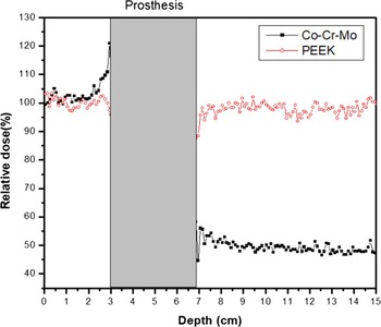
To examine the effect of PEEK, with the density of 1·32 g/cm3, dose distribution was calculated in the presence of this material as a prosthesis at the depth of 5 cm. The comparison of the relative dose for the PEEK and Co-Cr-Mo prosthesis (which had more effect on dose distribution) is shown in Figure 9.
Comparison of relative dose for PEEK and Co-Cr-Mo prosthesis.

It can be seen that PEEK had no perturbation due to backscattered electrons, because it is composed of low Z-number atoms (C-H-O) compared to Co-Cr-Mo and other current prostheses. The reason for this is that the pair production interaction is dominant for photons with energies higher than 10 MeV and electron is produced. The probability of this interaction is proportional to the square of atomic number (Z2). Therefore, it can be expected that Co-Cr-Mo alloy, with high Z-number atoms, would have the highest electron production with respect to the PEEK. The dose was not decreased after the PEEK; thus, the dose distribution was not changed because of less photon mass attenuation coefficient for low Z-number atoms of C, H and O with respect to the metals. It was found that the mean relative dose before and after of prosthesis is 99·2 and 97·1%, respectively. This means that PEEK is recommended to use instead of a metal implant. The treatment planning can be properly delivered to the patient using this substitute.
Conclusion
The calculated dose distributions showed that the Monte Carlo simulations allow taking into account the dose increase at two media interfaces. This information is very crucial because no commercial treatment planning system be currently able to perform such calculations. In this study, Monte Carlo simulations were carried out to determine the accurate dose perturbations which are caused by the various prosthesis during the radiotherapy with 15 MV photons from Siemens Primus linear accelerator. The calculations showed the important effect of the hip prosthesis composition, the position and thickness of the prosthesis within the irradiation field on the dose distribution. The significant dose increase at two media interfaces had been highlighted for Co-Cr-Mo prostheses that had the highest density among the other prosthesis. Indeed, the dose reduction had the most amount for this prosthesis due to more photon attenuation.
Without modifying and improving a new algorithm, there is no treatment planning system be able accurately to predict the dose distribution within high-density material. To prevent the dose uncertainty between treatment planning and Monte Carlo calculation, it is recommended to avoid the beams passing through these high-density implant materials, if possible. Otherwise, using the biomedical PEEK materials can overcome this perturbation dose. Since the new findings of this study showed that PEEK is a suitable material instead of metallic implants because its dose distribution was much less than metals and can be easily neglected. Thus, the treatment planning can be delivered the accurate prescribed dose to patients undergoing radiotherapy.
Acknowledgements
None.
Financial Support
None.