Introduction
Among fish parasites, Monopisthocotyla (Platyhelminthes) is a class of ectoparasites that is highly diverse in terms of the number of species (4,565 species) (Gibson Reference Gibson2025), as well as their morphology and ecology (Poulin Reference Poulin2002). Monopisthocotyls are known to be highly host-specific, infecting only one or a few species (Poulin Reference Poulin1992; Sasal et al. Reference Sasal, Desdevises and Morand1998). This attribute, associated with possible contiguous evolutionary events, has made them excellent models for cophylogenetic studies. However, the evolutionary processes that influence the interactions between monopisthocotyls and hosts are complex and dynamic, involving not only cospeciation but also other processes. Cophylogenetic studies between monopisthocotyls and their hosts have shown that coevolutionary patterns involve a supposedly high number of host-switch, duplications, and losses (Benovics et al. Reference Benovics, Desdevises, Šanda, Vukić and Šimková2019; Desdevises et al. Reference Desdevises, Morand, Jousson and Legendre2002; Huyse et al. Reference Huyse, Audenaert and Volckaert2003; Huyse et al. Reference Huyse, Oeyen, Larmuseau and Volckaert2017; Huyse and Volckaert Reference Huyse and Volckaert2005; Mendlová et al. Reference Mendlová, Desdevides, Civáňová, Pariselle and Šimková2012; Šimková et al. Reference Šimková, Serbielle, Pariselle, Vanhove and Morand2013). Therefore, to recognize possible coevolutionary processes at work in these parasite-host associations, it is necessary to have a robust knowledge of the diversity of the taxa involved in these associations.
Neotropical freshwater fish comprise the most diverse continental vertebrate fauna on Earth, with over 6,080 valid species (Albert et al. Reference Albert, Tagliacollo and Dagosta2020). South America is the epicenter of this remarkable diversity, not only for the Neotropical region but also for the world (Reis et al. Reference Reis, Albert, Di Dario, Mincarone, Petry and Rocha2016). Currently, 5,160 freshwater fish species have been identified in South America, with the orders Siluriformes and Characiformes standing out as the most diverse group, comprising 3,615 species (Reis et al. Reference Reis, Albert, Di Dario, Mincarone, Petry and Rocha2016). However, our knowledge of the parasites associated with this diversity is limited, as only approximately 10% of the diversity of freshwater fish belonging to various orders (i.e., Rajiformes, Lepidosireniformes, Osteoglossiformes, Characiformes, Siluriformes, Gymnotiformes, Cyprinodontiformes, Synbranchiformes, Perciformes) have been investigated for their parasites (Luque et al. Reference Luque, Pereira, Alves, Oliva and Timi2016). Currently, 586 species of monopisthocotyls are found in freshwater fish in South America. From this diversity, members of Urocleidoides Mizelle & Price, Reference Mizelle and Price1964 are remarkable for their wide host range, parasitizing fish from the orders Cyprinodontiformes, Characiformes, Gymnotiformes, and Siluriformes (Cohen et al. Reference Cohen, Kohn and Justo2013; Ebert et al. Reference Ebert, Osaki-Pereira, Pérez-Ponce de León and Da Silva2024; Ferreira et al. Reference Ferreira, Rodrigues, Cunha and Domingues2018; Freitas et al. Reference Freitas, Bezerra, Meneses, Justo, Viana and Cohen2021; Hasuike et al. Reference Hasuike, Scorsim, Arjona, Bellay, de Oliveira and Takemoto2024; Mendoza-Franco et al. Reference Mendoza-Franco, Caspeta-Mandujano, Salgado-Maldonado and Matamoros2015; Moreira et al. Reference Moreira, Scholz and Luque2015; Oliveira et al. Reference Oliveira, Da Silva, Vieira and Acosta2021; Santos Neto and Domingues, Reference Santos Neto and Domingues2023, Reference Santos Neto and Domingues2024; Yamada et al. Reference Yamada, Osaki-Pereira, Acosta, Ebert and Da Silva2024; Zago et al. Reference Zago, Yamada, Yamada, Franceschini, Bongiovani and Da Silva2020).
There are 59 valid species of Urocleidoides, from which 43 are exclusively found in Characiform fish belonging to the families: Anostomidae (11 species), Characidae (3 species), Crenuchidae (1 species), Ctenoluciidae (2 species), Curimatidae (8 species), Erythrinidae (12 species), Hemiodontidae (1 species), Lebiasinidae (1 species), and Parodontidae (4 species).
Studies on the phylogenetic relationships of some species of Urocleidoides have already been addressed, where the monophyly of Urocleidoides was not recovered using analyses based on partial 28S rDNA sequences. Zago et al. (Reference Zago, Yamada, Yamada, Franceschini, Bongiovani and Da Silva2020) recovered the monophyly of Urocleidoides species parasites of anostomids (Characiformes) in a polytomy with species of Urocleidoides from characiforms and gymnotiforms, Cacatuocotyle papilionis Zago, Franceschini, Muller & da Silva, 2018 reported from characids (Characiformes), and other dactylogyrids parasites of loricariids (Siluriformes). Oliveira et al. (Reference Oliveira, Da Silva, Vieira and Acosta2021) observed that U. uncinus Zago, Yamada, Yamada, Fanceschini, Bongiovani & da Silva, 2020, a gymnotiform parasite, appears as a sister group of dactylogyrids that parasitize characiforms, which include species of Urocleidoides from anostomid and parodontid fish, and C. papilionis. Santos Neto and Domingues (Reference Santos Neto and Domingues2023, Reference Yamada, Osaki-Pereira, Acosta, Ebert and Da Silva2024) recovered part of the results of Oliveira et al. (Reference Oliveira, Da Silva, Vieira and Acosta2021). However, with the inclusion of new taxa, these authors observed that some species of Urocleidoides parasites of erythrinids (U. brasiliensis Rosim, Mendoza-Franco & Luque, 2011, U. naris Rosim, Mendoza-Franco & Luque, 2011, U. macrosoma Santos Neto & Domingues, 2023) emerged as a sister group of species of Urocleidoides parasites of Gymnotiformes in a clade with C. papilionis and species of Diaphorocleidus. Ebert et al. (Reference Ebert, Osaki-Pereira, Pérez-Ponce de León and Da Silva2024) and Yamada et al. (Reference Yamada, Osaki-Pereira, Acosta, Ebert and Da Silva2024) recovered the topology of Santos Neto and Domingues (Reference Santos Neto and Domingues2023, Reference Santos Neto and Domingues2024), except by the position of C. papilionis, Diaphorocleidus spp. and Rhinoxenus spp. parasites of Characiformes that form a sister group clade of species of Urocleidoides parasites of erythrinids, which emerged as a sister group of species of Urocleidoides parasites of Gymnotiformes. Our understanding of the evolutionary relationships among Urocleidoides species is thus advancing as new data are systematically integrated into new analyses. These results are essential for assessing the historical associations between these parasites and their hosts. However, no study to date has employed a cophylogenetic approach to examine these interactions, and the coevolutionary patterns driving the relationship between these parasites and their hosts remain unknown.
In the present study, we describe four new species of Urocleidoides based on morphological and molecular data (28S rDNA), providing insights into the relationships between these new species and their congeners. Additionally, we apply a cophylogenetic approach in a study of the case to reconstruct the evolutionary processes that drive the diversification of Urocleidoides spp. and their characiform hosts.
Material and methods
Host collecting
Specimens of Erythrinus erythrinus (Bloch & Schneider), Hoplerythrinus unitaeniatus (Spix & Agassiz), Schizodon fasciatus (Spix & Agassiz), and Brachyhypopomus beebei (Schultz) were collected from May 2015 to November 2018. The collection was conducted using trawls, pull nets, and capture traps at five different locations in the Eastern Amazon region: Pratinha Community - Caeté River Basin, Bragança municipality, Pará, Brazil (01°5’2.49"S, 46°49’44. 67’’W); Igarapé Maratininga - Moju River Basin, municipality of Tailândia, Pará, Brazil (02°27’55.7’’S, 048°53’27.6’’W); Balneário Aracu - Guamá River Basin, municipality of Ourém, Pará, Brazil (01°34’1.02’’S, 47°9’52.35’’W); Peixe Boi River - Maracanã River Basin, municipality of Peixe Boi, Pará, Brazil (01°11’32.1’’S, 47°18’43.7’’W); and Lower Jari River, near the district of Jarilândia, municipality of Vitória do Jari, Amapá, Brazil (01°9’4.01”S, 52°0’53.22"W).
Parasitological procedures
The captured fish were euthanized using cerebral concussion. The gill arches of each host specimen were removed and placed in labeled vials containing heated water (~65°C). Each vial was vigorously shaken, and then the sediment and gills were fixed in 5% formalin for morphological studies or 96% ethanol for molecular characterization. In the laboratory, each vial content was examined using a LEICA S6D stereoscopic microscope, and the helminths found were removed from the gills or sediment using small needles and directed for morphological/molecular analysis. The monopisthocotylan specimens intended for studying the internal structures were stained with Gomori’s Trichrome (Boeger and Vianna Reference Boeger, Vianna and Thatcher2006; Humason Reference Humason1979) and mounted in Dammar Gum, and for the study of the sclerotized structures, the remaining specimens were mounted in Hoyer’s Medium or Grey & Wess (Boeger and Vianna Reference Boeger, Vianna and Thatcher2006; Humason Reference Humason1979). Measurements were taken according to the procedures of Mizelle and Klucka (Reference Mizelle and Klucka1953) and are presented in micrometers. Internal organs and other structures were measured in the dorsoventral view using Leica LAS© interactive measurement software. Lengths of curved or bent structures (i.e., anchors, bars, accessory parts) represent the straight-line distances between ends. The hooks were classified according to Mizelle and Price (Reference Mizelle and Klucka1953). The averages of the measurements were calculated from the minimum and maximum lengths and the number of structures measured (n). Illustrations of the species and their structures were prepared using a drawing tube on a microscope with differential interference contrast and phase-contrast optics. Authorship of the new taxa follows Recommendation 50.1 (Identity of authors) of the International Code of Zoological Nomenclature. Definitions of prevalence and average intensity were calculated according to Bush et al. (Reference Bush, Lafferty, Lotz and Shostak1997). The type specimens of the species presented in this study were deposited in the Helminthological Collection of the Oswaldo Cruz Institute (Portuguese acronym, CHIOC), Rio de Janeiro, state of Rio de Janeiro, Brazil. Acting in accordance with the regulations in article 8.5 of the amended 2012 version of the International Code of Zoological Nomenclature, details of the new taxa have been submitted to ZooBank.
DNA extraction, amplification, and sequencing
For identification, each parasite specimen submitted for molecular analysis was checked under a LEICA S6D stereoscopic microscope and divided using small needles. When the species was identified through haptoral structures, the anterior region of the body was placed in a 1.5-ml microtube with 96% ethanol and used for DNA extraction, and when the morphology of the male copulatory organ was used for identification, the posterior region of the body was used for extraction. The anterior or posterior region of the parasite’s body was flattened under coverslip pressure and mounted in Hoyer’s medium. These fragments were used as vouchers, referred to as hologenophores, according to Pleijel et al. (Reference Pleijel, Jondelius, Norlinder, Nygren, Oxelman, Schander, Sundberg and Thollesson2008). DNA was extracted using the Qiagen Dneasy® Blood and Tissue kit, according to the manufacturer’s protocol, with a final volume of 30 μl. DNA concentration was checked using a NanoDrop 2000 spectrophotometer (Thermo Fisher Scientific, Massachusetts, USA).
The partial region of the 28S rDNA gene was amplified using polymerase chain reaction (PCR) in two stages. In the first stage, the DNA was amplified with primer pairs 1200F (Littlewood et al. Reference Littlewood, Olson, Littlewood and Bray2001) and D2 (Wu et al. Reference Wu, Zhu, Xie and Li2006). In the second stage, nested PCR was carried out with primers C1 (Wu et al. Reference Wu, Zhu, Xie and Li2006) and D2, which amplified a fragment of ~800 bp. The amplification program was configured for an initial denaturation step of 94°C for 5 minutes, followed by 35 cycles of 94°C for 45 seconds, 50°C for 30 seconds, 72°C for 90 seconds, and a final extension of 72°C for 7 minutes. The semi-nested PCRs were conducted with 1 μl of the PCR product, diluted 1:1 in ultrapure water, and with the same amplification program described above. Sequencing was carried out using primers C1 and D2. The PCRs were carried out in a Matercycler® nexus (Eppendorff, Hamburg, Germany) with a final volume of 25 μl, with DreamTaq Green PCR Master Mix (2×) (Thermo Scientific Wilmington, USA) following the manufacturer’s recommendations. The amount of 0.5 mM of each primer and 3 μl of extracted DNA was used in the reactions. The PCR products were run on a 2% agarose gel stained with GelRed (Biotium Inc., Hayward, California, USA), and the quality of the DNA was assessed using an ultraviolet transilluminator. The amplified products were purified using the QIAquick PCR Purification Kit (Qiagen). Sequencing was carried out using the Big Dye® Terminator Cycle Sequencing Kit v.3.1 (Applied BiosystemsTM) on an ABI 3500 XL automatic sequencer (Applied Biosystems, Inc) at the Institute for Coastal Studies (Portuguese acronym, IECOS), based at the Federal University of Pará (Portuguese acronym, UFPA), Bragança Campus, Pará.
Phylogenetic analysis
Sequences of the partial 28S rDNA gene were successfully generated for the four new species and used to infer the phylogenetic analyses of the parasites. A BLAST search was conducted to check the similarity of the new sequences obtained in this study with other monopisthocotylan sequences deposited in the NCBI BioSystems database (Geer et al. Reference Geer, Marchler-Bauer, Geer, Han, He, He, Liu, Shi and Bryant2009). The sequences obtained were aligned with 22 species of Urocleidoides and 17 other Dactylogyridae species retrieved from GenBank (Table 1), with Ancyrocephalus percae (Ergens, 1966) and Ancyrocephalus paradoxus Creplin, 1839, used as outgroup. The phylogenetic analyses of the hosts were based on concatenated sequences of the COI, Cytb, RAG1, and RAG2 genes retrieved from GenBank (Table 2). The species Carassius auratus Linnaeus (Cypriniformes: Cyprinidae) was used to represent the outgroup in the phylogenetic reconstruction of the hosts. All the sequences were aligned using the MUSCLE algorithm implemented in Geneious v.7.1.3 (Kearse et al. Reference Kearse, Moir, Wilson, Stones-Havas, Cheung, Sturrock, Buxton, Cooper, Markowitz, Duran, Thierer, Ashton, Mentjies and Drummond2012). The final alignment of the host gene sequences was concatenated into a single matrix in the Mesquite v.3.7 (Maddison and Maddison Reference Maddison and Maddison2021).
List of the monopisthocotyls included in the phylogenetic analyses, with species data, host species, locality, GenBank accession numbers of the partial sequences of the 28S rDNA gene, and references. Newly obtained sequences are shown in bold. *Outgroup list

GenBank accession numbers of COI, Cytb, Rag1, and Rag2 gene DNA sequences from host fish and their Urocleidoides species detected in this study. A = Apareiodon; Ca. = Carassius; C. = Cyphocharax; E. = Erytrhinus; H. = Hoplerythrinus; L. = Leporinus; M. = Megaleporinus; P. = Pseudanos; P. = Parodon; R. = Rhytiodus; S = Schizodon; U. = Urocleidoides

The host-parasite associations in bold are based on the present study. The other host-parasite associations presented are based on the works of Zago et al. (Reference Zago, Yamada, Yamada, Franceschini, Bongiovani and Da Silva2020), Oliveira et al. (Reference Oliveira, Da Silva, Vieira and Acosta2021), Ebert et al. (2023), Santos Neto & Domingues (Reference Santos Neto and Domingues2023, Reference Santos Neto and Domingues2024), and Yamada et al. (Reference Yamada, Osaki-Pereira, Acosta, Ebert and Da Silva2024).
Bayesian Inference (BI) and Maximum Likelihood (ML) analyses were used for phylogenetic reconstruction of parasites, while the phylogenetic reconstruction of hosts utilized BI analysis. The JModelTest v.2.1.1 (Posada Reference Posada2008) was used to select the most suitable evolutionary model for the Akaike Information Criterion (AIC) for parasites analysis, while reconstruction of the host phylogeny followed the partitions recommended by PartitionFinder (Lanfear et al. Reference Lanfear, Calcott, Ho and Guindon2012), considering the codon subpartitions of the COI, Cytb, RAG1, and Rag2 genes. The partitions used were COI-1st, 3rd, Cytb-1st, RAG1- 1st, RAG2- 1st = GTR+G; Cytb-3rd, COI-2nd = HKY+I; Cytb-2nd = HKY+I+G; RAG2-3rd, RAG-3rd = GTR; RAG1- 2nd, RAG2-2nd = GTR+I. The GTR+R model was selected as the best evolutionary model for the parasite phylogeny. BI analyses were performed using MrBayes v.3.2 (Ronquist and Huelsenbeck Reference Ronquist and Huelsenbeck2003), with posterior probabilities estimated from 1 million generations. Two independent runs of four Markov Chain Monte Carlo (MCMC) chains were conducted, with the algorithms sufficient to keep the average standard deviation below 0.001 in each analysis. Trees were sampled every 1,000th generation, with diagnostics every 1,000th generation and a burn-in period covering the first 25,000 generations. The search for the ML tree was performed with bootstrap confidence determined by performing 1,000 replicates using PhyML v.3.0 implemented via the web server on the ATGC - Montpellier Bioinformatics Platform (http://www.atgcmontpellier.fr/phyml/) (Guindon et al. Reference Guindon, Dufayard, Lefort, Anisimova, Hordijk and Gascuel2010). The supports of the branches were categorized as weak, moderate, good, or strong support as proposed by Yang et al. (Reference Yang, He, Freyhof, Witte and Liu2006). Clade supports indicated by posterior probabilities values below 80% and bootstrap below 50% were not considered in the phylogenetic trees. The phylogenetic trees were visualized in FigTree v.1.4.3 (Rambaut Reference Rambaut2012) and edited using CorelDraw 2019©. The genetic divergence of the parasites was calculated using MEGA v.X (Kumar et al. Reference Kumar, Stecher, Li, Knyaz and Tamura2018).
Cophylogenetic analyses
To study the coevolutionary associations between monopisthocotylan species and their hosts, we used two methods widely used in historical reconstruction studies to assess the level of cophylogenetic signal and phylogenetic congruence in parasite-host associations. The Procrustean Approach to Cophylogeny (PACo) (Balbuena et al. Reference Balbuena, Míguez-Lozano and Blasco-Costa2013) is a global distance-based adjustment method performed with any pair of distance or dissimilarity matrices. Jane v.4 (Conow et al. Reference Conow, Fielder, Ovadia and Libeskind-Hadas2010) is a method based on reconstructing events and possible coevolutionary scenarios.
PACo was used to measure the cophylogenetic signal and degree of congruence between the phylogeny of the parasite with the phylogeny of the host and to identify the individual associations between parasite species and their hosts that contribute most to the cophylogenetic structure, as well as those that are most topologically incongruent. The analyses were performed with package paco (Hutchinson et al. Reference Hutchinson, Cagua, Balbuena, Stouffer and Poisot2017) in R v.4.1.3 (R Core Team 2025). To measure the strength of the cophylogenetic signal in PACo, we used the Procrustes R2 = 1 - m2xy (Legendre and Legendre 2012), according to Soares et al. (Reference Soares, Adriano, Domingues and Balbuena2023). Here, m2xy represents the sum of the squared residuals from the Procrustes superposition produced by PACo in symmetric mode, as described by Hutchinson et al. (Reference Hutchinson, Cagua, Balbuena, Stouffer and Poisot2017). Three files were used as input to run the analysis (datasets 1–3). Two of them correspond to the host and parasite phylogenies, which are transformed into distance matrices between taxa, and a binary host-parasite association matrix, which is constructed from the distribution of monopisthocotylan species in their hosts (Table 2), in which the presence of a given parasite species in a given host is coded as 1, while its absence is indicated by 0 (Balbuena et al. Reference Balbuena, Míguez-Lozano and Blasco-Costa2013). We followed the criteria set by Perez-Lamarque and Morlon (Reference Perez-Lamarque and Morlon2024) to determine cophylogenetic signal and phylogenetic congruence: a significant test with R2 < 0.25 indicates a cophylogenetic signal without phylogenetic congruence, while a R2 > 0.50 suggests strong support for phylogenetic congruence. The significance of host-parasite associations (P < 0.05) was assessed based on 10,000 permutations, following Soares et al. (Reference Soares, Adriano, Domingues and Balbuena2023).
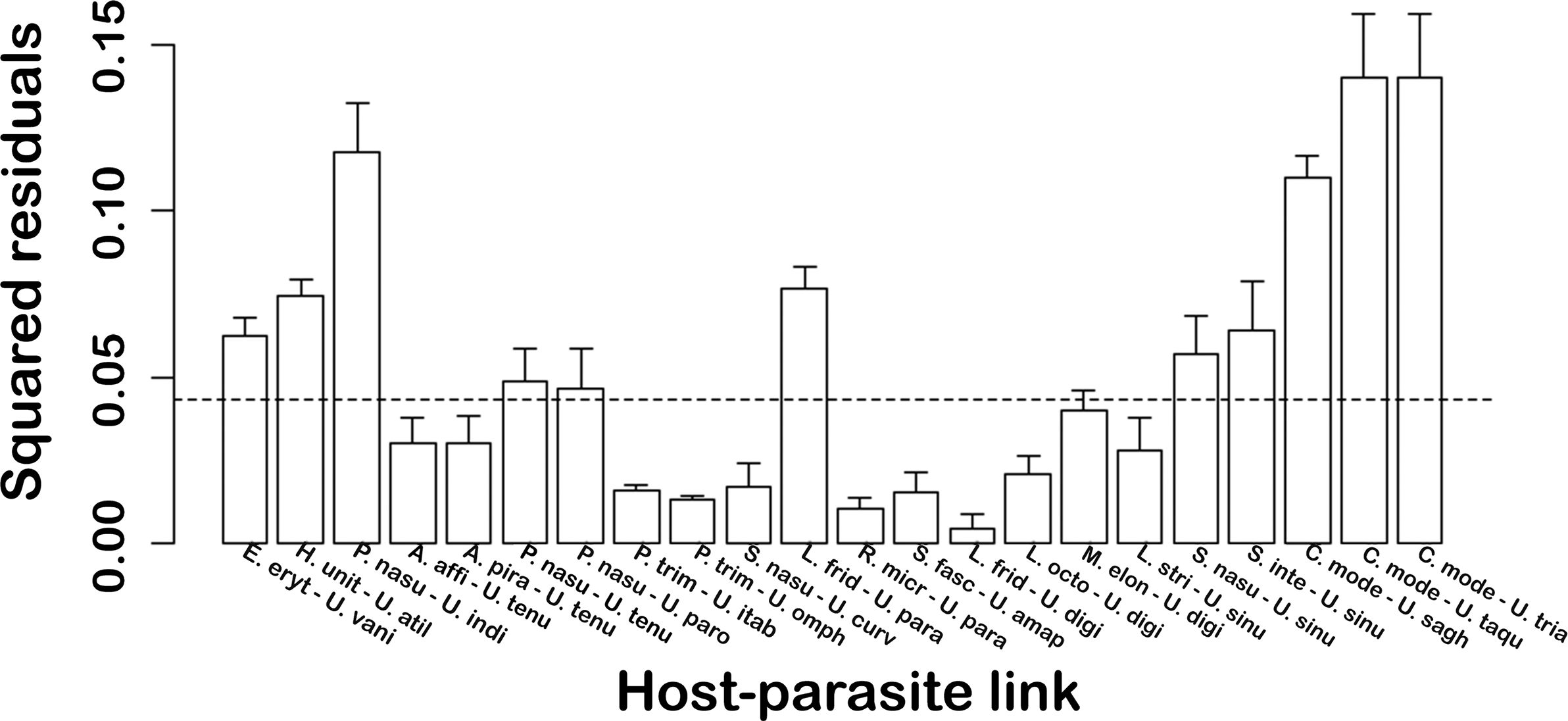
The contribution of each host-parasite link, whose 95% confidence intervals of the squared residuals of Procrustes fell below the median value, was probably due to cospeciation processes (Balbuena et al. Reference Balbuena, Míguez-Lozano and Blasco-Costa2013; Hutchinson et al. Reference Hutchinson, Cagua, Balbuena, Stouffer and Poisot2017).
In turn, Jane v.4 (Conow et al. Reference Conow, Fielder, Ovadia and Libeskind-Hadas2010) was used to determine the most plausible coevolutionary scenario for this parasite-host system and to quantify the prevalence of the different evolutionary events in determining the observed coevolutionary relationships. Jane reconciles host and parasite phylogenies using a polynomial-time dynamic programming algorithm combined with a genetic algorithm. This approach compares the two topologies by optimally mapping the parasite tree onto the host tree, assigning different costs to each of the five coevolutionary events: cospeciation, duplication, duplication followed by host-switch, loss, and failure to diverge (Conow et al. Reference Conow, Fielder, Ovadia and Libeskind-Hadas2010). We applied 10 event cost schemes, as described by Benovics et al. (Reference Benovics, Desdevises, Šanda, Vukić and Šimková2019) (Table 5), over 1,000 generations with a population size of 200, following Soares et al. (Reference Soares, Adriano, Domingues and Balbuena2023), to assess the influence of each type of coevolutionary event. The statistical significance of the global cost tests (P < 0.05) was assessed using 1,000 randomizations with Random Tip Mapping (input to analysis – data set 4).
Results
In this study, four new species (Urocleidoides simonecohenae n. sp., U. sphendonophorus n. sp., U. petinocleithrum n. sp., and U. amapaensis n. sp.) are described from hosts from the Eastern Amazon. In addition, the partial sequences of the 28s rDNA gene were obtained for the new species of Urocleidoides to reconstruct the phylogenetic relationships between the species of the genus. The hosts COI, Cytb, Rag1, and Rag2 gene sequences were retrieved from GenBank to reconstruct the phylogenetic relationships of the hosts studied herein (Table 2). The integration of molecular data from both parasites and their characiform hosts, alongside their interactions, provides significant insights into the evolutionary mechanisms associated with the diversification of Urocleidoides and their hosts.
Taxonomic summary
Class Monopisthocotyla Brabec, Salomaki, Kolisko, Scholz & Kuchta, 2023
Ordem Dactylogyridea Bychoswky, 1937
Family Dactylogyridae Bychowsky, 1933
Genus Urocleidoides Mizelle & Price, Reference Mizelle and Price1964
Urocleidoides simonecohenae Santos Neto, Domingues & Soares n. sp
Type host. Brachyhypopomus beebei (Schultz, 1944) (Gymnotiformes, Hypopomidae).
Type locality. Peixe-Boi River (Maracanã River Basin), municipality of Peixe-Boi, Pará, Brazil (01°11’33"S, 47°18’44"W).
Site of infestation. Gill filaments.
Prevalence of infection. 100% of 3 hosts examined.
Average intensity. 2.33 parasites per infected host.
Material deposited. Holotype (CHIOC No. 40835a); 6 paratypes (CHIOC No. 40835b–g); 1 hologenophore (CHIOC No. 40835h).
Molecular sequence data. 728 bp long partial 28S rDNA sequence data submitted to GenBank (accession number: PV984065).
Etymology. The specific name is in honor of Dra. Simone Chinicz Cohen, Instituto Oswaldo Cruz, Rio de Janeiro, Brazil, in recognition of her valuable contribution to the diversity of monopisthocotyls from South America.
Number of Zoobank. urn:lsid:zoobank.org:act:47DB574F-92ED-4142-9851-1ABF4F1C4DD0
Description. Based on 7 adult specimens, 3 mounted on Gomori’s Trichrome, 4 mounted on Hoyer’s medium. Body elongated, fusiform, total length excluding haptor 136 (108–185; n=6), total width at level of germarium 38 (31–48; n=6) (Figure 1a). Cephalic lobes poorly developed; three pairs of head organs; cephalic glands not observed. Eyes and accessory chromatic granules absent (Figure 1a). Mouth subterminal, midventral; pharynx subovate, muscular 13 (12–14; n=4) long, 11 (10–13; n=4) wide; esophagus short; two intestinal caeca, confluent posteriorly to gonads, lacking diverticula (Figure 1a). Gonads overlapping, testis dorsal to germarium. Genital pore, genital atrium, oviduct, uterus, Mehlis’ glands, eggs, not observed. Copulatory complex comprising male copulatory organ (MCO), accessory piece. MCO 69 (65–71; n=5) long, sclerotized, tubular, coiled counterclockwise, with approximately 1½ coils, base with sclerotized cap (Figure 1b); accessory piece sclerotized, non-articulated with base of MCO, with rounded proximal region, presenting a flap in the medial portion that serves as guide for the MCO, distal portion with tapered end (Figure 1b). Testis subspherical 19 (18–20; n=3) long, 18 (17–19; n=3) wide; vas deferens obscured by vitellaria; seminal vesicle sigmoid. Spherical prostatic reservoir (Figure 1a). Vagina single, vaginal pore medial, ventral; vaginal vestibule slightly sclerotized; vaginal canal sclerotized, sigmoid; seminal receptacle subspherical (Figure 1a). Vaginal sclerite 29 (24–37; n=5) long, with longitudinal superficial groove, thumb erect, point elongated, slightly curved (Figure 1c). Germarium bacilliform 19 (18–19; n=3) long, 8 (8–9; n=3) wide (Figure 1a). Vitellaria dense, extending from esophagus to confluence of intestinal caecum. Haptor trapezoidal 37 (29–57; n=6) long, 62 (42–88; n=8) wide. Anchors with distinct morphology (Figure 1i–j). Ventral anchor with developed superficial root; short deep root, rounded; slightly curved long shaft; curved point extending at level of tip of superficial root, base 8 (6–10; n=7), inner 20 (18–21; n=7) long, outer 19 (17–22; n=7) long (Figure 1i). Dorsal anchor with long superficial root, slightly depressed at distal surface; short deep root, rounded; point with ornaments as shredded sclerotized filaments, base 6 (5–7; n=6) long, inner 19 (14–21; n=6) long, outer 12 (10–13; n=6) long (Figure 1j). Ventral bar 27 (20–38; n=7) long, open V-shaped with slight anteromedial constriction, expanded ends (Figure 1g). Dorsal bar 27 (19–41; n=6) long, open U-shaped with rounded ends directed towards posterior region (Figure 1h). Hook pairs similar in morphology with shank divided into two subunits, erect thumb, evenly curved shaft, point; filament hook loop not observed. Hook pairs 1, 5 reduced in size 9 (8–11; n=7) long, shank with proximal dilation comprising 1/2 of length of shaft (Figure 1d); hook pairs 2–4, 7 22 (20–24; n=6) long, shank with proximal dilation comprising 2/3 of length of shaft (Figure 1e); hook pair 6 30 (25–40; n=7) long with erect thumb, elongated slightly curved shaft, delicate point, shank with proximal dilation comprising 1/5 of length of shaft, filament hook loop not observed (Figure 1f).
Urocleidoides simonecohenae n. sp. – a. ventral view, entire body; b. copulatory complex; c. vaginal sclerite; d. hooks of pairs 1 and 5; e. hooks of pairs 2, 3, 4, 7; f. hook pair 6; g. ventral bar; h. dorsal bar; i. ventral anchor; j. dorsal anchor. Figure scales – a: 50 μm scale; b–j: 25 μm scale.

Remarks. Urocleidoides simonecohenae n. sp. resembles U. nataliapasternakae Santos Neto & Domingues, Reference Santos Neto and Domingues2023 from B. brevirostris (Gymnotiformes: Hypopomidae), Urocleidoides cultellus Mendoza-Franco & Reina, Reference Mendoza-Franco and Reina2008 of Brachyhypopomus occidentalis (Gymnotiformes: Hypopomidae) and Urocleidoides carapus Mizelle, Kritsky & Crane, 1968 of G. carapo (Gymnotiformes: Gymnotidae) due to the absence of eyes and by having dorsal anchors with ornaments as shredded sclerotized filaments. However, U. simonecohenae n. sp. differs from U. nataliapasternakae by having an MCO with only 1½ rings (MCO with 2½ rings in U. nataliapasternakae), vaginal canal sigmoid (vaginal canal convoluted in U. nataliapasternakae) and an open ‘V’ shaped ventral bar with slight anteromedial constriction (rod-shaped ventral bar with widened ends in U. nataliapasternakae). Urocleidoides simonecohenae n. sp. can be distinguished from U. cutellus by possessing an MCO with 1½ rings (MCO with 5 rings in U. cutellus). The new species is easily distinguished from U. carapus by the presence of a vaginal sclerite (vaginal sclerite absent in U. carapus). Urocleidoides simonecohenae n. sp. also resembles U. ramentacuminatus Oliveira, Santos Neto & Domingues, 2020, which is found on Schizodon fasciatus, due to the presence of sclerotized shredded filaments in the dorsal anchor. However, the new species can be distinguished by having a vagina with a medial-ventral opening (marginal opening in U. ramentacuminatus), vaginal sclerite with a longitudinal groove, an erect thumb with a slightly curved point (lacking groove, a short rounded thumb with a straight point in U. ramentacuminatus), an accessory piece rounded and tapered (hook-shaped distal portion in U. ramentacuminatus) and lacking eyes (eyes present in U. ramentacuminatus).
Urocleidoides sphendonophorus Santos Neto, Domingues & Soares n. sp
Measurements (μm) of Urocleidoides sphendonophorus n. sp. parasites from the gills of Erythrinus erythrinus and Hoplerythrinus unitaeniatus from three localities

* Type locality **H. unitaeniatus host.
Type host. Erythrinus erythrinus (Bloch & Schneider, 1801) (Characiformes, Erythrinidae).
Other hosts: Hoplerythrinus unitaeniatus (Agassiz, 1829).
Type locality. Pratinha Community (Caeté River Basin), Bragança municipality, Pará, Brazil (01°5’2.49"S, 46°49’44.67"W).
Site of infestation. Gill filaments.
Prevalence of infection. 50% of 6 hosts examined.
Average intensity. 3 parasites per host.
Other host localities. Erythrinus erythrinus (Prevalence of infection: 50% of 2 hosts; Average intensity: 7), Igarapé Maratininga - Rio Moju, municipality of Tailândia, Pará, Brazil (02°27’55.7"S, 048°53’27. 6"W); Hoplerythrinus unitaeniatus (Prevalence of infection: 33% in 3 hosts; Average intensity: 4), Balneário Aracu - Rio Guamá, municipality of Ourém, Pará, Brazil (01°34’1.02"S, 47°9’52.35"W).
Material deposited. Holotype (CHIOC No. 40836a); 8 paratypes (CHIOC No. 40836b–e; 40837; 40838a–c); 9 vouchers (CHIOC No. 40839a–f; 40840a–c); 1 hologenophore (CHIOC No. 40841).
Molecular sequence data. 727 bp partial 28S rDNA sequence data submitted to GenBank (accession number: PV984066).
Etymology. The specific name (a noun) is from Greek (sphenodone = sling + phoros = bearing) and refers to the morphology of the vaginal sclerite, which is similar to a slingshot.
Number of Zoobank. urn:lsid:zoobank.org:act:F99B4987-63AE-40F5-B9D9-25239E012A04
Description. Based on 9 adult specimens, 3 mounted in Gomori’s Trichrome, 6 mounted in Hoyer’s medium. Body elongated, fusiform 182 (155–245; n=8) long, excluding haptor, total width at level of germarium 78 (57–107; n=8 (Figure 2a). Cephalic lobes (4) well-developed, 2 terminal, two bilateral; 4 pairs of head organs; cephalic glands not observed. Eyes present (2 pairs), posterior pair further apart than anterior, larger in size; accessory chromatic granules not observed (Figure 2a). Mouth subterminal, midventral; pharynx spherical, muscular 16 (12–19; n=9) long, 15 (12–17; n=9) wide; esophagus slightly elongated; two intestinal caeca, confluent posteriorly to gonads, lacking diverticula (Figure 2a). Gonads overlapping, testis dorsal to germarium. Genital pore, genital atrium, oviduct, Mehlis’ glands, uterus, eggs, prostatic reservoir not observed. Copulatory complex comprising MCO, accessory piece. MCO 18 (16–21; n=7) long, sclerotized, tubular, corkscrew-shaped with ten to eleven coils, counterclockwise, base with sclerotized cap (Figure 2b); accessory piece sclerotized, non-articulated with base of MCO, with enlarged proximal portion, distal portion bifurcate (Figure 2b). Testis ovate 18 (16–18; n=3) long, 15 (14–16; n=3) wide; vas deferens obscured by vitellaria; seminal vesicle sigmoid (Figure 2a). Vagina single, vaginal pore dextral, ventro-marginal; vaginal vestibule non-sclerotized; vaginal canal muscular, straight; seminal receptacle ovate 13 (n=1) long, 12 (n=1) wide (Figure 2a). Vaginal sclerite robust 33 (24–41; n=7) long, with distal region bifurcated in a ‘Y’ shape (Figure 2c). Germarium bacilliform 39 (36–42; n=2) long, 15 (12–19; n=2) wide (Figure 2a). Vitellaria dense, extending from esophagus to boundary of trunk, haptor (Figure 2a). Haptor trapezoidal 44 (40–52; n=9) long, 75 (60–92) wide. Anchors with distinct morphology (Figure 2i–j). Ventral anchor with well-developed superficial root, broad, depressed at distal surface; short deep root, rounded at distal surface; evenly curved shaft, point; point extending past level of tip of superficial root, base 14 (14–16; n=9), inner 36 (31–40; n=9), outer 41 (37–44; n=9) (Figure 2j). Dorsal anchor with poorly developed superficial root, with small fold at the base; short deep root, rounded; shaft elongated, slightly curved at axis; curved point extending past level of tip of superficial root, base 10 (10–11; n=5), inner 34 (30–37; n=5), outer 36 (30–39; n=5) (Figure 2i). Ventral bar 42 (33–51; n=9) long, 5 (4–7; n=9) wide, open U-shaped with expanded rounded ends (Figure 2g). Dorsal bar 37 (26–43; n=9) long, 21 (17–24; n=9) wide, U-shaped with well-developed posteromedial projection, tapering ends (Figure 2h). Hooks similar in morphology with shank divided into two subunits, erect thumb, elongated slightly curved shaft, delicate point; filamentous hook loop extending to near beginning of shank dilation. Hook pairs 1, 5 reduced in size 14 (10–16; n=7) long, shank with proximal dilation comprising 1/2 of length of shaft (Figure 2d); hook pairs 2, 3, 4, 6 17 (15–20; n=8) long, shank with proximal dilation comprising 1/2 of length of shaft (Figure 2e); pair 7 22 (19–24; n=8) long, shank with proximal dilation comprising 1/3 of length of shaft (Figure 2f).
Urocleidoides sphendonophorus n. sp. – a. ventral view, entire body; b. copulatory complex; c. vaginal sclerite; d. hook pairs 1 and 5; e. hook pairs 2, 3, 4, and 6; f. hook pair 7; g. ventral bar; h. dorsal bar; i. dorsal anchor; j. ventral anchor. Figure scale – a–i: 25 μm scale.

Remarks. Urocleidoides sphendonophorus n. sp. can be distinguished from other congeneric species by possessing a combination of the following features: robust ‘Y’-shaped vaginal sclerite, corkscrew-shaped MCO; dorsal bar with a well-developed posteromedial projection, and dorsal anchors with inconspicuous superficial roots. Additionally, this is the first description of a Urocleidoides species presenting a crokscrew-shaped MCO.
Urocleidoides petinocleithrum Santos Neto, Domingues & Soares n. sp
Type host. Erythrinus erythrinus (Bloch & Schneider, 1801) (Characiformes, Erythrinidae).
Type locality. Pratinha Community (Caeté River Basin), Bragança municipality, Pará, Brazil (01°5’2.49"S, 46°49’44.67"W).
Site of infestation. Gill filaments.
Prevalence of infection. 60% of 5 hosts examined.
Average intensity. 2 parasites per host.
Material deposited. Holotype (CHIOC No. 40832a); 5 paratypes (CHIOC No. 40832b–c; 40833 a–c); 1 hologenophore (CHIOC No. 40834).
Molecular sequence data. 734 bp partial 28S rDNA sequence data submitted to
GenBank (accession number: PV984068).
Etymology. The specific name (a noun) is from Greek (petino = flying + cleithrum = bar) and refers to the morphology of the dorsal bar, which bears a resemblance to a bird flying.
Number of Zoobank. urn:lsid:zoobank.org:act:1451E52D-A13E-45E5-90B7-1378E3C3A3D5
Description. Based on 6 adult specimens, 1 mounted on Gomori’s Trichrome and 5 mounted on Hoyer’s medium. Body elongated, fusiform 187 (145–257; n=5) long, excluding the haptor, total width at level of germarium 60 (41–80; n=6) (Figure 3a). Cephalic lobes poorly developed; three pairs of head organs; cephalic glands not observed. Eyes (2 pairs) present, posterior pair larger than anterior pair; anterior pair with few granules observed, accessory granules present and poorly distributed. Mouth subterminal, midventral; pharynx muscular, spherical 15 (12–17; n= 5) long, 16 (13–22; n=5) wide (Figure 3a); esophagus short; two intestinal caeca, confluent posteriorly to gonads, lacking diverticula (Figure 3a). Gonads overlapping, testis dorsal to germarium. Genital pore, genital atrium, oviduct, Mehlis’ glands, uterus, eggs, prostatic reservoir not observed. Copulatory complex comprising MCO, accessory piece. MCO 20 (20–21; n=3) long, sclerotized, tubular, corkscrew-shaped with approximately eight to nine coils, counterclockwise, base with sclerotized cap; accessory piece sclerotized, non-articulated with base of MCO, comprising variable distal sheath with medial fold (Figure 3b). Testis ovate 11 (n=1) long, 8 (n=1) wide; vas deferens obscured by vitellaria; seminal vesicle sigmoid (Figure 3a). Vagina single, vaginal pore dextral, ventro-marginal; vaginal vestibule non-sclerotized; vaginal canal muscular, sigmoid; seminal receptacle spherical 9 (n=1) long, 8 (n=1) wide (Figure 3a). Vaginal sclerite hook-shaped, 29 (18–35; n=3) long, with thumb poorly developed or inconspicuous, shaft, point slightly recurved (Figure 3c). Germarium bacilliform 19 (n=1) long, 8 (n=1) wide (Figure 3a). Vitellaria dense, extending from esophagus to boundary of trunk, haptor. Haptor subtrapezoidal 47 (40–52; n=6) long, 63 (42–82; n=6) wide (Figure 3a). Anchors with distinct morphology (Figure 3i–j). Ventral anchor with subtrapezoidal superficial root; subtriangular deep root, slightly rounded at distal surface; evenly curved shaft, point; point extending past level of tip of superficial root, base12 (10–15; n=6), inner 31 (27–36; n=6), outer 35 (30–40; n=6) (Figure 3i). Dorsal anchor with poorly developed superficial root, showing small fold at the base; short deep root, rounded; shaft elongated, straight; short, curved point, outer 34 (32–42; n=6) (Figure 3j). Ventral bar 45 (39–51; n=4) long, with anteromedial constriction, rounded ends (Figure 3g). Dorsal bar 36 (32–39; n=4) long, delicate, V-shaped, with small posteromedial projection (Figure 3h). Hook pairs 2, 3, 4, 6, and 7 22 (20–24; n=5) long similar in morphology with shank divided into two subunits, proximal dilation comprising 1/3 of length of shaft, erect thumb, lightly curved shaft, delicate point; filamentous hook loop extending to near beginning of shank dilation (Figure 3d); hook pairs 1, 5 reduced in size, without inflation, erect thumb, lightly curved shaft, delicate point; filamentous hook loop comprising 1/3 of the shank; hook pair 1 15 (14–17; n=4) long (Figure 3f); hook pair 5 14 (13–15; n=3) long (Figure 3e).
Urocleidoides petinocleithrum n. sp. – a. ventral view, entire body; b. copulatory complex; c. vaginal sclerite; d. hooks of pairs 2, 3, 4, 6. and 7; e. hook pair 5; f. hook pair 1; g. ventral bar; h. dorsal bar; i. ventral anchor; j. dorsal anchor. Figure scales – a–c, g–j: 25 μm scale; d–f: 10 μm scale.

Remarks. Urocleidoides petinocleithrum n. sp. resembles U. sphendonophorus n. sp. by having a corkscrew-shaped MCO, as well as by possessing the dorsal anchors with inconspicuous superficial roots and a small fold at the base. However, the new species differs from U. sphendonophorus n. sp. mainly by the morphology of the vaginal sclerite (hook-shaped sclerite in U. petinocleithrum n. sp., Y-shaped sclerite in U. sphendonophorus n. sp.), and also by the morphology of the dorsal bar (U-shaped dorsal bar with elongate posteromedial projection in U. sphendonophorus n. sp., delicate V-shaped dorsal bar with small posteromedial projection in U. petinocleithrum n. sp.). This is the second description of a Urocleidoides species presenting a crokscrew-shaped MCO.
Urocleidoides amapaensis Santos Neto, Domingues, Oliveira & Soares n. sp
Type host. Schizodon fasciatus Spix & Agassiz, 1829 (Characiformes, Anostomidae).
Type locality. Jari River, municipality of Vitória do Jari, state of Amapá, Brazil (01°9’4.01"S, 52°0’53.22"W).
Site of infestation. Gill filaments.
Prevalence of infection. 100% of 2 hosts examined.
Average intensity. 3 parasites per infected host.
Material deposited. Holotype (CHIOC No. 40831a); 4 paratypes (CHIOC No. 40831b–e); 1 hologenophore (CHIOC 40831f).
Molecular sequence data. 740 bp partial 28S rDNA sequence data submitted to GenBank (accession number: PV984067).
Etymology. The specific name refers to the Brazilian State of Amapá from which the species was collected.
Number of Zoobank. urn:lsid:zoobank.org:act:0E473C39-69DA-4533-AAC3-DCE114F35D50
Description. Based on 5 adult specimens, 1 mounted in Gomori’s Trichrome and 4 mounted in Hoyer’s medium. Body elongated, robust, 300 (235–374; n=5) long, excluding the haptor, total width at level of germarium 220 (114–317; n=5) (Figure 4a). Cephalic lobes poorly developed; three pairs of head organs; cephalic glands not observed. Eyes and accessory chromatic granules absent (Figure 4a). Mouth subterminal, midventral; pharynx muscular, ovate 35 (28–39; n=4) long, 28 (22–34; n=4) wide; esophagus short; two intestinal caeca, confluent posteriorly to gonads, lacking diverticula (Figure 4a). Gonads overlapping, testis dorsal to germarium. Genital pore, genital atrium, oviduct, Mehlis’ glands, uterus, eggs, prostatic reservoir not observed. Copulatory complex comprising MCO, accessory piece. MCO 33 (31–35; n=5) long, sclerotized, tubular, coiled counterclockwise, with approximately one coil (Figure 4b); accessory piece sclerotized, non-articulated with base of MCO, comprising variable distal sheath, located at distal region of MCO (Figure 4b). Testis ovate 31 (n=1) long, 24 (n=1) wide; vas deferens obscured by vitellaria; seminal vesicle sigmoid with medial dilation (Figure 4a). Vagina single, vaginal pore sinistral, ventro-marginal; vaginal vestibule non-sclerotized; vaginal canal muscular, sigmoid; seminal receptacle ovate (Figure 4a). Vaginal sclerite 26 (24–27; n=5) long, delicate, slender with a hook-shaped distal portion, short thumb, shaft without groove (Figure 4c). Germarium saccate 79 (n=1) long, 23 (n=1) wide (Figure 4a). Vitellaria dense, extending from the median portion of pharynx to boundary of trunk, haptor. Haptor trapezoidal 54 (48–59; n=5) long, 103 (95–109; n=5) wide. Anchors with distinct morphology (Figure 4j–k). Ventral anchor with divergent roots; superficial root developed, moderately elongated; deep root developed with rounded surface; evenly curved shaft, point; point extending past level of tip of superficial root, base 12 (10–13; n=5), inner 18 (18–19; n=5), outer 22 (20–24; n=5) (Figure 4j). Dorsal anchor with well-developed superficial root, subtriangular; deep root with rounded surface; shaft slightly recurved at axis; curved point extending past level of tip of superficial root, base 9 (8–10; n=4), inner 20 (17–22; n=4), outer 21 (18–23; n=4) (Figure 4k). Ventral bar narrow, open U-shaped, with rounded ends, 44 (34–55; n=5) long (Figure 4h). Dorsal bar open U-shaped with rounded ends 40 (29–44; n=5) long (Figure 4i). Hooks similar in morphology with shank divided into two subunits, slightly depressed thumb, elongated slightly curved shaft, delicate point; filamentous hook loop not observed. Hook pairs 2–4 30 (28–33; n=5) long, shank with proximal dilation comprising 1/5 of length of shaft (Figure 4g); hook pair 1, reduced in size 15 (14–16; n=4) long, shank with proximal dilation comprising 1/2 of length of shaft (Figure 4f); hook pair 5, reduced in size 10 (10–11; n=4) long, shank with proximal dilation comprising 2/3 of length of shaft (Figure 4d); hook pairs 6–7 22 (20–24; n=4) long, shank with proximal dilation comprising 1/3 of length of shaft (Figure 4e).
Urocleidoides amapaensis n. sp. – a. ventral view, entire body; b. copulatory complex; c. vaginal sclerite; d. hook pair 5; e. hooks of pairs 6 and 7; f. hook pair 1; g. hooks of pairs 2, 3, and 4; h. ventral bar; i. dorsal bar; j. ventral anchor; k. dorsal anchor. Figure scales – a: 50 μm scale; b, c: 10 μm scale; h–k: 30 μm scale; d–g: 20 μm scale.

Remarks. Urocleidoides amapaensis n. sp. resembles U. curvocuspidis Ebert, Osaki-Pereira, De León & Silva, 2024 parasite from the gills of Schizodon nasutus by lacking eye, and by the general morphology of the male copulatory organ and vaginal sclerite. However, they differ from one another mainly due to the position of the vaginal pore (sinistral in U. amapaensis n. sp. and dextral in U. curvocuspidis), morphology of the vagina (long, muscular in U. amapaensis n. sp. and short, sclerotized in U. curvocuspidis), and accessory piece (non-articulated to the MCO in U. amapaensis n. sp. and articulated to the MCO in U. curvocuspidis). The new species also is similar to U. falxus Zago, Yamada, Yamada, Franceschini, Bongiovani & Da Silva, Reference Zago, Yamada, Yamada, Franceschini, Bongiovani and Da Silva2020 parasite from the gills of Megaleporinus elongatus (Valenciennes) (Anostomidae) by the morphology of the copulatory complex (i.e., MCO with one coil; accessory piece non-articulated with base of MCO, comprising variable distal sheath, located at distal region of MCO), and position of the vaginal pore (i.e., sinistral). However, the new species can be differentiated from U. falxus mainly by possessing a vaginal sclerite delicate, slender with a hook-shaped distal portion, short thumb, without groove (vaginal sclerite with grooved shaft, erect thumb and robust, curved shaft in U. falxus), and lacking eyes (present in U. falxus).
Molecular data and phylogenetic inferences
Partial sequences of the 28S rDNA gene were obtained for U. simonecohenae n. sp. – 728 bp long, U. sphendonophorus n. sp. – 727 bp long, U. petinocleithrum n. sp. – 734 bp long, and U. amapaensis n. sp. – 740 bp long. After trimming the ends, the aligned 28S rDNA sequences had a length of 764 bp. The BI and ML analyses (28S rDNA) recovered topologies with posterior probabilities (PP) and bootstrap values (B) significant between Urocleidoides species that occur for different host families (Figure 5).
Molecular phylogeny estimated by Bayesian inference using partial 28S rDNA sequences from Neotropical dactylogyrids. Species newly sequenced for this study are shown in bold. The species name precedes the GenBank sequence ID. Bootstrap support values and posterior probabilities are given between the branches (bootstrap values < 50 and posterior probabilities < 80 not reported). Branch length scale bar indicates the number of substitutions per site.

The clade containing the Urocleidoides species is strongly supported by BI analysis (PP = 1, B = 83) and is divided into two major clades (Clades A and B) (Figure 5). Clade A with strongly support (PP = 1, B = 78) consisting of species of Urocleidoides that have been found parasitizing Characiformes from the families Erythrinidae (U. atilaiamarinoi, U. vanini), Parodontidae (U. parodoni, U. tenuis, U. indianensis), Anostomidae (U. sinus, U. digitabulum, U. curvocuspidis, U. amapaensis n. sp., U. paradoxus, U. itabocaensis, U. omphalocleithrum) and Curimatidae (U. triangulus, U. saghirus, U. taquariensis). Clade B is divided into two clades (Clades B1 and B2). Clade B1, presents a stronger support for both analyses (PP = 1, B = 100), grouping Urocleidoides species reported from Gymnotiformes hosts from the families Hypopomidae (U. simonecohenae n. sp., U. nataliaparternakae) and Gymnotidae (U. uncinus, U. carapus, U. gymnotus). Clade B2 (PP = 0.83, B = 58) presents a low support for both analyses and groups the Urocleidoides species occurring in erytrhinid hosts (U. naris, U. bulbophallus, U. brasiliensis, U. macrosoma, U. sphendonophorus n. sp., U. petinocleithrum n. sp.) and species of Rhinoxenus reported from Serrasalmidae, Cacatuocotyle spp. (Characidae) and species of Diaphorocleidus from the family Characidae (all parasites of Characiformes).
The Urocleidoides species in Clade A, which parasitize Characiformes fishes, and the species in Clade B, which parasitize both Gymnotiformes and Characiformes (Erytrhinidae), exhibit a genetic divergence ranging from 15% to 36.8% (111–282 bp) (Table 4). The genetic divergence between Urocleidoides spp. and Jainus spp. ranges between 15.4% and 28.5% (169–304 bp). The genetic divergence between Urocleidoides spp. (Clade A and B) and Rhinoxenus spp. diverge at rates between 18.5% and 33.3% (169–296 bp), and 16.9% and 31.4% (178–3014 bp), respectively. Urocleidoides spp. (Clade A) and Diaphorocleidus spp. diverged between 17.1% and 28.8% (163–572 bp), and Urocleidoides spp. (Clade B) diverged 15.2% and 32.1% (163–572 bp). Species of Urocleidoides that parasitize H. malabaricus (Characiformes: Erythrinidae) diverged genetically between 3.7% and 9.7% (29–87 bp). Urocleidoides sphendonophorus n. sp. and U. petinocleithrum n. sp. diverged 9.1% (81 bp) and when compared with the other species that parasitize erythrinids the divergence was 9.5% and 11.5% (85–102 bp). The Urocleidoides species that occur on anostomids diverged between 4.9% and 27.4% (35–174 bp), and U. amapaensis n. sp. diverged from the others between 7% and 15.8% (49–132 bp). The species that occur parasitizing Gymnotiformes diverged between 3% and 13% (36–107 bp). Urocleidoides simonecohenae n. sp. diverged between 10.1% and 13.3% (77–99 bp) from the other species that parasitize gymnotiformes. The Urocleidoides species that occur on parodontids diverged from 3.4% to 10.6% (70–164 bp), and the Urocleidoides species that occur on curimatids diverged from 7.4% to 15% (54–142 bp).
Paired genetic divergence of 28S rDNA sequences from Urocleidoides species. Upper triangular matrix (p-distance) shows number of nucleotide differences; lower triangular matrix shows differences in nucleotide percentage. Sequences obtained in this work are shown in bold. U. = Urocleidoides; R. = Rhinoxenus; D. = Diaphorocleidus; C. = Cacatuocotyle; J. = Jainus

Cophylogeny of a clade of species parasitizing Characiformes hosts
Given that our phylogenetic analysis rejected the monophyly of Urocleidoides, as species of Cacatuocotyle, Diaphorocleidus, and Rhinoxenus clustered together with Urocleidoides (Figure 5), we performed the cophylogenetic analysis using only a subset of Urocleidoides species belonging to a strongly supported clade (Clade A). This strategy was adopted to ensure that the cophylogenetic reconstruction would represent a single, coherent evolutionary lineage. To accomplish this, we conducted a separate Bayesian inference including only the Urocleidoides species assigned to Clade A (see Figure 5), using the same analytical settings described above in the ‘Materials and methods’ section.
Posterior probabilities indicated strong nodal support for all clades in the host phylogeny (PP = ≥ 0.95), as shown in Figure 6, except for the clade comprising Erythrinus erythrinus and Hoplerythrinus unitaeniatus, which received moderate support (PP = 0.90). For parasites, the new BI analysis, conducted exclusively on Urocleidoides species associated with Characiformes from Clade A (Figure 5), produced topologies identical to those in the previous section but with stronger posterior probability support in most clades, further reinforcing the robustness of the inferred phylogenetic relationships.
Tanglegram reflecting the application of the PACo (R2= 0.76, P < 0.01). Associations between 15 species of Characiformes from Neotropical region (left) and 15 species of Urocleidoides (right) – support values of posterior probabilities are given above the branches. The blue lines represent probable cospeciation events based on the sum of squared residuals.

Results of the cophylogenetic analyses with Jane for Urocleidoides and its hosts from the Erythrinidae, Parodontidae, Curimatidae, and Anostomidae families (corresponding to Clade A in Figure 5). Ten evolutionary models with different cost schemes for each evolutionary event were tested. The total costs and frequencies of each evolutionary event are shown for each model. Statistical significance of global cost tests (p < 0.05) was determined based on 1,000 random reconstructions with Random Tip Mapping

The tanglegram showing the associations between Urocleidoides species and their hosts revealed four distinct clades, each corresponding to a specific family of characiform hosts: Erythrinidae, Parodontidae, Curimatidae, and Anostomidae (Figure 6). Then, the global test conducted in PACo provided strong support for phylogenetic congruence (R2 = 0.76, P < 0.01 with 10,000 permutations). In PACo, examining the squared residuals of individual parasite-host associations showed that the confidence intervals for A. affinis - U. tenuis, A. piracicabae - U. tenuis, P. trimaculatus - U. itabocaensis, P. trimaculatus - U. omphalothecium, S. nasutus - U. curvocuspidis, R. microlepis - U. paradoxus, S. fasciatus - U. amapaensis n. sp., L. friderici - U. digitabulum, L. octofasciatus - U. digitabulum, and L. striatus - U. sinus, all groups remained within the median residual square value (Figure 7). This provides strong support for the associations among Parodontidae and Anostomidae species, suggesting cospeciation events with their corresponding Urocleidoides species (Figures 6–7).
Contribution of individual associations to overall phylogenetic congruence of Urocleidoides species and their Characiformes hosts. Columns represent the squared residual value of each association contributing to the total sum of squared residuals calculated by PACo for patristic distances derived from the phylogenies shown in Figure 6. Error bars correspond to confidence intervals of +95%. The median residual square is indicated by the dotted line. Parasite-host associations with 95% confidence intervals above the median are likely to represent non-congruent associations.

Using the Jane’s event-based method, a significant global signal of congruence was detected across the dataset (P < 0.01) (Table 5). Most simulations using models that attribute distinct costs to each individual evolutionary event have produced reconstructions with comparable coevolutionary proportions (Table 5). The simulations suggested that regardless of the models used, cospeciation events remained recurrent five-six (except in the cases of prohibitive and equal models). In contrast, loss events were likely the most frequent coevolutionary event in the system studied (6 out of 10 models), and duplication followed by host-switch. The host-switch adjusted TreeFitter model produced the lowest total cost (17) (see Table 5 for cost schemes), accounting for five-six cospeciation events, seven duplications, one-two duplications followed by host-switch, seven divergence failures, and eight-nine loss events (Figure 8). In contrast, the highest total costs were found in the sorting prohibitive and divergence failure prohibited models, suggesting that these scenarios are unlikely in the evolution of the parasites studied.
One of the possible cophylogenetic reconstructions generated by Jane. The black (independent) branches represent the host phylogeny. The blue (dependent) branches correspond to the parasite phylogeny.

Discussion
Recent studies have shown that the current knowledge about the diversity of Urocleidoides is still underestimated. In the last five years, 25 species have been described parasitizing fish from Brazilian continental waters (Ebert et al. Reference Ebert, Osaki-Pereira, Pérez-Ponce de León and Da Silva2024; Freitas et al. Reference Freitas, Bezerra, Meneses, Justo, Viana and Cohen2021; Hasuike et al. Reference Hasuike, Scorsim, Arjona, Bellay, de Oliveira and Takemoto2024; Oliveira et al. 2020, Reference Oliveira, Da Silva, Vieira and Acosta2021; Santos Neto and Domingues, Reference Santos Neto and Domingues2023, Reference Santos Neto and Domingues2024; Yamada et al. Reference Yamada, Osaki-Pereira, Acosta, Ebert and Da Silva2024; Zago et al. Reference Zago, Yamada, Yamada, Franceschini, Bongiovani and Da Silva2020;), suggesting that many species have yet to be described. Also, our knowledge of the evolutionary history of Urocleidoides lineages is restricted to a few works (Ebert et al. Reference Ebert, Osaki-Pereira, Pérez-Ponce de León and Da Silva2024; Hasuike et al. Reference Hasuike, Scorsim, Arjona, Bellay, de Oliveira and Takemoto2024; Oliveira et al. Reference Oliveira, Da Silva, Vieira and Acosta2021; Santos Neto and Domingues, Reference Santos Neto and Domingues2023, Reference Santos Neto and Domingues2024; Yamada et al. Reference Yamada, Osaki-Pereira, Acosta, Ebert and Da Silva2024; Zago et al. Reference Zago, Yamada, Yamada, Franceschini, Bongiovani and Da Silva2020), and studies on the historical associations of this parasite-host system are still unknown. In this paper, we describe four new species of Urocleidoides parasitizing members of Characiformes (Anostomidae and Erythrinidae) and Gymnotiformes (Hypopomidae), which enhances our understanding of the diversity of this group in the Neotropical region. Furthermore, we present a phylogenetic hypothesis based on molecular data (28S rDNA) to infer relationships between Urocleidoides species. Additionally, for the first time, a cophylogenetic analysis was conducted to investigate the evolutionary relationships between some Urocleidoides (Figure 5, Clade A) and their Characiformes hosts (Erythrinidae, Parodontidae, Curimatidae, and Anostomidae) (Figure 6).
The proposal of four new species in this study is supported by various lines of evidence from an integrative taxonomy approach. Key morphological features, particularly in haptoral structures (anchors, bars) and reproductive organs (vaginal sclerite, copulatory complex, and vaginal opening position), validate the new species. Genetic divergence among the new species ranges from 9.1% to 22.9% (Table 4), with the lowest interspecific divergence between U. petinocleithrum and U. sphendonophorus (9.1%) and the highest divergence between U. amapaensis and U. sphendonophorus (22.9%), consistent with expected levels for distinct species within the Dactylogyridae (Oliveira et al. Reference Oliveira, Da Silva, Vieira and Acosta2021; Santos Neto and Domingues, Reference Santos Neto and Domingues2023, Reference Santos Neto and Domingues2024; Zago et al. Reference Zago, Yamada, Yamada, Franceschini, Bongiovani and Da Silva2020;).
The non-monophyly of Urocleidoides has already been shown in previous studies (Ebert et al. Reference Ebert, Osaki-Pereira, Pérez-Ponce de León and Da Silva2024; Oliveira et al. Reference Oliveira, Da Silva, Vieira and Acosta2021; Santos Neto and Domingues, Reference Santos Neto and Domingues2023, Reference Santos Neto and Domingues2024; Yamada et al. Reference Yamada, Osaki-Pereira, Acosta, Ebert and Da Silva2024). Our phylogenetic tree results for parasites partially align with the hypotheses of Zago et al. (Reference Zago, Yamada, Yamada, Franceschini, Bongiovani and Da Silva2020), Oliveira et al. (Reference Oliveira, Da Silva, Vieira and Acosta2021), Santos Neto and Domingues (Reference Santos Neto and Domingues2023, Reference Santos Neto and Domingues2024), and Ebert et al. (Reference Ebert, Osaki-Pereira, Pérez-Ponce de León and Da Silva2024), supporting the non-monophyly of Urocleidoides species. In our 28S rDNA phylogenetic analysis, one clade is formed by Urocleidoides species that parasitize Characiformes (Anostomidae, Curimatidae, Erythrinidae, and Parodontidae), and another clade formed by Urocleidoides species from Gymnotiformes (Hypopomidae, Gymnotidae). Our results align with those reported by Yamada et al. (Reference Yamada, Osaki-Pereira, Acosta, Ebert and Da Silva2024). In our analysis, only Urocleidoides species in Clade A were strongly supported. In contrast, species associated with Clade B showed weak support (Figure 5), leading us to focus our cophylogenetic analysis solely on Clade A, which is related to Characiformes.
The phylogeny constructed exclusively for Urocleidoides species associated with Characiformes yielded a topology identical to that of the general phylogeny (Clade A, Figure 5). In this case, species were grouped into distinct clades, each comprising parasites of hosts from the families Erythrinidae, Parodontidae, Curimatidae, and Anostomidae, respectively (Figure 6). In contrast, the phylogeny of Characiform host species parasitized by Urocleidoides exhibited interfamily relationships that align with those reported by Betancur-R et al. (Reference Betancur-R, Wiley, Arratia, Acero, Bailly, Miya, Lecointre and Orti2017) and Melo et al. (Reference Melo, Sidlauskas, Near, Roxo, Ghezelayagh, Ochoa and Oliveira2022). Therefore, the phylogenies obtained were deemed robust enough for cophylogenetic analyses.
The results of PACo (R2 = 0.76, P < 0.01) and Jane (P < 0.01, in all models) provide strong statistical support for phylogenetic congruence sensu Perez-Lamarque and Morlon (Reference Perez-Lamarque and Morlon2024), showing that the phylogeny of the parasites is strongly associated with the phylogeny of the hosts. This suggests the occurrence of cospeciation events, a clear pattern when looking at the tanglegram (Figure 6), which revealed four distinct clades of Urocleidoides, each associated with a specific host family (Erythrinidae, Parodontidae, Curimatidae, and Anostomidae). These findings indicate that, in general, the diversification of the parasites may have occurred in parallel with the diversification of their hosts, a typical pattern of cospeciation. This scenario is also corroborated by Jane’s results, which point to cospeciation as a relevant process at the family level (Figure 8).
Boeger and Kritsky (Reference Boeger and Kritsky1997) suggested that in monopisthocotyls, cospeciation is more likely to occur at higher taxonomic levels of hosts, such as families and genera. This hypothesis was later supported by Desdevises et al. (Reference Desdevises, Morand, Jousson and Legendre2002), who further argued that phylogenetic associations between monopisthocotyls and their hosts are driven by historical factors, including immunological and morphological barriers, acting at these taxonomic levels. Meanwhile, Braga et al. (Reference Braga, Razzolini and Boeger2014), based on their studies of interaction networks between gill monopisthocotyls and fish in the Neotropical region, proposed that the restricted composition of monopisthocotylan faunas is influenced by both host phylogenetic relationships and geographical distribution. Our findings partially align with these perspectives, as each Characiform family harbors a distinct assemblage of Urocleidoides, while closely related hosts share some parasite species.
Monopisthocotyls are known to have high host specificity (Braga et al. Reference Braga, Razzolini and Boeger2014; Poulin Reference Poulin1992), which suggests that cospeciation plays an important role in the evolution of these associations. However, several cophylogenetic studies involving monopisthocotyls and their hosts indicate that events such as host-switch, divergence failure and duplication are more common than cospeciation in these interactions (Benovics et al. Reference Benovics, Desdevises, Šanda, Vukić and Šimková2019; Desdevises et al. Reference Desdevises, Morand, Jousson and Legendre2002; Huyse et al. Reference Huyse, Audenaert and Volckaert2003; Mendlová et al. Reference Mendlová, Desdevides, Civáňová, Pariselle and Šimková2012; Míguez-Lozano et al. Reference Míguez-Lozano, Rodríguez-González and Balbuena2017; Seidlová et al. Reference Seidlová, Benovics and Šimková2022; Šimková et al. Reference Šimková, Morand, Jobet, Gelnar and Verneau2004; Zietara and Lumme Reference Zietara and Lumme2002). Our results are in line with these patterns. Among the 10 models tested in Jane (Table 5), the ‘Host-switch-adjusted TreeFitter’ had the lowest total cost (17), suggesting that the evolution of the parasite-host system studied here is best explained by a combination of cospeciation, duplication, failure to diverge, and host-switch.
However, in all models analyzed using Jane (Table 5), the reconstructions consistently suggested recurrent cospeciation events (5 to 6 instances), indicating that a significant portion of the associations may have originated through joint speciation. The specific host-parasite associations that exhibited the lowest total sum of squared residuals calculated in PACo (e.g., A. affinis – U. tenuis, A. piracicabae – U. tenuis, P. trimaculatus – U. itabocaensis, L. friderici – U. digitabulum; see Figure 7 for additional associations) further reinforce that, within the Parodontidae and Anostomidae families, the congruence between host and parasite phylogenies is particularly robust. This suggests that phylogenetic host tracking, or cospeciation, plays a more important role in shaping associations within these groups.
Melo et al. (Reference Melo, Sidlauskas, Near, Roxo, Ghezelayagh, Ochoa and Oliveira2022) proposed that the diversification of Anostomidae species is closely linked to ecomorphological factors; that is, environmental pressures have driven morphological adaptations in these fishes, accelerating their speciation. Conversely, other studies have suggested that speciation in monopisthocotylan parasites may be more strongly driven by phylogenetic tracking of host traits rather than by strict host-parasite cospeciation (Blasco-Costa et al. Reference Blasco-Costa, Hayward, Poulin and Balbuena2021; Soares et al. Reference Soares, Adriano, Domingues and Balbuena2023).
Soares et al. (Reference Soares, Adriano, Domingues and Balbuena2023), for example, used a Cophylospace framework to show that the diversification of monopisthocotyls parasitizing marine catfish along the Atlantic coast of South America is more closely associated with the morphological characteristics of their hosts, driven by environmental pressures, than with strict host-parasite cospeciation. A similar pattern appears to emerge in our findings on Anostomidae and Urocleidoides. In this case, host diversification seems to have been driven by ecomorphological factors (sensu Melo et al. Reference Melo, Sidlauskas, Near, Roxo, Ghezelayagh, Ochoa and Oliveira2022), with parasites tracking these evolutionary changes, consistent with a model of coadaptive codiversification as defined by Clayton et al. (Reference Clayton, Bush and Johnson2015). Our results also align with the patterns reported by Blasco-Costa et al. (Reference Blasco-Costa, Hayward, Poulin and Balbuena2021) for Ligophorus spp. (monopisthocotyls) infecting gray mullets (Mugilidae) in the Mediterranean and Black Seas, further supporting the notion that host ecological and morphological traits play a crucial role in shaping parasite diversification.
The predominance of loss and failure to diverge events suggests that, while cospeciation is an important factor, the evolution of these parasites is not exclusively driven by host speciation. This pattern suggests that, throughout the evolutionary history of Urocleidoides in their hosts, there have been multiple instances of local extinction or the disappearance of specific lineages within particular Characiformes species. Vanhove et al. (Reference Vanhove, Pariselle, Van Steenberge, Raeymaekers, Hablützel, Gillardin, Hellemans, Breman, Koblmüller, Sturmbauer, Snoeks, Volckaert and Huyse2015), in their study on cichlids and monopisthocotyls, suggested that the high frequency of loss events is attributed to several factors, including the overdispersed nature of the parasites and the small, fluctuating population sizes. Other factors, such as the physiological resistance of the host (Ricklefs et al. Reference Ricklefs, Fallon and Bermingham2004) or a decrease in the host population (de Castro and Bolker Reference De Castro and Bolker2005), can also result in loss events. The wide dispersal of Urocleidoides with species occurring only in one host species and other colonizing more than one host can contribute to the fragmentation of Urocleidoides populations and, consequently, increase the number of loss events. However, failure to diverge events can be challenging to differentiate from a recent host-switch before speciation on the newly colonized host (Banks and Paterson Reference Banks and Paterson2005). Miguez-Lozano et al. (2017) in their study, noted that Jane’s analysis does not differentiate between failure to divergence events and host-switch, which may inflate the number of divergence failure events.
Several studies suggest that host-switch is an important association event between monopisthocotyls and their hosts (Blasco-Costa et al. Reference Blasco-Costa, Míguez-Lozano, Sarabeev and Balbuena2012; Desdevises et al. Reference Desdevises, Morand, Jousson and Legendre2002; Huyse and Volckaert Reference Huyse and Volckaert2005; Mendlová et al. Reference Mendlová, Desdevides, Civáňová, Pariselle and Šimková2012; Zietara and Lumme Reference Zietara and Lumme2002). In the Urocleidoides- Characiformes system analyzed, the occurrence of duplication followed by host-switch appears to be frequent, indicating that certain Urocleidoides may have diversified by exploiting novel hosts within the same phylogenetic group. The present study observed that host-switch events were less frequent, occurring between hosts at different taxonomic levels, such as hosts of different genera within the same host family. Wendt et al. (Reference Wendt, Malabarba, Braga, Boeger, Landis and Carvalho2022) reported that species of Characithecium demonstrate a capacity to colonize a wide range of host species indicating a broader ecological flexibility. This finding indicates that Characithecium can readily infect compatible hosts based on ecological fitting. It implies that the evolutionary history of the hosts, alongside subsequent dispersal events, has enabled Characithecium to establish in new environments and across different host species. Alda et al. (Reference Alda, Mendoza-Franco, Hanson-Regan, Reina, Bermingham and Torchin2025) studied the phylogeographic patterns of monopisthocotyls parasitizing Astyanax species (Characiformes, Characidae) in Central America and Mexico. They found that geographical factors significantly influence the diversification and distribution of these parasites, affecting host dispersal, isolating populations, and facilitating host-switch due to the ecological adaptability of Astyanax species. In this context, the phylogenetic contiguity within the Characiformes probably allows Urocleidoides species to have access to distant hosts, since this monophyletic group of fishes has diversified exclusively within the Neotropical region.
Conclusion
The present study increases the diversity of Urocleidoides to 60 valid species. The non-monophyly of the genus is supported by phylogenetic analyses using sequences of the partial 28S rDNA, with the addition of the four species described herein. Some of the clades of Urocleidoides species appeared closely related to their hosts at the family or order levels. Our findings indicate a complex evolutionary scenario in which cospeciation has played a significant role; however, it has also been accompanied by significant loss events, failure to diverge, and host-switch. This suggests that the diversification of Urocleidoides in Characiformes hosts did not occur in a constrained manner but rather involved episodes of adaptation and potential colonization of new hosts over time. This pattern is consistent with the evolutionary trajectories observed in many monopisthocotyls, where periodic niche expansion balances the process of coevolutionary specialization.
This study will inspire future coevolutionary investigations incorporating new taxonomic data and molecular sequences of Urocleidoides species and their hosts, aiming at a future comprehensive understanding of the diversification of this parasite group in the Neotropical region.
Acknowledgements
The authors thank the members of the Laboratório de Sistemática e Coevolução (UFPA) for assistance during the collecting trips, Simone Cohen (Fiocruz) for providing information from the database on South American freshwater fish Monopisthocotyls/Polyopisthocotyls for consultation, Dr. Simoni Santos da Silva (UFPA) and Dr. Edson Aparecido Adriano (Federal University of São Paulo) for allowing us to use their laboratory facilities for DNA extraction and amplification. We also thank the anonymous reviewers for their contribution to this manuscript.
Financial support
The present study was partly supported by research grants from the Conselho Nacional de Desenvolvimento Científico e Tecnológico, CNPq – Brazil (CNPq - PROTAX 001/2015-440526/2015-9) and Fundação Amazônia de Amparo a Estudos e Pesquisas, FAPESPA – Brazil (ICAAF 017-2018) to M.V. Domingues. J.F. Santos Neto was supported by a doctoral scholarship from the Coordenação de Aperfeiçoamento de Pessoal de Nível Superior, CAPES – Brazil (grant# 88882.460123/2019-01), and G.B. Soares was supported by a Postdoctoral fellowship granted #2022/15973-3, Fundação de Amparo à Pesquisa do Estado de São Paulo (FAPESP). M.V. Domingues received research productivity grants from the Conselho Nacional de Desenvolvimento Científico e Tecnológico (grants #309896/2019-3 and 307657/2022-1).
Declaration of generative AI and AI-assisted technologies in the writing process
During the preparation of this work, the authors used DeepL for translation and Grammarly in order to check the grammar, spelling, and readability of captions. After using this tool/service, the authors reviewed and edited the content as needed and take full responsibility for the content of the publication.
Credit authorship contribution statement
João F. dos Santos Neto: Conceptualization, Data curation, Formal analysis, Investigation, Methodology, Software, Validation, Visualization, Writing – original draft, review and editing. Geusivam B. Soares: Conceptualization, Methodology, Software, Formal analysis, Validation, Visualization, Writing – review and editing. Marcus V. Domingues: Conceptualization, Funding acquisition, Project administration, Resources, Supervision, Validation, Visualization, Writing – review and editing.
Competing interests
The authors declare that they have no known competing financial interests or personal relationships that could have appeared to influence the work reported in this paper.
Ethical standard
All applicable institutional, national, and international guidelines for the care and use of animals were followed. Specimens were collected under the license for collection of biological material (43381) granted by the Instituto Chico Mendes de Conservação da Biodiversidade (ICMBio).
Data availability
Datasets 1–4 required to perform all the analyses are deposited on Zenodo (https://doi.org/10.5281/zenodo.15716024).