When sudden cardiac death occurs in athletes, it receives widespread publicity and raises important questions, as these individuals have usually undergone pre-participation screening. In a study of 1929 normotensive 15–34-year-old athletes, five patients (0.26%) had a dilated aortic root. Among them, four were basketball players and one was a volleyball player. All of these individuals were tall, but only two had other features of Marfan syndrome.Reference Kinoshita, Mimura, Obayashi, Katsukawa, Onishi and Yamazaki 1 In a recent study that collected data over 10 years from the databases of the US National Registry of Sudden Death in Athletes and the National Collegiate Athletic Association, there were 182 sudden deaths out of 4,052,369 participants and 64 of them were considered to be due to cardiac disease (six per year). There were three deaths due to aortic rupture (4.6% of cardiovascular causes).Reference Maron, Haas, Murphy, Ahluwalia and Rutten-Ramos 2
Biophysics of exercise and aortic rupture
During maximal aerobic exercise, there is an increase in systolic blood pressure, usually up to 140–180 mmHg, and rarely up to 210 mmHg, with concomitant decrease in diastolic blood pressure due to peripheral vasodilation, with the net outcome being a modest increase in mean pressure. With isometric exercise, however, there is a significant increase in systolic and diastolic blood pressure, occasionally to massive proportions – for example, up to 300–500 mmHg with weight lifting. The maximum wall stress that has been found experimentally to be tolerated by aortic strips is 1000 kPa. To put this into clinical perspective, although patients with an aortic root diameter of 4 cm and a blood pressure of 120 mmHg have a wall stress of 110 kPa, patients with aortic root diameters of 6 cm and blood pressure of 200 mmHg have a wall stress of 850 kPa, close to the wall rupture limit.Reference Koullias, Modak, Tranquilli, Korkolis, Barash and Elefteriades 3 It is obvious from the above that individuals with a dilated aorta are at increased risk of aortic dissection and rupture under conditions of heavy exercise, especially isometric exercise.
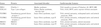
Several inherited conditions are known to predispose to aortic dilation, especially of the aortic root and ascending aorta. Prominent among these are autosomal-dominant connective tissue disorders such as Marfan syndrome, Loeys–Dietz syndrome, and Ehlers–Danlos syndrome (Table 1).
Inherited autosomal-dominant aortic conditions.

AAA=abdominal aortic aneurysm; AI=aortic insufficiency; MR=mitral regurgitation; MVP=mitral valve prolapse; PDA=patent ductus arteriosus; TAA=thoracic aortic aneurysm
Marfan syndrome
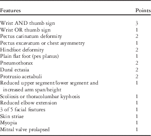
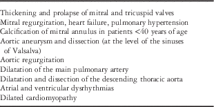
Marfan syndrome was first described by Antoine-Bernard Marfan more than a century ago. It has an incidence of ~1:5000Reference Judge and Dietz 4 and is inherited in an autosomal-dominant manner. Approximately 25% of cases are due to de novo mutations. Individuals with Marfan syndrome have long limbs and tall stature – “dolichostenomelia”. Owing to their height, there is likely some degree of self-selection among these individuals for participation in sports, where being tall gives a competitive advantage; thus, there appears to be an increased incidence of Marfan syndrome among basketball and volleyball players. The diagnosis of Marfan syndrome requires the presence of several clinical criteria, commonly called the Ghent Criteria, which have been revised in 2010.Reference Loeys, Dietz and Braverman 5 The diagnostic criteria involve the following five major categories: family history, cardiac evaluation, eye examination, systemic score, and genetic testing. The diagnosis is made when criteria are met for at least two categories, and seven points are required for the systemic category to be satisfied (Table 2). Cardiovascular features are listed in Table 3. Marfan syndrome is due to mutations in the FBN1 gene,Reference Dietz, Cutting and Pyeritz 6 located at chromosome locus 15q21.1,Reference Kainulainen, Pulkkinen, Savolainen, Kaitila and Peltonen 7 encoding the glycoprotein fibrillin 1,Reference Sakai, Keene and Engvall 8 which is the principal component of the extracellular matrix microfibril. There is increased aortic stiffness and decreased aortic distensibility in patients with Marfan syndrome, which is present in childhood and becomes progressively worse with age. Aortic stiffness – as measured by MRI – correlates with the rate of aneurysm progression and adverse outcome in patients with Marfan syndrome.Reference Nollen, Groenink and Tijssen 9 In addition, there is evidence that fibrillin 1 plays an important role in the regulation of the transforming growth factor β signalling pathway. Fibrillin 1 has a homology with latent transforming growth factor β binding proteins, thus suggesting a role in the activation of transforming growth factor β and initiation of a downstream signalling cascade.Reference Neptune, Frischmeyer and Arking 10 This pathogenic mechanism may account for the other manifestations of Marfan syndrome that are not easily explained by extracellular matrix defects, such as long bone overgrowth, myxomatous changes of the mitral valve, lung pathology, and dural ectasia. The relationship between fibrillin 1 and transforming growth factor β signalling cascade also explains the similarities between Marfan syndrome and Loeys–Dietz syndrome, which is mostly due to mutations in genes encoding the transforming growth factor β receptors – TGFBR1 and TGFBR2.Reference Loeys, Chen and Neptune 11
Systemic score calculation for Marfan’s syndrome.

Cardiovascular features of Marfan’s syndrome.

The diagnosis and management of Marfan syndrome require a multi-disciplinary approach including specialists from genetics, orthopedics, cardiology, ophthalmology, neurology, and psychology.
After a clinical diagnosis of Marfan syndrome is established, monitoring for progression or development of new cardiovascular manifestations is essential. At least yearly assessment of aortic root and thoracic aortic growth and appearance of evidence of dissection is recommended. In addition, assessment of mitral valve and myocardial function is important. Marfan patients may be prone to ventricular dysrhythmias, and 24-hour Holter monitoring might be indicated. In patients who are followed-up carefully with serial echocardiograms, the risk of sudden death appears to be minimal, whereas the risk from ventricular dysrhythmias may reach 4%. The risk appears to be increased in patients with a dilated left ventricle and prolongation of the QT interval.Reference Yetman, Bornemeier and McCrindle 12
Medical therapy has included medications that achieve a reduction in blood pressure with or without reduction in heart rate, such as β-blockers, angiotensin-converting enzyme inhibitors, angiotensin receptor blockers, or calcium channel blockers. β-blockers have been the most commonly used drugs and probably work through both negative inotropic and chronotropic effects. A small randomised trial showed that fewer patients treated with a β-blocker (propranolol) reached the end points of aortic insufficiency, aortic dissection, cardiovascular surgery, congestive heart failure, and death. The normalised rate of aortic dilatation was lower in the treated group than in the control group (0.023 versus 0.084 per year, p<0.001).Reference Shores, Berger, Murphy and Pyeritz 13 Approximately 10–20% of patients are intolerant to β-blockers because of fatigue, asthma, or depression.
Treatment with angiotensin receptor blockers has theoretical advantages, as these drugs have been shown to inhibit transforming growth factor β and to reduce aortic growth in a mouse model.Reference Habashi, Judge and Holm 14 A recent, large, randomised, double-blind, placebo-controlled European trial, the Marfan-Sartan trial, however, showed no additional benefit in reduction of aortic root dilatation rate of dual drug therapy (β-blocker and losartan) over standard therapy with β-blockers alone.Reference Milleron, Arnoult and Ropers 15 A different, large, randomised controlled trial comparing atenolol versus losartan in children and young adults with Marfan syndrome showed no difference in the 3-year rates of aortic root surgery, aortic dissection, and death between the two groups.Reference Lacro, Dietz and Sleeper 16 Both drugs appeared to be beneficial. Surgical therapy is recommended in patients who have already developed or are considered at high risk for aortic dissection. Elective surgery to replace the aortic root is indicated in adults with maximum aortic diameter >5.0 cm.Reference Gott, Greene and Alejo 17 Earlier surgical intervention is recommended for patients with a rate of increase >0.5–1.0 cm/year. Composite replacement of the aortic root and valve (Bentall procedure) was the initial standard procedure, and has shown excellent results.Reference Gott, Greene and Alejo 17 , Reference Bentall and De Bono 18 Mortality for elective surgery was 1.9%, for urgent surgery 2.6%, and for emergency surgery within 24 hours of surgical consultation 11.7%.Reference Gott, Greene and Alejo 17
Owing to the risks of thromboembolism and need for anticoagulation, which is especially problematic in young patients and pregnant women, valve-sparing procedures, initially pioneered by David,Reference Bentall and De Bono 18 have gained popularity and now are considered the standard of care. The 10-year survival of the Bentall procedure and the David procedure are 90.5 versus 96.3%, respectively, with the latter having fewer thromboembolic and haemorrhagic events.Reference Price, Magruder and Young 19 , Reference David, David, Manlhiot, Colman, Crean and Bradley 20
Loeys–Dietz syndrome
Loeys–Dietz syndrome is a relatively recently described condition characterised by aortic aneurysms, generalised arterial tortuosity, hypertelorism, bifid uvula and/or cleft palate, and generalised systemic organ involvement.Reference Loeys, Chen and Neptune 11 It is also inherited in an autosomal-dominant manner.
Multiple similarities exist between Loeys–Dietz syndrome and Marfan syndrome, the main one being the propensity for aneurysms of the ascending aorta. It is believed that the pathogenesis of the syndrome is due to the altered signalling of transforming growth factor β. There are now thought to be four types of Loeys–Dietz syndrome, related to mutations in four different genes, TGFBR1, TGFBR2, SMAD3, and TGFB.Reference Loeys, Chen and Neptune 11 , Reference Loeys, Schwarze and Holm 21 – Reference Cleuziou, Eichinger, Schreiber and Lange 26 The first two are the most common. They are characterised by severe craniofacial features, and patients are at particularly high risk for rupture of the aorta and other smaller arteries, even at young age and at smaller dimensions than other aortopathies. In addition, other CHD are common, including bicuspid aortic valve, patent ductus arteriosus, and atrial septal defects. Mitral valve prolapse and mitral regurgitation are also common. Left ventricular hypertrophy and impaired left ventricular systolic function have also been reported. Arrhythmias including atrial fibrillation affect a significant number of patients.
A mutation in any of the above-mentioned genes in combination with arterial aneurysm or dissection, or family history of documented Loeys–Dietz syndrome, is sufficient to make the diagnosis.
The aggressive nature of aortic and other vessel disease in Loeys–Dietz syndrome mandates close monitoring and appropriate medical and surgical management.Reference Loeys, Chen and Neptune 11 , Reference Loeys, Schwarze and Holm 21 , Reference van de Laar, Oldenburg and Pals 22 The threshold for aortic root replacement is much lower than Marfan syndrome. The adult threshold for surgery is an aortic root of >4.0 cm, or rapid growth of >0.5 cm/year. In addition, severe craniofacial features and family history of early dissection should influence decision regarding timing of intervention. A valve-sparing procedure should be preferred, especially in young patients and successful valve-sparing procedures have been performed in children younger than 1 year of age.Reference Boileau, Guo and Hanna 25 Valve-sparing procedures are contraindicated in the presence of leaflet perforations, bicuspid aortic valve with extensive calcification or dysfunction, or acute dissection in unstable patients.Reference Patel, Arnaoutakis and George 27
In young patients, it is recommended that careful observation with echocardiography at least every 6 months is recommended until the aortic root reaches 4.0 cm. In the postoperative state, very frequent echocardiographic monitoring every 2–3 months is recommended for early re-dissection.
Medical management includes β-blockers, angiotensin-converting enzyme inhibitors, or angiotensin receptor blockers. The latter may be particularly beneficial because of their effects on the transforming growth factor β signalling cascade. The recommended dose of losartan is 2.0 mg/kg/day for children and 100 mg/day for adults.Reference MacCarrick, Black and Bowdin 28 Owing to the nature of the disease, serial assessment of all portions of the aorta and its branches with MRI – head to pelvis – is recommended.Reference Valverde, Simpson and Beerbaum 29
Ehlers–Danlos syndrome
Ehlers–Danlos syndrome is a heterogeneous disorder related to mutations in the collagen genes, with an estimated prevalence of 1:5000 to 1:25,000 births.Reference Steinman, Royce and Superti-Furga 30 The relative frequencies of different Ehlers–Danlos syndrome types are not known precisely, but classic and hypermobile types account for >90%. In comparison, vascular Ehlers–Danlos syndrome likely accounts for <5% of Ehlers–Danlos syndrome patients (incidence of <1: 100,000). Vascular Ehlers–Danlos syndrome is due to mutations in the COL3A1 gene. Vascular catastrophes are frequently the first major manifestation of vascular Ehlers–Danlos syndrome. There is no established medical management. Treatment with both endovascular and open surgical techniques is effective, although there is an increased rate of vascular complications due to extreme fragility of the vessels. Previous reports have estimated operative mortality in vascular reconstruction in vascular-type patients to be from 20 to 65%; however, with careful approach including hypothermic circulatory arrest for central aortic operations and elective surgical repair, operative mortality may be decreased to 8%.Reference Brooke, Arnaoutakis, McDonnell and Black 31
CHD
Bicuspid aortic valve, a very common condition that is present in ~1–2% of the population, appears to be heritable, and is frequently associated with dilated ascending aorta, usually at the mid-ascending aortic level. The degree of aortic dilatation does not always correlate with the degree of aortic valve stenosis. Aortic dissection and rupture have been described in patients with bicuspid aortic valve.
Patients with tetralogy of Fallot and with truncus arteriosus have been known to have dilated ascending aortas, sometimes to impressive dimensions. Patients with transposition of the great arteries, especially after arterial switch operation have been known to develop dilatation of the neo-aortic root.Reference Yetman and Graham 32
Bicuspid aortic valve
The frequency of dilation of the aorta in patients with bicuspid aortic valve is up to 50%,Reference Tzemos, Therrien and Yip 33 and there is an incidence of dissection or rupture of 8% among those with dilation.Reference Davies, Kaple and Mandapati 34 The previous terminology of “post-stenotic dilatation” has been misleading, in the sense that a large majority of patients with bicuspid aortic valve have dilatation of the ascending aorta independent of aortic valve stenosis. In fact, bicuspid aortic valve and thoracic aortic aneurysms have been found independent of each other with increased frequency in the same family, with a pattern suggestive of autosomal-dominant inheritance.Reference Loscalzo, Goh, Loeys, Kent, Spevak and Dietz 35 There is also an increased incidence of dilatation of the ascending aorta and dissection in patients with Turner syndrome,Reference Matura, Ho, Rosing and Bondy 36 as well as in individuals with previous coarctation repair.Reference Oliver, Gallego, Gonzalez, Aroca, Bret and Mesa 37 It is recommended that all patients with bicuspid aortic valve undergo frequent evaluation with echocardiography and, if indicated, with MRI or CT angiography. As the point of maximal dilatation is usually in the mid-ascending aorta, axial imaging techniques may be especially useful in older patients who lack good echocardiographic windows.Reference Beroukhim, Roosevelt and Yetman 38 It is also suggested to screen all first-degree relatives of a patient with bicuspid aortic valve with echocardiography, given the high possibility of identifying another affected family member.
The recommendations for surgical management of patients with bicuspid aortic valve and ascending aortic aneurysm are similar to those for patients with Marfan syndrome, as aortic dissection occurs at a similar aortic diameter irrespective of whether the dilation is due to Marfan syndrome or bicuspid aortic valve. Surgical intervention for the aortic root is indicated at a diameter of 5.0 cm.Reference Bonow, Carabello and Chatterjee 39 A valve-sparing procedure should be preferred if the aortic valve functions normally and is expected to have a longevity for at least 10 years; however, if the indication for surgical intervention is valvar dysfunction, concomitant ascending aortic replacement should be considered, given the high likelihood of repeat surgical intervention in such patients.Reference Borger, Preston and Ivanov 40 In patients with Turner syndrome, aortic dissection has been reported to occur at smaller diameters. An aortic root size of 2 cm/m2, which can be <3 cm absolute diameter given the short stature of these patients, has been suggested as the indication for intervention.Reference Matura, Ho, Rosing and Bondy 36
Tetralogy of Fallot and other conotruncal anomalies
The presence of ascending aortic dilation in patients with tetralogy of Fallot has long been recognised, and the incidence has been reported to be as high as 48%.Reference Jonsson, Ivert and Brodin 41 Histological abnormalities of the aortic media in patients with tetralogy of Fallot are similar to that seen in patients with Marfan syndrome and have been noted as early as a few days of age.Reference Tan, Davlouros, McCarthy, Gatzoulis and Ho 42 Risk factors for progressive aortic dilation include the presence of pulmonary atresia, right aortic arch, history of an aortopulmonary shunt, male sex, and the presence of a 22q11 deletion.Reference Niwa 43 Marked ascending aortic dilation has been found in 15% of adults with repaired tetralogy of Fallot, with the need for surgical intervention in 12%.Reference Niwa 43
A yearly examination that includes an imaging modality is recommended in patients with tetralogy of Fallot with increased aortic diameter. The risk of dissection appears to increase above an aortic diameter of 55 mm; however, a recent, large study reviewing all hospitalisations in Texas over 13 years questioned the increased risk for dissection in conotruncal anomalies. From 37.9 million hospitalisations from 1999–2012, there were 12,016 cases of thoracic aortic dissection, and 214 of these cases occurred in patients with CHD. The most common lesions were bicuspid aortic valve (42%), atrial septal defect (21%), aortic coarctation (7%), ventricular septal defect (6%), and patent ductus arteriosus (4%). Only six patients with conotruncal anomalies – three with tetralogy of Fallot, two with D-transposition of the great arteries, and one with truncus arteriosus – were admitted with thoracic aortic dissection. Statistical analysis showed a significant association between thoracic aortic dissection and bicuspid aortic valve (odds ratio 10, 95% confidence interval 8.2–13), but not with coarctation of the aorta or any conotruncal lesion.Reference Frischhertz, Shamszad, Pedroza, Milewicz and Morris 44
Exercise and follow-up recommendations for patients with aortic pathology
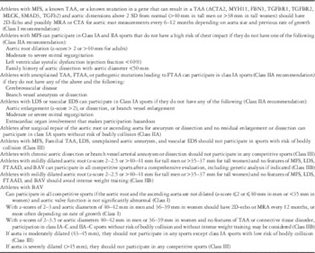
A recent American Heart Association/American College of Cardiology document summarised the recommendations for exercise participation in athletes with heart disease.Reference Braverman, Harris, Kovacs and Maron 45 The recommendations for athletes with dilatation of the aorta are depicted in Table 4. As a reminder, for the purposes of exercise recommendations, sports can be classified according to their degree of static – I: low, II: moderate, and III: high – and dynamic – A: low, B: moderate, and C: high – components. At one end of the scale are class 1A activities such as golf and bowling and at the other end are the 3C activities such as speed skating and triathlons.
Exercise and follow-up recommendations in patients with aortic pathology.

BAV=bicuspid aortic valve; CTA=computerised thoracic arteriogram; EDS=Ehlers–Danlos syndrome; FTAA=familial thoracic aortic aneurysm; FTAAD=familial thoracic aortic aneurysm or dilation; LDS=Loeys–Dietz syndrome; MFS=Marfan syndrome; MRA=magnetic resonance angiography; TAA=thoracic aortic aneurysm; 2D=two dimensional
Acknowledgements
The author thank Robert Ardinger, MD, for critically reviewing this manuscript and making useful comments.
Financial Support
This research or review received no specific grant from any funding agency or from commercial or not-for-profit sectors.
Conflicts of Interest
None.
Ethical Standards
The authors assert that all referenced work contributing to this review complies with the ethical standards of biomedical or medicolegal investigation.