Early pubertal development in girls portends heightened risk for a number of adverse health outcomes. These include all-cause mortality and deaths attributable to cardiovascular disease and cancer, incident coronary heart disease, breast cancer, uterine fibroid tumors, and in adolescence, early emergence of risk factors for heart disease, such as increased blood pressure, progressive weight gain, insulin insensitivity, and an atherogenic lipid profile (e.g., Adair & Gordon-Larsen, Reference Adair and Grodon-Larsen2001; Bernstein, Reference Bernstein2002; Cooper et al., Reference Cooper, Ephross, Weinberg, Baird, Whelan and Sandler1999; Golub et al., Reference Golub, Collman, Foster, Kimmel, Rajpert-De Meyts and Reiter2008; Hsieh, Trichopoulos, Katsouyanni, & Yuasa, Reference Hsieh, Trichopoulos, Katsouyanni and Yuasa1990; Jacobsen, Oda, Knutsen, & Fraser, Reference Jacobsen, Oda, Knutsen and Fraser2009; Lakshman et al., Reference Lakshman, Forouhi, Sharp, Luben, Bingham and Khaw2009; Marshall et al., Reference Marshall, Spiegelman, Goldman, Manson, Colditz and Barbieri1998; Remsberg et al., Reference Remsberg, Demerath, Schubert, Chumlea, Sun and Siervogel2005). Problems of psychological adjustment are also common among girls experiencing early puberty, as evidenced by low self-esteem and depressive symptomatology (Ge, Conger, & Elder, Reference Ge, Conger and Elder1996; Graber, Lewinsohn, Seeley, & Brooks-Gunn, Reference Graber, Lewinsohn, Seeley and Brooks-Gunn1997; Graber, Seeley, Brooks-Gunn, & Lewinsohn, Reference Graber, Seeley, Brooks-Gunn and Lewinsohn2004; Hayward et al., Reference Hayward, Killen, Wilson, Hammer, Litt and Kraemer1997; Kaltiala-Heino, Marttunen, Rantanen, & Rimpela, Reference Kaltiala-Heino, Marttunen, Rantanen and Rimpela2003; Stice, Presnell, & Bearman, Reference Stice, Presnell and Bearman2001), body image concerns and eating disorders (Attie & Brooks-Gunn, Reference Attie and Brooks-Gunn1989; Kaltiala-Heino, Rimpel, Risannen, & Rantanen, Reference Kaltiala-Heino, Rimpel, Rissanen and Rentanen2001; Janna, Kaltiala-Heino, Kovisto, & Rantanen, 2003), affiliation with deviant peers, truancy, and experimentation with drugs and alcohol (e.g., Caspi & Moffitt, Reference Caspi and Moffitt1991; Deardorff, Gonzales, Christopher, Roosa, & Millsap, Reference Deardorff, Gonzales, Christopher, Roosa and Millsap2005; Kim, Smith, & Palermiti, Reference Kim, Smith and Palermiti1997; Mezzich et al., Reference Mezzich, Tarter, Giancola, Lu, Kirisci and Parks1997; Romans, Martin, Gendall, & Herbison, Reference Romans, Martin, Gendall and Herbison2003; Silbereisen, Petersen, Albrecht, & Kracke, Reference Silbereisen, Petersen, Albrecht and Kracke1989; Stice, Presnell, & Bearman, Reference Stice, Presnell and Bearman2001). Compared to later developing girls, those who mature earliest begin dating at a younger age, have earlier sexual intercourse and more sexual partners, date older boys, and report earlier first cohabitation; these behaviors also contribute to risk for sexually transmitted diseases, early pregnancy, and teenage childbearing (e.g., Deardorff et al., Reference Deardorff, Gonzales, Christopher, Roosa and Millsap2005; Dunbar, Sheeder, Lezotte, Dabelea, & Stevens-Simon, Reference Dunbar, Sheeder, Lezotte, Dabelea and Stevens-Siimon2008; Hoier, Reference Hoier2003; Jorm, Christensen, Rodgers, Jacomb, & Easteal, 2004; Kim & Smith, 1999; Kim et al., Reference Kim, Smith and Palermiti1997; Lam, Shi, Ho, Stewart, & Fan, Reference Lam, Shi, Ho, Stewart and Fan2002; Mezzich et al., Reference Mezzich, Tarter, Giancola, Lu, Kirisci and Parks1997; Miller et al., Reference Miller, Norton, Curtis, Hill, Schvaneveldt and Young1997; Phinney & Jensen, 1998).
These sequelae of early puberty underscore the public health significance of pubertal timing and, hence, of understanding its determinants. In addition to well-established biological antecedents, such as nutritional status, body weight, and adiposity (Ellis, Reference Ellis2004; Ellis & Essex, Reference Ellis and Essex2007; Lassek & Gaulin, Reference Lassek and Gaulin2007; Lipson, Reference Lipson and Ellison2001; Sloboda, Hart, Doherty, Pennell, & Hickey, Reference Sloboda, Hart, Doherty, Pennell and Hickey2007), psychosocial factors and aspects of the family environment have emerged as significant predictors of pubertal development. In an influential paper published nearly two decades ago, Belsky, Steinberg, and Draper (Reference Belsky, Steinberg and Draper1991) drew on life history models to posit an evolutionary theory of socialization having implications for reproductive maturation. These authors hypothesized a certain plasticity of pubertal timing that is molded by early cues to future prospects, particularly prospects germane to reproduction. They suggested that qualities of the rearing environment not only shape expectations of future material resources but also girls' expectations for experiencing enduring and supportive marital and family relationships as adults. These in turn were thought to somehow modulate pubertal timing (possibly through changes in body weight and associated neuroendocrine mechanisms), such that investment in prepubertal growth and development is curtailed in favor of accelerated progression to reproductive maturity when, in the face of early adversity, girls internalize family life and marriage as unpredictable, fragile, and fractious. Subsequent research largely confirmed the model's novel predictions regarding pubertal development, and this work has been the subject of comprehensive empirical and theoretical review by Ellis (Reference Ellis2004, Reference Ellis, Ellis and Bjorklund2005; also Ellis, Shirtcliff, Boyce, Deardoff, & Essex, Reference Ellis, Shirtcliff, Boyce, Deardorff and Essex2011 [this issue]).
It is now well established that pubertal development tends to occur earlier in girls who are raised without a father than among those reared in traditional two-parent families, and this association may be strongest when the father's absence is first experienced in early childhood or persists over an extended number of years (Ellis & Garber, Reference Ellis and Garber2000; Hoier, Reference Hoier2003; Maestripieri, Roney, DeBias, Durante, & Spaepen, Reference Maestripieri, Roney, DeBias, Durante and Spaepen2004; Moffitt, Caspi, Belsky, & Silva, Reference Moffitt, Caspi, Belsky and Silva1992; Quinlan, Reference Quinlan2003; Romans et al., Reference Romans, Martin, Gendall and Herbison2003; Surbey, Reference Surbey, Zigler and Bercovitch1990; Wierson, Long, & Forehand, Reference Wierson, Long and Forehand1993). In addition, and largely independent of father absence, family environments prone to discord, harsh parenting, or lacking in close interpersonal relations are associated with girls' earlier pubertal development (Belsky et al., Reference Belsky, Steinberg, Houts, Friedman, DeHart and Cauffman2007; Ellis & Garber, Reference Ellis and Garber2000; Ellis, McFayden-Ketchum, Dodge, Petit & Bates, 1999; Graber, Brooks-Gunn, & Warren, Reference Graber, Brooks-Gunn and Warren1995; Kim et al., Reference Kim, Smith and Palermiti1997; Kim & Smith, Reference Kim and Smith1998; Moffitt et al., Reference Moffitt, Caspi, Belsky and Silva1992; Quinlan, Reference Quinlan2003; Romans et al., Reference Romans, Martin, Gendall and Herbison2003; Steinberg, Reference Steinberg1988; Tither & Ellis, Reference Tither and Ellis2008), whereas girls mature more slowly when reared in affectionate, cohesive families (Ellis et al., Reference Ellis, McFadyen-Ketchum, Dodge, Pettit and Bates1999; Ellis & Essex, Reference Ellis and Essex2007). These relations are documented in both short- and long-term longitudinal investigations, in retrospective studies, and among studies employing diverse indices of family environment. Nonetheless, effect sizes are typically small, and in investigations employing multiple family measures, pubertal timing may be predicted inconsistently across indicators.
Although the conceptual framework articulated by Belsky et al. (Reference Belsky, Steinberg and Draper1991), Ellis (Reference Ellis2004), and others (e.g., Chisholm, Reference Chisholm1999) implies a causal relation between aspects of the family environment and pubertal development, genetic factors afford alternative interpretations. For instance, behavioral correlates of early puberty, such as sexual precocity and adjustment problems, might presage later marital and family difficulties among women who reach reproductive competence at a young age. If variation in pubertal timing is heritably transmitted over generations, early pubertal maturation in daughters could reflect shared genetic influences that occasioned the accelerated reproductive development of their mothers and then, indirectly, the adversities of family environment to which the daughters were later exposed (i.e., gene–environment correlation; Moffitt et al., Reference Moffitt, Caspi, Belsky and Silva1992; Surbey, Reference Surbey, Zigler and Bercovitch1990). Genetic factors are known to play an important role in reproductive development, and a sentinel indicator of pubertal maturation in females, age at menarche, correlates moderately across generations (e.g., Brooks-Gunn & Warren, Reference Brooks-Gunn and Warren1988; Damon, Damon, Reed, & Valadian, 1962; Graber et al., Reference Graber, Brooks-Gunn and Warren1995; Kaprio et al., Reference Kaprio, Rimpela, Winter, Viken, Rimpela and Rose1995; Towne et al., Reference Towne, Czerwinski, Demerath, Blangero, Roche and Siervogel2004), with an estimated heritability of about 50% (e.g., Doughty & Rodgers, Reference Doughty, Rodgers, Rodgers, Rowe and Miller2000; Kirk, Blomberg, Duffy, Heat, Owens, & Martin, Reference Kirk, Blomberg, Duffy, Health, Owens and Martin2001; Rowe, Reference Rowe, Rodgers, Rowe and Miller2000; Snieder, MacGregor, & Spector, Reference Snieder, MacGregor and Spector1998; Towne et al., 2005). Nonetheless, the few studies reporting data on mothers' pubertal development suggest that absence of the father and qualities of the family environment predict girls' menarcheal ages even after adjusting for correlated variation in maternal age at menarche (Campbell & Udry, Reference Campbell and Udry1995; Ellis & Essex, Reference Ellis and Essex2007; Graber et al., Reference Graber, Brooks-Gunn and Warren1995; Surbey, Reference Surbey, Zigler and Bercovitch1990).
A second genetic possibility is suggested by accumulating evidence of Gene × Environment interaction, in which DNA sequence variation (polymorphisms) of biologically relevant genes have been found to moderate environmental influences on such prominent behavioral phenotypes as childhood temperament, early cognitive abilities, juvenile and adult antisocial behavior, and depression (Caspi et al., Reference Caspi, McClay, Moffitt, Mill, Martin and Craig2002, Reference Caspi, Sugden, Moffitt, Taylor, Craig and Harrington2003, Reference Caspi, Williams, Kim-Cohen, Craig, Milne and Poulton2007; Cicchetti, Rogosch, & Sturge-Apple, Reference Cicchetti, Rogosch and Sturge-Apple2007; Moffitt, Caspi, & Rutter, Reference Moffitt, Caspi and Rutter2005; Scheese, Voelker, Rothbart, & Posner, 2007; Taylor & Kim-Cohen, Reference Taylor and Kim-Cohen2007). Analogously, differences in family environments could conceivably affect the pace of pubertal maturation more strongly in some girls than others owing to heritable variation in susceptibility to rearing influences, a possibility anticipated in the seminal paper of Belsky et al. (Reference Belsky, Steinberg and Draper1991, p. 630). The developmental theory prompting this Special Section of Development and Psychopathology, referred to as “differential susceptibility” (Belsky, Reference Belsky1997; Belsky & Pluess, Reference Belsky and Pluess2009) or “biological sensitivity to context” (Boyce & Ellis, Reference Boyce and Ellis2005), is premised on the observation that individuals differ in their responsiveness to varying environmental conditions. With respect to reproductive maturation, for instance, Belsky, Bakermans-Kranenburg, and van IJzendoorn (Reference Belsky, Bakermans-Kranenburg and van IJzendoorn2007) found maternal sensitivity and maternal harsh control in childhood to predict pubertal onset in opposite directions among children exhibiting, as infants, high versus low negative emotionality. In addition, Ellis, Shirtcliff, et al. (Reference Ellis, Shirtcliff, Boyce, Deardorff and Essex2011 [this issue]) have shown the tempo of pubertal development to covary with quality of parent–child relationships more strongly among adolescents who exhibit heightened cardiac-autonomic and adrenocortical reactions to laboratory stressors (high biological sensitivity to context) than among less reactive counterparts. Because it is likely that individual differences in susceptibility to rearing influences are at least partly heritable (just as infant temperament and stress-related physiologic reactivity, which are putative markers of susceptibility, are known to be heritable; Manuck & McCaffery, in press; Saudino, Reference Saudino2005), investigation is warranted into sources of genetic variation that might moderate family environmental effects on pubertal timing. Although according well with the models of differential susceptibility, as well as existing studies of Gene × Environment interactions, the genetic modulation of psychosocial influences on pubertal development has not been examined previously to our knowledge or in relation to specific gene polymorphisms.
Unfortunately, little is known yet regarding sources of molecular variation that may underlie differences in pubertal timing. Physiologically, onset of menstruation (menarche) occurs with advancing maturation of the hypothalamic–pituitary–ovarian axis, following a gradual increase in pulsatile release of the pituitary-derived gonadotropins (follicle-stimulating hormone, luteinizing hormone) and heightened exposure of tissues to the gonadal steroid, estrogen (DiVall & Radovick, Reference DiVall and Radovick2008, Reference DiVall and Radovick2009). Because the biological actions of estrogen are largely mediated through activation of intracellular estrogen receptors, variation in estrogen receptor genes could contribute to heritable influences on pubertal development. Common polymorphisms of the gene encoding estrogen receptor-α (ESR1) have been associated with menarcheal ages of adolescent girls in northwestern Greece (Stavrou, Zois, Ioannidis, & Tsatsoulis, Reference Stavrou, Zois, Ioannidis and Tsatsoulis2002) and, in interaction with other genetic variations, among Chinese (premenopausal) and Spanish (postmenopausal) women (Long et al., Reference Long, Xu, Zhao, Liu, Shen and Liu2005; Mendoza et al., Reference Mendoza, Moron, Quereda, Vazquez, Rivero and Martinez-Astorquiza2008; Xu, Long, Li, & Deng, Reference Xu, Long, Li and Deng2005). In four other investigations, however, ESR1 variation was unrelated to age at menarche (Boot et al., Reference Boot, van Der Sluis, de Muinck Keizer-Schrama, van Meurs and Krenning2004; Gorai et al., Reference Gorai, Tanake, Inada, Morinaga, Uchiyama and Kikuchi2003; Mitchell et al., Reference Mitchell, Farin, Stapleton, Tsai, Tao and Smith-DiJulio2008; Weel et al., Reference Weel, Uitterlinden, Westendorp, Burger, Schuit and Hofman1999). These inconsistencies could stem from an absence of true association; methodological differences among studies, including limited statistical power; unexamined gene–gene interaction (epistasis); or an interaction of ESR1 variation with unmeasured environmental moderators (gene–environment interaction).
In sum, although pubertal maturation among girls may be accelerated or retarded by characteristics of family relationships prevailing in childhood (father absence, conflict, cohesion), it remains unclear whether such associations reflect direct psychosocial effects, correlated genetic influences on pubertal timing, or possibly, an interaction of family environment with relevant genetic variation. In this regard, the purpose of the present study was to further evaluate associations of menarcheal age with family relationship variables and polymorphisms of ESR1, as seen in a study cohort of 455 middle-aged women. Specifically, we examined (a) whether age at menarche and ESR1 polymorphisms covaried with reported qualities of participants' early rearing environments and (b) in exploratory analysis, whether any association of menarcheal age with these family variables may have differed in magnitude as a function of allelic variation in ESR1.
Method
Participants
Study data were derived from a sample of 550 non-Hispanic Caucasian women who had participated in the University of Pittsburgh Adult Health and Behavior (AHAB) project between 2001 and 2005. The AHAB project provides a registry of behavioral and biological measurements on midlife community volunteers recruited via mass-mail solicitation from communities of southwestern Pennsylvania (principally Allegheny County). Registry data include sociodemographic measurements; indices of personality and temperament; psychiatric history and symptomatology; aspects of social and cognitive functioning (e.g., social networks, cognitive abilities); health-impairing attributes of habit and life style; physiological measurements germane to cardiovascular, autonomic, metabolic, immune, and central nervous system functioning; and DNA extracted for the study of genetic variation associated with registry phenotypes (cf. Bleil, Gianaros, Jennings, Flory, & Manuck, Reference Bleil, Gianaros, Jennings, Flory and Manuck2008; Halder, Muldoon, Ferrell, & Manuck, Reference Halder, Muldoon, Ferrell and Manuck2007; Halder et al., 2008, Reference Halder, Marsland, Cheong, Muldoon, Ferrell and Manuck2010; Hall et al., Reference Hall, Muldoon, Jennings, Buysse, flory and Manuck2008; Manuck, Phillips, Gianaros, Flory, & Muldoon, Reference Manuck, Phillips, Gianaros, Flory and Muldoon2010). AHAB participants were 30–54 years of age, with no clinical history of atherosclerotic cardiovascular disease, chronic kidney or liver disease, cancer treatment within the preceding year, major neurologic disorders, schizophrenia, or other psychotic illness. Other AHAB study exclusions included pregnancy and use of insulin, glucorticoid, antiarrhythmic, psychotropic, or prescription weight-loss medications. Data collection occurred over multiple laboratory sessions, and informed consent was obtained in accordance with approved guidelines of the University of Pittsburgh institutional review board.
Although the AHAB registry includes measurements on a small proportion of African American women, study analyses were limited to the larger Caucasian cohort to mitigate confounding by sample heterogeneity and race/ethnicity differences in allele frequencies of ESR1 polymorphisms (e.g., Kardia, Chu, & Sowers, Reference Kardia, Chu and Sowers2006). Unfortunately, separate analysis of African American women alone was not feasible here, as the number of completed data protocols that also met other family criteria listed below was too small (62) to detect genetic associations of plausible effect size. In addition, our measure of early family environment (described below) did not segment childhood by participant age or with respect to any alteration in family (parental) composition that may have happened during childhood, but instead, asked participants only to report globally on family relationships while “growing up.” This instrument was therefore insensitive to variation or change in family constellation because, in these instances, the family unit referenced in self-report is ambiguous, or at least indeterminate. For this reason, as well as to avoid confounding any association of menarcheal age and reported family relationships with differences in family structure (e.g., father absence), we restricted our analyses to women who had been raised by their two biological parents through at least the first 14 years of life. This resulted in exclusion of 73 participants who had only ever lived with a single parent (8), were adopted or raised by a nonbiological parent (20), or whose parents had divorced (45). An additional seven participants were excluded for missing data on one or more nongenetic study variables to yield a working sample of 470 women.
Genotyping
Genomic DNA was isolated from peripheral white blood cells using the PureGene kit (Gentra Systems, Minneaopolis, MN). The two single nucleotide polymorphisms (SNPs) of ESR1 examined in prior research on pubertal timing, rs9340799 and rs2234693, were genotyped in this investigation. These SNPs also figure prominently in genetic studies of other estrogen-dependent conditions, such as osteoporosis, breast cancer, and atherosclerotic cardiovascular disease (e.g., Boyapati et al., Reference Boyapati, Shu, Ruan, Cai, Smith and Wen2005; Cai et al., Reference Cai, Shu, Jin, Dai, Wen and Cheng2003; Kunnas, Laippala, Penttilla, Lehtimaki, & Karhunen, Reference Kunnas, Laipala, Pentilla, Lehtmaki and Karhunen2000; Lehtimaki et al., Reference Lehtimaki, Kunnas, Mattila, Perola, Penttila and Koivula2002; Modugno et al., Reference Modugno, Zmuda, Potter, Cai, Ziv and Cummings2005; Onland-Moret, van Gils, Roest, Grobbee, & Peters, Reference Onland-Moret, van Gils, Roest, Grobbee and Peeters2005; Schuitt et al., Reference Schuit, Oei, Witteman, van Kessel, van Meurs and Nijhuis2004; Shearman et al., Reference Shearman, Cupples, Demissie, Peter, Schmid and Karas2003; Shin et al., Reference Shin, Kang, Nishio, Lee, Park and Kim2003; Wedren et al., Reference Wedren, Lovmar, Humphreys, Magnusson, Melhus and Syvanen2004), where they are often labeled for the restriction enzymes by which they are recognized (XbaI and PvuII) or identified by their respective locations at 351 and 398 base pairs 5′ to exon 2 of the ESR1 gene. The two ESR1 polymorphisms were genotyped by standard restriction enzyme digestion, with resolution on 2% agarose gels and ethidium bromide staining (Castagnoli, Maestri, Bernardi, & Del Senno, Reference Castagnoli, Maestri, Bernardi and Del Senno1987).
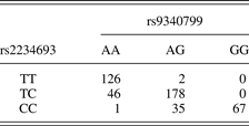
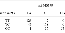
Four hundred sixty-one participants (98%) were genotyped successfully for rs9340799 and 457 (97%) for rs2234693. Only women for whom genotypes of both SNPs were available were included in statistical analyses, leaving a final study sample of 455 participants (Table 1). Frequencies of the rs9340799 A and G alleles were 62% and 38%, respectively, and of the rs2234693 T and C alleles, 53% and 47%, respectively. In the absence of selection pressures, nonrandom mating, migration, or other disturbing influences, the distribution of genotypes at a given locus should remain constant over generations, as defined by the Hardy–Weinberg equilibrium (for a two-allele locus, the expected distribution of genotypes is p 2 + 2pq + q 2, where p and q denote the frequencies of the two alleles). The distribution of ESR1 genotypes in our sample did not depart from equilibrium (rs9340799: AA = 173, AG = 215; GG = 67; rs2234693: TT = 128, TC = 204, CC = 103; χ 21s < 0.10, ns). The two SNPs were also in strong linkage disequilibrium (D′ = 0.98, r 2 = .67), a measure of nonrandom association among alleles of more than one polymorphism, which is consistent with the proximity of these loci.
Distribution of estrogen receptor-α genotypes
Because genetic data derived from individuals of common race/ethnicity may still exhibit stratification, we tested for possible genetic substructure in this sample. Fifteen additional, genome-spanning SNPs (rs1022106, rs1335995, rs1439564, rs1502812, rs1860300, rs548146, rs705388, rs715994, rs720517, rs722743, rs730899, rs734204, rs9059966, rs1328994, and rs1485405) were genotyped for analysis using the program STRUCTURE (Falush, Stephens, & Pritchard, Reference Falush, Stephens and Prichard2003; Pritchard, Stephens, & Donnelly, Reference Pritchard, Stephens and Donnelly2000). A model with admixture, uncorrelated allele frequencies, individual alpha parameters, and independent F statistic (fixation index) for all subpopulations was run separately assuming one, two, or three subpopulations. For each model, we used a burn-in of 40,000 simulations, followed by 80,000 repetitions, and compared the likelihoods of models fitting the data. Evidence of stratification could be inferred if the likelihood of data fitting a model with greater than or equal to two subpopulations was greater than that of a model with 1 population. However, no evidence of genetic substructure was detected (log probabilities for k = 1, 2, and 3 subpopulations were −6675.7, −6740.1, and −6741.9, respectively), and therefore no further adjustments were made for stratification.
Menarcheal age
The first menstrual period is a highly salient event in girls' lives and a well-recollected developmental milestone. Studies of the reliability of retrospectively reported menarcheal age yield remarkably similar correlations with contemporaneously recorded age at menarche, whether assessed over recall intervals of short and long duration (4–5 years: rs = .81–.83; 17 years: r = .75; 19 years: r = .78; 29 years: r = .79) (Bergsten-Brucefors, Reference Bergsten-Brucefors1976; Damon et al., Reference Damon, Damon, Reed and Valadian1969; Koprowski, Coates, & Bernstein, Reference Koprowski, Coates and Bernstein2001; Livson & McNeill, 1969; Must et al., Reference Must, Phillips, Naumova, Blum, Harris and Dawson-Hughes2002). In the AHAB project, women were asked to report their age at first menstruation as a component of a medical history interview administered by project nurses. Reported age at menarche averaged 12.6 ± 1.4 years, with a mean recall interval of 32.4 ± 6.7 years. The average menarcheal age recorded here was similar to the 12.8 years reported in the National Health Examination Survey (NHES) for White girls studied in 1963–1970, whose years of birth would have overlapped but slightly preceded the average of the present sample (Anderson, Dallal, & Must, Reference Anderson, Dallal and Must2003). In addition, the cumulative proportion of AHAB participants attaining menarche by ages 12 (47%), 13 (78%), 14 (92%), and 15 (96%) years closely match corresponding NHES values at the same ages (43%, 73%, 91%, and 98%, respectively).
Quality of the family environment
Degree of conflict and interpersonal warmth in participants' early family environments were indexed by the conflict and cohesion subscales of the widely used Family Environment Scale (FES; Moos & Moos, Reference Moos and Moos1981), as modified by Plomin, McClearn, Pedersen, Nesselroade, and Bergeman (Reference Plomin, McClearn, Pedersen, Nesselroade and Bergeman1988) to assess self-reported childhood rearing environments among adults. Scale modifications included conversion of the FES response format from true–false to 5-point Likert scales (anchored by end ratings of strongly disagree and strongly agree) and shortening each scale from an original nine items to the five items having the strongest factor loadings for the scale. Conflict Scale items reference competition, criticism, expressed anger, and fighting within the family, whereas Cohesion Scale items reference agreeable relationships among family members, mutual assistance, attention and support, and shared family spirit. The five items of each scale were averaged for each subject and the resulting score distributions standardized. In the current sample, internal reliabilities (α) of the conflict and cohesion scales were 0.86 and 0.89, respectively, and the two scales correlated inversely (r = −.66). For primary analyses, an overall measure of Quality of Family Environment (QFE) was calculated by reverse scoring the Conflict Scale (so that higher scores indicated low family conflict), then averaging the two scales and standardizing the sample distribution of QFE scores. The conflict and cohesion scales were also examined separately.
Measures of both experienced and perceived environmental exposures often reflect nontrivial genetic influences (Kendler & Baker, Reference Kendler and Baker2007) and, with respect to the FES scales, minor heritable variation has been found on retrospective reports of both family conflict and cohesion (Plomin et al., Reference Plomin, McClearn, Pedersen, Nesselroade and Bergeman1988). The latter findings are consistent with other biometric studies using the same or analogous scales (Deater-Deckard, Fulker, & Plomin, Reference Deater-Deckard, Fulker and Plomin1999; Jacobson & Rowe, Reference Jacobson and Rowe1999; Jang, Vernon, Liveley, Stein, & Wolf, Reference Jang, Vernon, Liveley, Stein and Wolf2001; Krueger, Markon, & Bouchard, Reference Krueger, Markon and Bouchard2003; Plomin, McClearn, Pedersen, Nellelroade, & Bergman, Reference Plomin, McClearn, Pedersen, Nesselroade and Bergeman1989). Recently, Krueger et al. (Reference Krueger, Markon and Bouchard2003) reported on the heritability of a composite measure of “perceived cohesion versus conflict in the family environment,” as derived from multiple environmental scales (on which FES cohesion scores loaded positively and FES conflict scores loaded negatively) in the Minnesota Study of Twins Reared Apart. The 16% of phenotypic variance attributable to heritable variation in that study could be fully explained by genetic covariance with two higher order dimensions of personality, negative emotionality and constraint, as measured by the Multidimensional Personality Questionnaire (MPQ; Tellegen, Reference Tellegen1982). Scores on an abbreviated version of the MPQ, the 155-item MPQ—Brief Form (MPQ-BF; Patrick, Curtin, & Tellegen, Reference Patrick, Curtin and Tellegen2002), were also available in the AHAB registry. To evaluate whether any association of menarcheal age with FES-assessed family environment might be accounted for by correlated variation in these personality dimensions, MPQ-BF traits of constraint and negative and positive emotionality were included in the present study. MPQ-BF scales have good internal consistency, and the three broad traits examined here correlate strongly (rs = .94–.98) with their corresponding MPQ factors.
Results
Sample characteristics
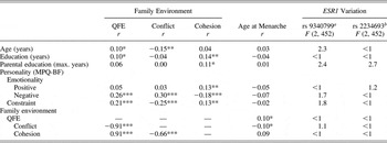
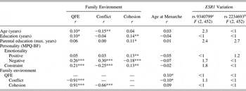
Participants had a mean age of 45.1 ± 6.6 years and were well educated on the whole, averaging 15.9 ± 2.9 years of schooling. By level of educational attainment, 38% lacked a college diploma, 36% held a Bachelors degree, and 26% had completed graduate or professional training. Consistent with secular trends, women reported their parents having somewhat fewer years of schooling than themselves (highest parental education: M = 13.9 ± 3.2 years). At the time of study participation, 73% of women were married and an equivalent proportion (76%) was employed full or part time. As shown in Table 2, participant age and personal and parental education were variously related to self-reported early family environment. Our overall measure of family environment (QFE) correlated negatively with age and participant education, whereas Conflict scores separately correlated negatively with age, and family Cohesion scores covaried positively with both personal and parental education. However, these sample characteristics were independent of menarcheal age and, when tested for association with ESR1 polymophisms by one-way analysis of variance (Table 2), were unrelated to ESR1 variation.
Correlations among study variables and main effects of ESR1 variation on participant age, personal and highest parental education, personality traits, and early family environment
Note: ESR1, estrogen receptor-α; QFE, quality of family environment; MPQ-BF, Multidimensional Personality Questionnaire, Brief Form.
ars9340799 genotypes: AA, AG, GG.
brs2234693 genotypes: CC, CT, TT.
*p < .05. **p < .01. ***p < .001.
QFE, ESR1 variation, and menarcheal age
Table 2 shows a modest, but significant, inverse association between QFE and menarcheal age (r = .10). Consistent with prior literature, women who reported growing up in families characterized by less conflict and greater cohesion experienced menarche at a later age than those raised in families with more conflict and less cohesion. Age at menarche also correlated negatively with Conflict scores alone (r = −.10) and positively with Cohesion scores (r = .09, p = .05).
Table 2 also summarizes the simple effects of ESR1 polymorphisms on QFE and family conflict and cohesion scales separately, none of which proved significant. As a result, we next tested whether polymorphic variation in ESR1 predicted age at menarche or moderated the association of menarcheal age with QFE. We entered participants' QFE scores in the first step of hierarchical linear regression, followed by ESR1 genotype (Step 2), and finally, the multiplicative interaction of QFE and genotype (Step 3). Effects of genotype were tested on an additive model of genetic influence and analyses run separately for each ESR1 polymorphism (coded −1, 0, 1 for genotypes AA, AG, and GG of rs9340799 and TT, TC, and CC of rs2236693, respectively). Subsequent analyses produced virtually identical results when including demographic factors (age, personal and highest parental education) as covariates, so that for simplicity these variables are not considered in analyses summarized below.
ESR1 polymorphisms
rs9340799
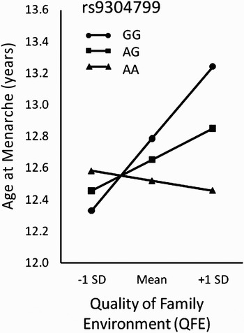
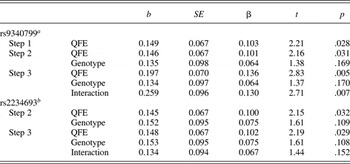
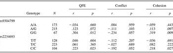
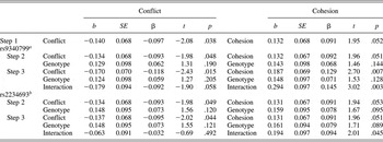
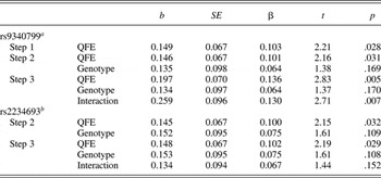
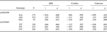
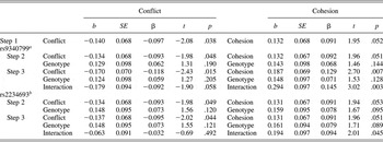
Regression outcomes are presented in Table 3. Consistent with the bivariate correlations reported above, age at menarche was predicted modestly by QFE (b = 0.149, SE = 0.067, t = 2.21, p = .028), but was unrelated to genotype of rs9340799. However, the association of menarcheal age with QFE varied significantly by genotype, as indicated in the interaction term of the final regression model (b = 0.259, SE = 0.096, t = 2.71, p = .007). As illustrated in Figure 1, QFE scores, plotted to one standard deviation above and below the sample mean, covaried with menarcheal age more strongly in women homozygous for the rs9340799 G allele than among those of other genotypes. Bivariate correlations within the three genotypes of this polymorphism (Table 4) show approximately 9% of the variance in menarcheal age explained by QFE scores among GG homozygous women (r 67 = .304, p = .012). The corresponding correlation among women carrying one G allele (AG heterozygotes) only approached significance (r 215 = .123), and no association at all obtained among participants homozygous for the alternate A allele (r 173 = −.034). Correlations calculated for the separate conflict and cohesion scales (also listed in Table 4) showed menarcheal age to covary positively with family cohesion (r 67 = .319, p = .009) and negatively, albeit marginally, with family conflict (r 67 = −.234, p = .057) among GG homozygotes. In contrast, age at menarche was unrelated to either dimension of family functioning in participants of heterozygous genotype and those homozygous for the A allele. These genotype-dependent associations were corroborated in parallel regression analyses (Table 5), where genotype of rs9340799 predicted menarcheal age significantly in interaction with reported family cohesion (b = 0.294, SE = 0.097, t = 3.02, p = .003), but did so only weakly in interaction with family conflict (b = −0.179, SE = 0.094, t = −1.90, p = .058). When the two family predictors and their corresponding interaction terms were entered together in a further regression analysis, moreover, the interaction of rs9340799 genotype and cohesion remained significant (b = 0.303, SE = 0.124, t = 2.36, p = .019), suggesting that this relationship is independent of correlated variation in family conflict.Footnote 1
Menarcheal ages estimated from the interaction of the rs9304799 genotype and Quality of Family Environment (QFE).

Summary of regression analyses testing the effects of QFE, ESR1 genotypes, and their interaction on age at menarche (N = 455)
Note: QFE, quality of family environment; ESR1, estrogen receptor-α.
ars9340799 genotypes: AA, AG, GG.
brs2234693 genotypes: TT, TC, CC.
Correlation of menarcheal age with QFE and with family conflict and cohesion scores separately within genotypes of ESR1 polymorphisms
Note: QFE, quality of family environment; ESR1, estrogen receptor-α.
Summary of regression analyses testing the effects of family conflict and cohesion, ESR1 variation, and their interaction on age at menarche (N = 455)
Note: ESR1, estrogen receptor-α.
ars9340799 genotypes: AA, AG, GG.
brs2234693 genotypes: TT, TC, CC.
rs2234693
It is interesting that our second ESR1 polymorphism showed a similar pattern of within-genotype correlation between QFE and age at menarche (genotype: CC, r = −.218, p = .026; CT, r = −.053, ns; TT, r = −.044, ns), even though the corresponding interaction term in regression was not significant for this polymorphism (b = 0.134, SE = 0.094, t = 1.44, p = .152) (Table 3). Like rs9304799, menarcheal age correlated positively with cohesion scores alone (r = .218, p = .027) and inversely (and again weakly) with reported family conflict (r = −.192, p = .051) among women homozygous for the less common rs2234693 C allele. Among participants of TT or TC genotype, menarcheal age was unrelated to either cohesion or conflict scores. In parallel regression models (Table 5), rs2234693 genotype predicted age at menarche in interaction with family cohesion (b = 0.194, SE = 0.097, t = 2.01, p = .045), but not family conflict (b = −0.063, SE = 0.091, t = −0.69, p = .492). Lke rs9340799, further analysis showed genotype of rs2234693 to predict menarcheal age in interaction with family cohesion even in a model including both family predictors and their respective interaction terms (Genotype × Cohesion: b = 0.276, SE = 0.130, t = 2.13, p = .034).
Role of personality
Earlier we cited evidence that heritable variation in a composite measure of family “cohesion versus conflict” (which incorporated the FES scales used here) could be accounted for by correlated variation in two MPQ-assessed dimensions of personality, negative emotionality and constraint (Krueger et al., Reference Krueger, Markon and Bouchard2003). As shown in Table 1, negative emotionality correlated positively and constraint negatively with both QFE and Conflict Scale scores in the current study, whereas scores on the Cohesion Scale covaried inversely with negative emotionality and positively with both constraint and positive emotionality. Despite these associations, none of the personality dimensions correlated significantly with age at menarche. In addition, substituting trait measures for the family environment scales in regression analyses predicting menarcheal age revealed no significant interactions between genotypes of the two ESR1 polymorphisms and the three personality dimensions. Finally, entering the trait measures as covariates on reanalysis of family QFE, conflict and cohesion scores did not alter the previously reported interactions of these variables with ESR1 variation. Thus, QFE continued to predict age at menarche in interaction with rs9340799 genotype (b = 0.259, SE = 0.096, t = 2.69, p = .007), as did cohesion scores alone (b = 0.291, SE = 0.097, t = 2.99, p = .003), and family cohesion continued to predict menarcheal age in interaction with genotypes of rs2234693 (b = 0.189, SE = 0.097, t = 1.96, p = .051).
Discussion
In this study of 455 midlife women reared to adolescence in stable, two-parent families, participants who described their families as cohesive and lacking in conflict tended to reach menarche at a later age than women who reported being reared in families prone to discord and lacking cohesion. This association is consistent with previous studies, both retrospective and longitudinal, in which analogous dimensions of family relationship similarly predicted daughters' pubertal timing (Ellis et al., Reference Ellis, McFadyen-Ketchum, Dodge, Pettit and Bates1999; Ellis & Garber, Reference Ellis and Garber2000; Graber et al., Reference Graber, Brooks-Gunn and Warren1995; Kim et al., Reference Kim, Smith and Palermiti1997; Kim & Smith, Reference Kim and Smith1998; Moffitt et al., Reference Moffitt, Caspi, Belsky and Silva1992; Romans et al., Reference Romans, Martin, Gendall and Herbison2003; Steinberg, Reference Steinberg1988; Wierson et al., Reference Wierson, Long and Forehand1993). In addition, the association of early family environment with age at menarche in our study was moderated by polymorphic variation in ESR1, a gene of hypothesized association with reproductive maturation. Thus, quality of the family environment (assessed here by composite of the conflict and cohesion scales of the FES) covaried positively with menarcheal age among women homozygous for the minor alleles of rs9304799 (GG genotype) and rs2234693 (CC), but not among women of other ESR1 genotypes. Moreover, this genotype-dependent association between family environment and age at menarche was principally attributable to variation in family cohesion. The main effect of family relationships on menarcheal age here is consistent with prior literature; its modulation by ESR1 variation is also consistent with predictions from differential susceptibility and accords with other research showing heritable “person variables,” such as infant negative emotionality and stress reactivity, to moderate environmental influences on pubertal timing (Belsky et al., Reference Belsky, Steinberg, Houts, Friedman, DeHart and Cauffman2007; Ellis, Shirtcliff, et al., Reference Ellis, Shirtcliff, Boyce, Deardorff and Essex2011 [this issue]).
In girls, the onset of menses defines a sentinel event in the sequence of physiological and anatomic changes initiating puberty. Because these changes are occasioned by heightened exposure of reproductive and other tissues to the ovarian hormone, estrogen, it is plausible that variation in genes encoding estrogen receptors affect the pacing of pubertal development. When activated by estrogen, estrogen receptors (of which there are two, ER-α and ER-β) migrate to the cell nucleus, where they exert transcriptional control of estrogen-dependent genes by binding to estrogen response elements within gene regulatory sequences. In this way, estrogen (like other steroid hormones) can promote or repress the expression of genes specifying an array of cellular proteins. Genetic variation that alters estrogen receptor activity could therefore modulate a range of estrogen-sensitive phenotypes, including pubertal maturation. At present, though, it is unclear how the ESR1 polymorphisms genotyped here might influence girls' reproductive development. The absence of allele-dependent effects on menarcheal age among all study participants (i.e., the absence of a genetic “main effect”), albeit in agreement with all but one previous study, actually suggests a more complex etiology (Boot et al., Reference Boot, van Der Sluis, de Muinck Keizer-Schrama, van Meurs and Krenning2004; Gorai et al., Reference Gorai, Tanake, Inada, Morinaga, Uchiyama and Kikuchi2003; Hong et al., 2005; Long et al., Reference Long, Xu, Zhao, Liu, Shen and Liu2005; Mendoza et al., Reference Mendoza, Moron, Quereda, Vazquez, Rivero and Martinez-Astorquiza2008; Mitchell et al., Reference Mitchell, Farin, Stapleton, Tsai, Tao and Smith-DiJulio2008; Stavrou et al., Reference Stavrou, Zois, Ioannidis and Tsatsoulis2002; Weel et al., Reference Weel, Uitterlinden, Westendorp, Burger, Schuit and Hofman1999). Given the present findings, elucidating underlying biological mechanisms would also require understanding how ESR1 variation might variably influence pubertal development as a function of the quality of family relationships.
One possible mechanism could involve effects on pubertal development related to variation in body weight, which may be associated with differing family environments. For instance, certain ESR1 genotypes might promote a proportionately greater weight gain among girls who respond to family adversity by overeating, thus advancing menarcheal age in these genetically susceptible girls via the well-established relationship between adiposity and age at menarche. Unfortunately, we do not have data on participants' childhood weight to address this possibility, but note that controlling for weight in several prior studies did not attenuate associations of family variables with pubertal timing (Campbell & Udry, Reference Campbell and Udry1995; Ellis & Essex, Reference Ellis and Essex2007; Graber et al., Reference Graber, Brooks-Gunn and Warren1995; Moffitt et al., Reference Moffitt, Caspi, Belsky and Silva1992).
Little is known about the functionality of ESR1 polymorphisms. Because rs9340799 and rs2234693 are both located in a noncoding region of the gene (intron 1), they do not produce estrogen receptor proteins of differing structure (unless, hypothetically, through alternative splicing of the primary mRNA transcript), but might be in linkage disequilibrium with functional variation elsewhere in ESR1. Introns are also known occasionally to affect rates of gene transcription, and it is noteworthy that the C allele of rs2234693 forms part of a binding site for a transcription factor, B-myb, that has been shown to enhance transcriptional efficiency in an in vitro assay system (Herrington et al., Reference Herrington, Howard, Brosnihan, McDonnell, Li and Hawkins2002). In addition, B-myb is itself activated by estrogen, which might further augment ESR1 expression to influence cellular activity in pathways underlying hormone-sensitive interactions (Santen, Reference Santen1998). It is conceivable, for instance, that variation in ER-α signaling in certain hypothalamic nuclei (arcuate, anteroventral periventricular nucleus) modulates reawakening of the gonadotropin-releasing hormone (GnRH) pulse generator in puberty. Such modulation might act on the pubertal decline in estrogen-dependent inhibition of luteinizing hormone (and, hence, GnRH) or contribute more directly to activation of GnRH neurons. Both processes are regulated critically by the protein, kisspeptin, and the expression of kisspeptin is induced by gonadal steroids, as mediated by ER-α (Smith, Clifton, & Steiner, Reference Smith, Clifton and Steiner2006). Until it is demonstrated that the molecular variation of rs2234693 actually alters receptor availability in tissues expressing ER-α, however, the in vivo functionality of this polymorphism remains uncertain.
Whatever the biological mechanisms, is the magnitude of association between early family environment and menarcheal age observed here developmentally meaningful? QFE scores correlated positively, but only modestly, with pubertal timing across all women (r = .10), and the spread of QFE defined by a departure of one standard deviation above and below the sample mean was associated with a difference of only about 3.5 months in participants' age at menarche. Yet the difference in estimated menarcheal age over the same range of QFE variation among women of an “environmentally sensitive” ESR1 genotype, such as those homozygous for the G allele of rs9340799 (as depicted in Figure 1), was 1 year. It is interesting that adult ovarian function is also established more slowly among those who reach menarche latest. Longitudinal observations show girls reaching menarche at ages <12, 12–12.9, and ≥13 years to experience 50% ovulatory menstrual cycles at 1, 3, and 4.5 years, respectively, following menarche (Apter and Vikho, Reference Apter and Vihko1983). Thus, developmental variation indexed to small differences in menarcheal age may be magnified on further progression to reproductive maturity, as in the transition from primarily anovulatory to reliably ovulatory cycling. Distal sequelae of early and late menarche may be linked also to the range of variation in menarcheal ages predicted here by the interaction of ESR1 variation and early family environment. A 1-year delay in menarche reduces breast cancer risk by 10% to 20% (Bernstein, Reference Bernstein2002), and in a recent prospective study of women with mean age at baseline similar to the present sample, a 1-year delay in menarche lowered total, coronary, and stroke mortality risk, respectively, by 4.5%, 6.0%, and 8.6% over an average follow-up of 11.1 years (Jacobsen et al., Reference Jacobsen, Oda, Knutsen and Fraser2009; see also Lakshman et al., Reference Lakshman, Forouhi, Sharp, Luben, Bingham and Khaw2009).
A previous report that paternal promiscuity, parental divorce, father absence, and menarcheal age were predicted by length variation in a trinucleotide (GGC) repeat polymorphism of the androgen receptor gene suggested that family instability and pubertal timing might be genetically correlated (Comings, Muhleman, Johnson, & MacMurray, Reference Comings, Muhleman, Johnson and MacMurray2002; but see also Jorm et al., Reference Jorm, Christensen, Jacomb and Easteal2004) Here, in contrast, genetic variation in ESR1 was unrelated to family relationship variables, but instead moderated associations between early family environment and age at menarche. This observation is consistent with Belsky's hypothesis that girls differ genetically in their susceptibility to rearing effects on pubertal maturation (Belsky, Reference Belsky, Rodgers, Rowe and Miller2000, Reference Belsky, Ellis and Bjorklund2005; Belsky et al., Reference Belsky, Steinberg, Houts, Friedman, DeHart and Cauffman2007), as well as models of context-dependent development (differential susceptibility) that are less explicit regarding genetic determinants of individuals' relative sensitivity to the environment (Boyce & Ellis, Reference Boyce and Ellis2005; Ellis, Shirtcliff, et al., Reference Ellis, Shirtcliff, Boyce, Deardorff and Essex2011 [this issue]). It is interesting that all strands of theorizing on the origins of differential susceptibility have invoked evolutionary considerations, speculating on the circumstances in which variation in the capacity to adjust development in response to environmental conditions (i.e., developmental plasticity) would tend to be favored or rejected by natural selection (Belsky et al., Reference Belsky, Steinberg and Draper1991; 2005; Boyce & Ellis, Reference Boyce and Ellis2005; Ellis, Boyce, Belsky, Bakermans-Kranenburg, & van IJzendoorn, Reference Ellis, Boyce, Belsky, Bakermans-Kranenburg and van IJzendoorn2011 [this issue]; Ellis, Jackson, & Boyce, Reference Ellis, Jackson and Boyce2006). When specific sources of genetic variation (gene polymorphisms) are found to underlie this variation (as the present findings might imply), an evolutionary account of differential susceptibility requires explaining how the implicated polymorphisms are perpetuated in a reproducing population. This is not a trivial consideration, as selection ordinarily acts to reduce genetic variation, not promote it, and if the latter, will maintain alternate alleles in their respective frequencies at a given locus over generations only if their bearers experience, on balance, equal reproductive fitness (the currency of selection).
Differential susceptibility, as defined by Belsky and Pluess (Reference Belsky and Pluess2009) and Ellis, Boyce, et al. (Reference Ellis, Shirtcliff, Boyce, Deardorff and Essex2011 [this issue]), is present when regression lines reflecting associations between a phenotype and environmental factor crossover when plotted for different variants of a susceptibility marker (here, genotypes of a polymorphism), such that the line of greater slope (plasticity) predicts both higher and lower phenotypic values at opposite poles of an environmental gradient than an alternate variant (genotype). This is illustrated in Figure 1, where QFE scores are not only associated more strongly with menarcheal age among women homozygous for the rs9304799 G allele but also tend to predict both later and earlier age at menarche at higher and lower QFE, respectively, compared, for instance, to AA homozygotes. Life history variables, like pubertal maturation, that are related to reproductive timing can respond to natural selection if they affect lifetime reproductive outcomes, such as the number and quality of offspring (Chisholm & Burbank, Reference Chisholm and Burbank2001). Consider that early menarche presages a number of adverse sequelae, including problems of mood and conduct in adolescence, early metabolic changes that heighten risk for cardiovascular disease, and in adulthood, breast cancer, diseases of heart and vasculature, and premature mortality (e.g., Bernstein, Reference Bernstein2002; Cooper et al., 2999; Ge et al., Reference Ge, Conger and Elder1996; Golub et al., Reference Golub, Collman, Foster, Kimmel, Rajpert-De Meyts and Reiter2008; Hayward et al., Reference Hayward, Killen, Wilson, Hammer, Litt and Kraemer1997; Jacobsen et al., Reference Jacobsen, Oda, Knutsen and Fraser2009; Kaltiala-Heino, Kosmen, & Rempela, Reference Kaltiala-Heino, Kosunen and Rimpela2003; Lakshman et al., Reference Lakshman, Forouhi, Sharp, Luben, Bingham and Khaw2009; Marshall et al., 2998; Remsberg et al., Reference Remsberg, Demerath, Schubert, Chumlea, Sun and Siervogel2005; Stice et al., Reference Stice, Presnell and Bearman2001). If, in addition, child-bearing risks and the other health-related consequences of early menarche are mirrored in a lower reproductive fitness (and oppositely for delayed menarche), so that age at menarche predicts lifetime reproductive success, in the context of our findings selection would theoretically favor the G allele of rs9304799 among women raised in a positive family environment but would favor the A allele among those reared under less propitious family circumstances. That is, if a genotype conferring greater plasticity results in reproductive advantage at one end of the environmental gradient to which it is sensitive and relative disadvantage at the other, net fitness over all environments (though more variable) might approximate that of an alternative genotype associated with lesser sensitivity to environmental variation. Because neither genotype is optimal in all environments, both will tend to be preserved (Stearns, Reference Stearns1992).
In contrast to the argument from competing advantage, proponents of differential susceptibility models have not viewed early and later pubertal development as a predictor of lifetime reproductive success so much as potentiating different reproductive strategies that adapt individuals to the varied circumstances of their development. Thus, girls whose early life experiences encourage little expectation of a materially secure, stable, and emotionally supportive marital and family life in adulthood may trade off continued growth and development for accelerated sexual maturation, early childbearing, and a limited reliance on paternal investment in child care and parenting (Belsky et al., Reference Belsky, Steinberg and Draper1991; Chisholm, Reference Chisholm1999; Ellis, Reference Ellis2004). Presumably, the higher risk of early sexual development and childbearing are offset by a potentially longer reproductive life span, or perhaps this “early and fast” reproductive strategy simply offers the best prospect against omens of an uncertain future. Conversely, girls experiencing stable, warm, and cohesive family relationships in early childhood may anticipate the same for themselves as adults and pursue a reproductive strategy entailing slower pubertal development and deferred childbearing, premised on expectations of a secure marital bond and high paternal investment in child rearing (Belsky et al., Reference Belsky, Steinberg and Draper1991; Ellis, Reference Ellis2004, Reference Ellis, Ellis and Bjorklund2005). However, this reasoning alone does not explain how genetic variation underlying interindividual variability in responsiveness to environmental differences would be maintained in a population. If the function of developmental plasticity is to maximize the adaptive responses (reproductive strategies) of individuals born into family environments of differing qualities, good or bad, selection might be expected to favor genetic variation enabling such plasticity and, over time, supplant genotypes promoting a less flexible developmental phenotype.
However, if a capacity to adapt to differing environments carries potential costs, as well as benefits, or does so in certain circumstances, it is conceivable that phenotypes of both greater and lesser sensitivity to the environment might prove equally fit overall and, therefore, persist in ratios of prevalence defined by whatever circumstances condition cost and benefit. That sensitivity to environmental influences could entail costs is recognized explicitly in differential susceptibility models (see Ellis, Boyce, et al., Reference Ellis, Shirtcliff, Boyce, Deardorff and Essex2011 [this issue]). For instance, Belsky (Reference Belsky1997, Reference Belsky, Ellis and Bjorklund2005) argues that responsiveness to environmental differences should be favored where conditions of early life provide reliable cues to circumstances that are likely to prevail at maturity but, absent such predictability, parents' reproductive fitness would be better served by conceiving offspring of varying, less flexible phenotypes matched to particular environmental conditions (“diversified bet-hedging”). By this reasoning, individual differences in sensitivity to environmental cues (and, by implication, their genetic etiologies) will be sustained in a population hosting environments of both predictability and unpredictability. In a second model, Boyce and Ellis (Reference Boyce and Ellis2005) propose that individuals vary in their “biological sensitivity to context” and have indexed this variability in children via autonomic and adrenocortical reactivity to standardized laboratory stressors (Boyce et al., 1995; Ellis, Shirtcliff, et al., Reference Ellis, Shirtcliff, Boyce, Deardorff and Essex2011 [this issue]). The tempo of pubertal development among high stress-reactive children was delayed and accelerated in families of high- and low-quality parent–child relationships, respectively, relative to children exhibiting less stress reactivity (less biological sensitivity to context). Heightened reactivity may be seen as promoting developmental outcomes best adapted to rearing conditions of either advantage or adversity, but the same reactivity to stress that supports conditional adaptation is also known to promote stress-related psychiatric and physical disorders, including pathologies associated with premature mortality (e.g., atherosclerotic cardiovascular disease; Boyce & Ellis, Reference Boyce and Ellis2005; Manuck, Reference Manuck1994; Manuck, Marsland, Kaplan, & Williams, Reference Manuck, Marsland, Kaplan and Williams1995). Thus, Ellis, Boyce, et al. (Reference Ellis, Shirtcliff, Boyce, Deardorff and Essex2011 [this issue]) suggest that, although fitness benefits may accrue from context-dependent biological sensitivity in the most positive and negative rearing environments, the majority of children, who are raised in normative family circumstances, may instead fare better if they are less sensitive to context.
These several propositions frame differential susceptibility in an evolutionary context and, in explaining how developmental phenotypes of more and less plasticity may coexist through a balance of fitness benefits and costs, they also help explain how specific genetic variation associated with these differences might be retained in a reproducing population. Accounting for the persistence of a given gene polymorphism (such as ESR1 variation) via fitness effects of reproductive strategies that are entrained by differential susceptibility to varying childhood environments is still, though, only a theoretical possibility. Such speculation may be critiqued, if applied in the present context, for privileging a particular phenotype (pubertal timing) out of the myriad physiological events affected by estrogen signaling, so that any single instance of Gene × Environment interaction involving the estrogen receptor (or other molecule of similarly extensive biological action) is most likely a byproduct of other regulatory processes (Manuck, Reference Manuck2010; Stearns, Reference Stearns1992). In contrast, this objection is mitigated if timing of reproductive maturation predicts womens' later reproductive histories, as proposed in the foregoing evolutionary models.
To our knowledge, it is not known whether women's lifetime reproductive success is either correlated with age at menarche or, as a result of environmentally contingent (facultative) differences in reproductive strategy, equivalent in women who reached sexual maturity earlier versus later. The question, at least as an evolutionary proposition, may also prove difficult to adjudicate in modern societies, where contraception now frequently renders childbearing an active decision rather than a byproduct of sexual activity. In addition, where menarche has been studied in relation to reproductive outcomes in nonindustrialized societies, results may be confounded by dissimilar social and family structures and a proportionately greater impact of other environmental influences on pubertal development, such as variation in nutritional status, health, and body weight. Among the rural Kipsigis of Kenya, for instance, early maturing girls bear more living offspring over their reproductive lives and command a higher bride price than girls who reach menarche later (Borgerhoff Mulder, Reference Borgerhoff Mulder, Betzig, Borgerhoff Mulder and Turke1988, Reference Borgerhoff Mulder1989). Similarly, in the Pumé foragers of Venezuela, earlier age at first reproduction predicts higher completed fertility unless the mothers are exceptionally young at the birth of their first child (<14 years, when risks of infant mortality outweigh the benefits of early childbearing; Kramer, Reference Kramer2008). Psychosocial influences on menarcheal age have not been modeled in these foraging populations, however, and owing to differences in family constellation, rearing practices, and cultural norms, might differ appreciably from those observed in study samples drawn from industrialized, Western societies.
In regard to the present findings, we may also ask to what extent our observations really reflect an interaction of genetic and environmental variation. Thus, it is noteworthy that personality traits previously shown to explain heritable variation in reported family conflict and cohesion, although correlated in expected directions with FES scales here, did not account for the interactive effects of ESR1 variation and family environment on age at menarche. This would seem to credit speculation that our findings reflect Gene × Environment interaction, even though our measures of early family environment are self-reported and retrospective. However, should any other heritable, but unmeasured, determinants of reported family relationships exist in this sample or when using these instruments, our measurement of the family “environment” would merit at least a partly genetic interpretation. Where genetic and nongenetic influences on ostensibly environmental measurements cannot be partitioned by study design (as may be achieved in certain twin and adoption studies), then, inferring Gene × Environment interaction remains ambiguous. As a result, genotype-dependent associations of early family environment with menarcheal age in the present study could conceivably entail interactive effects involving other genes (epistasis), strictly environmental factors (Gene × Environment interaction), or both.
Limitations and Conclusion
Finally, we point out several methodological and interpretive limitations of this study. Although a convenient starting point, retrospective report cannot substitute for prospective investigation and reliance on adults' self-reported early environments is less informative than observational and contemporaneous family measurements. In addition, findings reported elsewhere suggest that father–daughter relationships predict pubertal timing uniquely or more strongly than mother–daughter relationships, positive family relationships are more predictive than negative relationships, and the frequency of father–daughter interactions (positive and negative) predicts later pubertal development (Ellis et al., Reference Ellis, McFadyen-Ketchum, Dodge, Pettit and Bates1999; Ellis & Garber, Reference Ellis and Garber2000; Moffitt et al., Reference Moffitt, Caspi, Belsky and Silva1992). Unfortunately, the FES conflict and cohesion scales we administered do not index parent-specific behaviors or frequency of interactions and, although there was a slight indication that menarcheal age might covary more reliably with family cohesion than with conflict within “environmentally sensitive” ESR1 genotypes, common method variance may have rendered these scales too highly correlated (inversely) to detect differential effects. Our findings are also restricted to observations on non-Hispanic Caucasian women, so that their generalizability to other populations is unknown, and to limited variation within a single gene. Menarche is a reliably measured event, but progression through puberty encompasses other development milestones, including notably the appearance of secondary sexual characteristics. In sum, subsequent research would benefit from studies of longitudinal design that entail observational measurement of the family environment, broader participant sampling, greater assessment of relevant genetic variation, and inclusion of multiple indicators of pubertal timing. At present, our findings afford initial evidence that familial influences on the pace of pubertal maturation vary in magnitude among individuals and do so in relation to genetic differences plausibly implicated in pubertal development.