Implications
This study demonstrates an increased oxidative damage (measured by a significant increase of the markers inducible nitric oxide synthase (iNOS) and nitrotyrosine (NITT)) in the liver of intensively reared beef cattle receiving moderate copper (Cu) supplementation. This oxidative damage could be considered as the initial pathological change in the animal before further damage will occur, and thus, the markers iNOS and NITT could be used as early indicators of subclinical Cu toxicity in cattle under these nutritional conditions.
Introduction
Copper (Cu) is an essential metal associated with several enzymes (either as a co-factor or as an allosteric component) that acts as an electron transfer intermediate in redox reactions (Uauy et al., Reference Uauy, Olivares and Gonzalez1998) therefore being vital for the normal function of the organisms. Cu in the liver is accumulated bound to metallothioneins (MT), proteins involved in the detoxification of essential and non-essential metals, storage of essential metals and sequestration of reactive oxygen species (ROS) and reactive nitrogen species (RNS; López-Alonso et al., Reference López-Alonso, Prieto, Miranda, Castillo, Hernández and Benedito2005; Yoshida et al., Reference Yoshida, Saegusa, Fukuda, Akama and Owada2005; Formigari et al., Reference Formigari, Irato and Santon2007). Cu also forms part of the antioxidant defense system of the cells due to its essential role in the Cu–Zn superoxide dismutase (SOD) and caeruloplasmin (Pan and Loo, Reference Pan and Loo2000). Thus, Cu deficiency in cattle is associated with increased oxidative damage (Ward and Spears, Reference Ward and Spears1997; Gengelbach and Spears, Reference Gengelbach and Spears1998; Cerone et al., Reference Cerone, Sansinanea, Streitenberg, García and Auza2000).
However, the same properties that make Cu essential, when in excess also generate free radicals that can be seriously deleterious to cells (Rana, Reference Rana2008). Hepatic toxicity, caused by Cu overload, is hypothesized to result from redox properties of Cu and is connected with the formation of ROS (Britton, Reference Britton1996; Luza and Speisky, Reference Luza and Speisky1996). ROS are highly reactive and are capable of damaging all biological macromolecules like proteins, lipids and DNA (Grune et al., Reference Grune, Reinheckel, Joshi and Davies1995) that can lead to apoptosis (Wätjen and Beyersmann, Reference Wätjen and Beyersmann2004; Banasik et al., Reference Banasik, Lankoff, Piskulak, Adamowska, Lisowska and Wojcik2005; Xu et al., Reference Xu, Ji and Xu2006).
Traditionally, cattle were thought to be relatively tolerant to Cu accumulation and as Cu deficiency is considered one of the main nutritional disorders in grazing cattle (Ramirez et al., Reference Ramirez, Mattioli, Tittarelli, Giuliodori and Yano1998), cattle diets are regularly supplemented with high Cu concentrations. This supplementation can be up to 35 mg Cu/kg dry matter (DM), the maximum level of Cu supplementation for cattle established by the European Union (Commission Regulation 1334/2003/EC), well above general physiological requirements (10 mg/kg DM; National Research Council (NRC), 2000). The relatively wide margin for Cu supplementation is because in cattle and ruminants in general, Cu nutritional requirements do not depend exclusively on dietary Cu concentrations. These requirements are highly dependent on Cu availability (Cu from cereals in concentrate feeds is more available than from forage; NRC, 2000) and the presence of other dietary elements that can influence Cu absorption and metabolism. The use of elevated Cu supplementation in cattle has been justified in most cases in view of the interference of Cu with other micronutrients. The main Cu antagonists in cattle are molybdenum (Mo) and sulfur (S), due to the formation of thiomolybdates in the rumen that could inhibit Cu absorption (Suttle, Reference Suttle2010); but also other minerals such as iron (Fe) and zinc (Zn) (Kendall et al., Reference Kendall, Illingworth and Telfer2001). However, in recent years an increase in the number of episodes of Cu toxicity has been reported in cattle reared under intensive systems (Bidewell et al., Reference Bidewell, David and Livesey2000; VLA, 2001). Cu oversupplementation has been associated with subclinical hepatopathy and increase in liver enzymes without clinical signs of toxicity (Laven et al., Reference Laven, Livesey, Offer and Fountain2004). It is widely accepted that diagnosis of chronic Cu toxicity is complicated and that health risk assessment cannot be based solely on Cu chemical analyses (Walker, Reference Walker1998; Kakkar and Jaffery, Reference Kakkar and Jaffery2005). However, little attention was paid to the liver tissue from a histological point of view and when it does, the studies focused in rat tissue or ruminants that were experimentally induced clinical Cu intoxication (Gooneratne et al., Reference Gooneratne, Howell and Gawthorne1979; Kumaratilake and Howell, Reference Kumaratilake and Howell1987; Alexandrova et al., Reference Alexandrova, Petrov, Georgieva, Kessiova, Tzvetanova, Kirkova and Kukan2008).
In a recent study carried out by our research group, it was found that the addition of 15 mg/kg Cu to a standard concentrate feed, the commonly used Cu supplementation in cattle intensive production systems in NW Spain and also in many European countries, was not justified to maintain an adequate Cu status in the animals or to improve productive parameters. On the contrary, a high percentage of animals which had been fed this Cu-supplemented diet had hepatic Cu concentrations that could be associated to subclinical Cu toxicity (García-Vaquero et al., Reference García-Vaquero, Miranda, López-Alonso, Castillo and Benedito2011).
The aim of this paper is to evaluate at a histopathological level the effect of the most commonly used Cu supplementation (15 mg/kg DM) in the liver of intensively reared beef cattle. This was done by a histochemistry evaluation of (i) the antioxidant capacity in the liver – by the determination of MT and SOD expression – as well as (ii) the possible induction of oxidative damage – by the determination of inducible nitric oxide synthase (iNOS), nitrotyrosine (NITT), malondialdehyde (MDA) and 8-oxoguanine (8-oxo) – that (iii) could increase apoptotic cell death – determined by cytochrome-c, caspase 1 (casp1) and terminal deoxynucleotidyl transferase dUTP nick end labeling (TUNEL).
Material and methods
Animals
A total of 10 animals were randomly selected (non-supplemented group, n = 5; Cu-supplemented group, n = 5) from a previous experiment to evaluate the need of Cu supplementation in intensively reared beef cattle. The animals were maintained in a typical commercial Spanish feedlot during the growing (from 12 to 24 weeks) and finishing periods (from 24 to 36 weeks). The liver samples of each animal were collected at the time of slaughter at the end of the finishing period when animals were ∼9 months old. Both groups of animals received the same concentrate feed in which additional 15 mg Cu sulphate/kg DM were added to the concentrate of the animals of the Cu-supplemented group. The diet of both groups was analyzed for Cu and its main antagonists (Mo, S, Fe and Zn) during growing and finishing periods (n = 20). There were no significant statistical differences in diet composition in any of the chemicals analyzed, except in the case of Cu (5 ± 1 mg/kg DM and 8 ± 3 mg/kg DM in non-supplemented; 23 ± 8 mg/kg DM and 26 ± 10 mg/kg DM in Cu-supplemented group during growing and finishing periods, respectively). Both diets showed Mo concentrations (mg/kg DM) of 1.8 ± 0.6 and 1.7 ± 0.6 during growing and finishing periods; S (% DM) 0.09 ± 0.02 and 0.09 ± 0.01, Fe (mg/kg DM) 193 ± 54 and 265 ± 43 and Zn (mg/kg DM) 59 ± 12 and 62 ± 26 during growing and finishing periods, respectively. NRC (2000) has established that Cu requirements in growing and finishing beef cattle that can vary largely on the concentration of dietary Mo and S. The general, recommended Cu concentration in beef cattle diets is 10 mg Cu/kg DM if the diet does not exceed 0.25% S and 2 mg Mo/kg DM. In addition, to fulfill these requirements, the ratio Cu : Mo in the diet of this study was always above 2 : 1 considered as the lower limit acceptable in cattle (Raisbeck et al., Reference Raisbeck, Siemion and Smith2006) and the concentrations of Fe and Zn (the other main Cu antagonist) in the concentrate diet met the nutritional requirements of beef cattle (50 to 1000 and 30 to 500 mg/kg DM, respectively; NRC, 2000). Taking everything into account, the levels of S, Mo, Fe and Zn were in a range in which significant Cu antagonism was not expected.
Mean ± standard deviation liver Cu concentrations in the animals of this study (mg/kg wet weight (ww)) were 56 ± 4.5 in non-supplemented and 136 ± 9 in Cu-supplemented cattle. All the animals of the Cu-supplemented group showed hepatic Cu concentrations above the range of normality (25 to 100 mg/kg ww; Puls, Reference Puls1994), but these Cu concentrations were not associated with toxicity in cattle (>150 mg/kg ww; Perrin et al., Reference Perrin, Schiefer and Blakley1990; Gummow, Reference Gummow1996). More details on animal care and handling, clinical evaluation of the animals, and minerals in and the chemical composition and balance of the diet are available in García-Vaquero et al. (Reference García-Vaquero, Miranda, López-Alonso, Castillo and Benedito2011).
Sample collection and tissue processing
Liver samples from the right external lobe were collected at slaughter at the end of the finishing period when animals were ∼9 months old. Samples were then stored in formaldehyde, dehydrated according to standard procedures and embedded in paraffin by the fully automatic Shandon Excelsior Tissue Processor Histokinette and cut in serial 6 μM thick sections for immunohistochemistry.
Sections were rehydrated and underwent heat-induced antigen retrieval in a microwave oven in citrate buffer at pH 6.0 for 10 min in the case of casp1 and 30 min for MT or tetraethylene glycol buffer at pH 9.1 for 10 min for iNOS and 30 min for NITT. They were incubated in 1.5% H2O2 in tris buffered saline (TBS)/Nonidet P-40 to quench endogenous peroxidase (TBS: 0.05 M Tris, pH 7.4, 0.15 M NaCl; with 0.01% Nonidet P-40; Sigma-Aldrich, Saint Louis, MO, USA, code N6507). The sections were then preincubated with 10% goat serum (In vitro, Fredensburg, Denmark, 04009-1A) in TBS/Nonidet P-40 for 30 min at room temperature in order to block nonspecific binding.
Immunohistochemistry
Sections were incubated overnight at 4°C with one of the following primary antibodies: mouse anti-horse metallothionein (MT-I + II) 1 : 50 (DAKO, Copenhagen, Denmark, code M0639); mouse anti-human Cu–Zn superoxide dismutase (Cu/Zn-SOD) 1 : 50 (Biogenesis, UK, code 8474-9702); rabbit anti-mouse iNOS 1 : 100 (Alexis Biochemicals, San Diego, CA, USA, code 210-503-R050; as a marker of nitric oxide production); NITT 1 : 200 (Alpha Diagnostics Int.; San Antonio, TX, USA, code NITT 12-A; as a marker for peroxynitrite-induced nitration of tyrosine residues); rabbit anti-mouse MDA 1 : 100 (Alpha Diagnostics Int.; San Antonio, TX, USA, code MDA 11-S; as a marker of byproducts of fatty acid peroxidation); mouse anti-8-oxoguanine (8-oxo) 1 : 100 (Chemicon, Temecula, CA, code MAB 3560; as a marker of free radical-induced base modification in the DNA); rabbit anti-horse cytochrome-c (cyto-c) 1 : 100 (Cell Signaling Technology, USA, code 4272; as a marker for apoptotic signaling); and rabbit anti-mouse ICE/casp1 1 : 100 (Santa Cruz, USA, code sc-1218R; as a marker of apoptosis).
The primary antibodies were detected by using biotinylated mouse anti-rabbit immunoglobulin (Ig)G 1 : 400 (Sigma-Aldrich, Copenhagen, Denmark, B3275) or biotinylated goat anti-mouse IgM 1 : 10 (Jackson ImmunoResearch Lab., West Baltimore Pike, West Grove, PA, USA, code 115-065-020) or biotinylated goat anti-mouse IgG 1 : 200 (Sigma-Aldrich, USA, code B8774) for 30 min at room temperature. These secondary antibodies were detected by streptavidin–biotin–peroxidase complex (StreptABComplex/HRP, DAKO, Glostrup, DK, code K377 and Perkin Elmer Inc., Waltham, MA, code NEL 750001EA) for 30 min at room temperature and tyramide signal amplification (TSA)-kit (NEN, Life Science Products, USA, code NEL700A) prepared according to the manufacturer's recommendations. The immunoreactions was visualized using 0.015% H2O2 in 3,3′-diaminobenzidine tetrahydrochloride (DAB)/TBS (TBS: 0.05 M TRIS, pH 7.4, 0.15 M NaCl) for 10 min at room temperature. In order to evaluate the extent of nonspecific binding in the immunohistochemical experiments, control sections were incubated in absence of primary antibody, absence of secondary antibody and absence of DAB. Results were considered only if these controls were negative.
In situ detection of DNA fragmentation (TUNEL)
Terminal deoxynucleotidyl transferase (TdT)-mediated deoxyuridine triphosphate (dUTP)-biotin nick end labeling (TUNEL) was performed using the Fragment End Labeling (FragEL) Detection Kit (Calbiochem, San Diego, CA, USA, code QIA33). The FragEL kit contains all the materials used below and each step was performed according to the manufacturer's recommendations. Rehydrated sections were incubated with 20 μg/ml proteinase K for 20 min to strip off nuclear proteins. After immersion in equilibration buffer for 20 min, the sections were incubated with TdT and biotin-labeled deoxynucleotids (dNTP-biotin) in a humified chamber at 37°C for 1.5 h. This was followed by a wash buffer and the stop solution for 5 min at room temperature to stop the reaction. After washing in TBS and incubation in blocking buffer for 10 min, the sections were incubated with peroxidase-streptavidin for 30 min and afterwards DAB was used as chromogen. The sections were counterstained with methyl-green. Negative control sections were treated similarly, but incubated in the absence of TdT enzyme or dNTP-biotin or peroxidase-streptavidin. TUNEL-stained tissue sections were compared with tissue sections stained for the apoptotic markers cyto-c and casp1. In addition, morphologic criteria for apoptosis (e.g. cell shrinkage, formation of apoptotic bodies, membrane blebbing, pyknotic nuclei) were evaluated.
Cell counts
Cell counts were carried out in 0.39 mm2 areas around central veins (CV) and portal triads (PT; 0.78 mm2 for each animal) in 6 μM liver sections, which allowed statistical analysis. To this end, positively stained cells, defined as cells with staining of the soma (cytoplasm), or in the case of 8-oxo and TUNEL cells with nuclear staining, were counted. The cells counts were made in a randomized and blinded manner by the same independent attending person, who was unaware of the animal's identity.
Statistical analysis
All statistical analyses were performed using SPSS for Windows v. 15.0. The results were evaluated by an ANOVA to check for the influence of the region within the liver (CV v. PT) and Cu supplementation (Cu-supplemented v. non-supplemented) on positive cells of the different staining distribution. In all cases, the criterion for statistical significance was P < 0.05. The significance of correlations between the positive cell counts of the different stainings and liver Cu concentrations were calculated using Pearson correlation analysis.
Results
MT and SOD expression
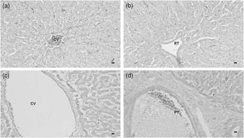
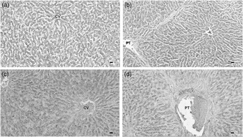
In all liver sections from both non-supplemented and Cu-supplemented cattle, MT expression was diffuse all over the hepatic parenchyma, with no clear positive (+) cells, so it was not possible to make the cell counts (Figure 1).
Metallothionein (MT-I + II) in matched areas of central veins (CV) and portal triads (PT) in the liver. In all liver sections from both non-supplemented (a, b) and Cu-supplemented cattle (c, d), MT expression was diffuse all over the hepatic parenchyma, with no clear + cells. Scale bars, 50 μM.

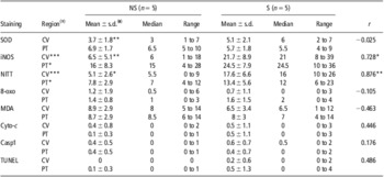
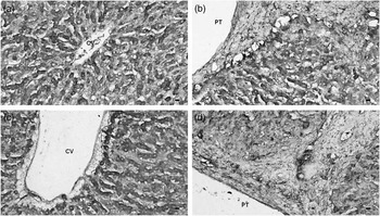
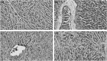
The animals from the non-supplemented group showed significantly more SOD+ cells around the PT than CV, however, this difference was not appreciated in the animals from the Cu-supplemented group (Table 1). In all calves, the SOD+ cells were distributed all over the hepatic parenchyma. No special association of the + cells was appreciated around any particular liver structure such as vessels or connective tissue in CV and PT, resulting in an identical image in both groups of animals (Figure 2). No statistically significant correlations were appreciated between the number of SOD+ cells and Cu concentrations in the liver (Table 1).
Cellular counts of SOD, iNOS, NITT, 8-oxo, MDA, cyto-c, casp1 and TUNEL+ cells from CV and PT from NS and S animals and Pearson correlations with liver Cu concentrations

SOD = superoxide dismutase; iNOS = inducible nitric oxide synthase; NITT = nitrotyrosine; 8-oxo = 8-oxoguanine; MDA = malondialdehyde; cyto-c = cytochrome-c; casp1 = caspase 1; TUNEL = terminal deoxynucleotidyl transferase dUTP nick end labeling; CV = central veins; PT = portal triads; NS = non-supplemented; S = Cu-supplemented; Cu = copper.
Results are shown as mean ± standard deviation, median and range + cells per 0.39 mm2 and Pearson correlation coefficients (r).
*P < 0.05, **P < 0.01, ***P < 0.001 indicate statistically significant correlations between the cellular counts and liver Cu concentrations in r column and differences in cell counts between (†) NS and S animals in CV and PT regions and between (‡) CV and PT within the same group of animals (S or NS).
Cu–Zn superoxide dismutase (SOD) immunoreactivity in matched areas of central veins (CV) and portal triads (PT). The SOD+ cells were distributed all over the hepatic parenchyma, without any special association around vessels or connective tissue in central veins and portal triads in both non-supplemented (a, b) and Cu-supplemented animals (c, d). Scale bars, 50 μM.

Oxidative stress
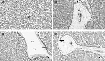
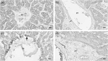
Non-supplemented animals showed significantly higher iNOS and NITT+ cells in the PT than in the CV. However, in the Cu-supplemented animals, the differences in the cell counts between both regions could no longer be appreciated since the number of + cells significantly increased in both regions. This increase was higher around the CV (71% for both iNOS and NITT) than in the PT (32% and 42% for iNOS and NITT, respectively; Table 1). The iNOS (Figure 3) and NITT (Figure 4) + cells in the non-supplemented animals were occasional hepatocytes, Kupffer cells and macrophages localized immediately around the central vessel of the CV. As Cu load increased in the Cu-supplemented group, the number and immunoreactivity of the cells increased in this same location. However, in the PT the + cells were localized as dispersed + cells all over the field around the PT area, whereas in Cu-supplemented animals the + cells were placed immediately close to the connective tissue of the PT.
Inducible nitric oxide synthase (iNOS) immunoreactivity in matched areas of central veins (CV) and portal triads (PT) of the liver of both non-supplemented (a, b) and Cu-supplemented (c, d) animals. The iNOS+ cells in the central veins of the non-supplemented animals (a) were a few cells localized immediately around the central vessel and in the Cu-supplemented animals (c) the number and immunoreactivity of the cells increased in the same location. In the portal triads of the non-supplemented animals (b) the + cells were more dispersed than in the animals of the Cu-supplemented group (d) in which the + cells were closer to the connective tissue of the portal triads. Arrows are pointing out some + cells. Scale bars, 50 μM.

Nitrotyrosine (NITT) immunoreactivity in matched areas of central veins (CV) and portal triads (PT) of the liver of both non-supplemented (a, b) and Cu-supplemented (c, d) animals. The NITT+ cells followed the same distribution as +iNOS. In the central veins of the non-supplemented animals (a) were a few cells localized immediately around the central vessel and in the Cu-supplemented animals (c) the number and immunoreactivity of the cells increased in the same location. In the portal triads of the non-supplemented animals (b) the + cells were more dispersed than in the animals of the Cu-supplemented group (d) in which the + cells were closer to the connective tissue of the portal triads. Arrows pointing out some + cells. Scale bars, 100 μM.

There were no statistically significant differences in 8-oxo and MDA+ cells between CV and PT in both groups of animals and also no differences with Cu supplementation (Table 1). In the case of 8-oxo (Figure 5) and MDA (Figure 6), the + cells were distributed all over the parenchyma without any special association to any liver structure in CV and in PT areas of both non-supplemented and Cu-supplemented animals.
8-oxoguanine (8-oxo) immunoreactivity in matched areas of central veins (CV) and portal triads (PT) of the liver. The + cells were distributed all over the parenchyma without any special association to any structure in central veins and in portal triad areas of both non-supplemented (a, b) and Cu-supplemented animals (c, d). Scale bars, 50 μM.

Malondialdehyde (MDA) immunoreactivity in matched areas of central veins (CV) and portal triads (PT). The + cells were distributed all over the parenchyma without any special association to any structure in central veins and in portal triad areas of both non-supplemented (a, b) and Cu-supplemented animals (c, d). Scale bars, 50 μM.

There were statistically significant positive correlations between iNOS and NITT+ cells and the liver Cu concentration but no significant correlations were appreciated in the case of 8-oxo or MDA+ cells (Table 1).
Apoptotic cell death
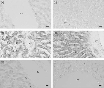
There were no statistically significant differences in TUNEL, cyto-c and casp1+ cells between CV and PT or non-supplemented and Cu-supplemented cattle as well as between both groups of animals in each hepatic area (Table 1). The + cells of all these markers were basically undetectable. The immunoreactivity for cyto-c and casp1 was predominantly punctuate suggesting the mitochondrial localization of the protein (Woo et al., Reference Woo, Hakem, Elia, Hakem, Duncan, Patterson and Mak1999), whereas the number of casp1 and cyto-c+ cells considered as those with cytosolic diffuse staining patterns were rare and were in good agreement with the reduced number of TUNEL+ cells (Figure 7).
Cytochrome c (cyto-c; a, b) and caspase 1 (casp1; c, d) immunoreactivity and TUNEL staining (e, f) in matched areas of central veins (CV) and portal triads (PT). The + cells were basically undetectable, and the immunoreactivity for cyto-c and casp1 was predominantly punctuate suggesting the mitochondrial localization of the protein, thus the considered casp1 and cyto-c+ cells were those with cytosolic diffuse staining patterns. The cyto-c, casp1 and TUNEL+ cells were rare and did not show any special association to any structure in central veins and in portal triad areas of both non-supplemented and Cu-supplemented animals. Scale bars, 100 μM.

There were no statistically significant correlations between TUNEL, cyto-c and casp1+ cells and the Cu concentration in the liver of the animals of this study (Table 1).
Discussion
The results of our study, with significantly higher SOD, iNOS and NITT+ cells around the PT compared with the CV in the non-supplemented animals points out the antioxidative role of copper in the hepatic cell. As these animals have an adequate Cu status – liver Cu concentrations (56 ± 4.5 mg Cu/kg ww) and hematological parameters were within the normal ranges described for cattle (see more at García-Vaquero et al., Reference García-Vaquero, Miranda, López-Alonso, Castillo and Benedito2011) – the higher SOD activity as well as the higher oxidative damage measured by the iNOS and NITT immunoreactivity in the PT could indicate a higher metabolic activity in this region in a normal liver. Because of the characteristic blood flow within the hepatic lobules – oxygenated blood, nutrients and toxins arrive first into the hepatic lobules through the vessels of the PT (Ross and Pawlina, Reference Ross and Pawlina2006) – cells around the PT act as the first defense line against toxins entering the liver.
At higher Cu hepatic loads in the animals of the Cu-supplemented group (liver Cu concentrations of 136 ± 9 mg/kg ww, well above the normal range of 25 to 100 mg/kg ww described by Puls (Reference Puls1994) for cattle) an increase of the oxidative damage, measured as a higher iNOS and NITT immunoreactivity, was observed in PT but especially around the CV. This distribution of iNOS and NITT+ cells agreed with histological studies in sheep in which at early stages of hepatic copper accumulation the greatest intensity of Cu staining granules was noticed in the cells of the centrolobular zones, particularly adjacent to CV. As Cu loading increased the number and size of Cu granules increased and the positive reaction extended through the midlobular zones toward the PT (Kumaratilake and Howell, Reference Kumaratilake and Howell1987). The fact that both iNOS and NITT followed the same distribution within the liver is in good agreement with some studies that demonstrated that tyrosine nitration of proteins is a process localized in close proximity to iNOS and to enzymes capable of peroxidative chemistry and ROS production (Druzhyna et al., Reference Druzhyna, Musiyenko, Wilson and LeDoux2005; Heijnen et al., Reference Heijnen, van Donselaar, Slot, Fries, Blachard-Fillion, Hodara, Lightfoot, Polydoro, Spielberg, Thomson, Regan, Crapo and Ischiropoulos2006).
Despite the increase of iNOS and NITT, no significant effect of Cu supplementation was appreciated for MDA, 8-oxo, cyto-c, casp1 and TUNEL. Studies in the liver of rats reported increased oxidative stress damage of proteins and protein degeneration caused by Cu (Alexandrova et al., Reference Alexandrova, Petrov, Georgieva, Kessiova, Tzvetanova, Kirkova and Kukan2008), lipid peroxidation expressed as MDA activity or immunoreactivity (Zhang et al., Reference Zhang, Noordin, Rahman and Haron2000; Ozcelik and Uzun, Reference Ozcelik and Uzun2009) and DNA damage (Gaetke and Chow, Reference Gaetke and Chow2003; Alexandrova et al., Reference Alexandrova, Kebis, Mislanova and Kukan2007) that finally could lead to apoptotic cell death (Rana, Reference Rana2008). Lipid peroxidation produced by ROS is considered to be the most likely mechanism to damage proteins, rather than a direct action of ROS generated by Cu or other metals overload (Yamada et al., Reference Yamada, Sogawa, Suzuki, Izumi, Agui and Matsumoto1992; Ogihara et al., Reference Ogihara, Ogihara, Miki, Yasuda and Mino1995). However, in these studies animals/cells were treated with high Cu doses. In cattle of this study, ROS generated by Cu induced protein damage, that could be considered as one of the first pathological alterations in the liver even when liver Cu concentrations were not associated with those considered as toxic. Despite the oxidative damage appreciated in proteins, the Cu overload of the liver was not enough to cause cell and tissue injury, reflected in the no significant effects of Cu on the oxidative stress markers of the other macromolecules analyzed and the almost non-detectable apoptotic cell death. More studies in cattle under different Cu dietary conditions will be necessary to better understand the role of iNOS and NITT and to appreciate other oxidative stress damages that could lead to apoptotic cell death and the development of clinical Cu toxicity.
In spite of the Cu dosed animals in our study showing more oxidative damage in the liver, Cu supplementation did not improve their antioxidative protection. SOD activity in the Cu-supplemented animal did not differ from that in the controls. Controversial results have been found in animals exposed to high Cu doses and other oxidative toxics. Decreased SOD activity was appreciated in the liver and/or serum of Cu loaded rats (Roy et al., Reference Roy, Mandal, Sen and Biswas2009) that could be due to an increase in protein degradation (Kwon et al., Reference Kwon, Lee, Floyd and Park1998) or nuclear damage caused by Cu toxicity that could affect gene expression, and thus, a decrease of SOD activity (Mauriz et al., Reference Mauriz, Molpeceres, García-Mediavilla, González, Barrio and González2007; Yu et al., Reference Yu, Li, Deng and Mao2008). Thus, according to these authors, the non-increased SOD activity in the Cu-supplemented animals in this study could be explained by the increased protein damage that could lead to a higher SOD degradation. However, other studies showed elevated concentrations of hepatic SOD in the liver of cattle due to increased levels of ROS produced by toxics such as ethionine (Abd Ellah et al., Reference Abd Ellah, Okada, Goryo, Oishi and Yasuda2009).
The incipient oxidative damage found in the liver of the Cu-supplemented animals in our study could be explained by the very low capacity of cattle to accumulate Cu safely bound to MT. The poor immunoreactivity for MT and the lack of differences between non-supplemented and Cu-supplemented calves in this study are in good agreement with previous toxicological studies in cattle showing that Cu is a poor inducer of MT synthesis. Although it has been demonstrated that Cu can induce MT synthesis in laboratory animals, this only happens when administrated in a very large dose (i.e. intraperitoneal injections) and variations of dietary Cu have little effect on liver MT expression until the levels are extremely high (Mercer, Reference Mercer1997). The marked differences between animal species to Cu toxicity seem to be highly related to their ability to synthesize MT and accumulate Cu bound to MT. Thus, Cu toxicity is reduced in the liver of species such as pig and dog in which most of the hepatic Cu is bound to MT (Bremner and Beattie, Reference Bremner and Beattie1990) and is very high in others, as sheep and cattle, that have a limited capacity to induce MT synthesis in response to increased dietary Cu intake even at very high concentrations (Bremner and Beattie, Reference Bremner and Beattie1995; López-Alonso et al., Reference López-Alonso, Prieto, Miranda, Castillo, Hernández and Benedito2005).
Conclusions
Under the conditions of this study, which are the conditions of the cattle raised in intensive systems in NW Spain and also many European countries, routinely Cu supplementation increased the risk of the animals to undergo subclinical Cu toxicity, with no significant changes in the Cu storage capacity and the antioxidant defensive system evaluated by MT and SOD expression, but with a significant and important increase of oxidative damage measured by iNOS and NITT. The results of this study indicated that iNOS and NITT could be used as early markers of initial pathological changes in the liver caused by Cu supplementation in cattle, although more studies in cattle under different levels of Cu supplementation are needed.
Acknowledgments
This work was supported by the Xunta de Galicia (Spain) through contract 07MRU030261PR and by a research grant María Barbeito awarded to M. García-Vaquero by the Dirección Xeral de Investigación, Desenvolvemento e Innovación from Xunta de Galicia and to FSE (Fondo Social Europeo) funds through Programa de Recursos Humanos, do Plan Galego de Investigación, Desenvolvemento e Innovación Tecnolóxica de Galicia. The authors thank Dr B. Gutiérrez and L. Casanova Iglesias for samples collection and technical assistance, respectively.