Introduction
Solitary fibrous tumour is a soft tissue tumour of mesenchymal origin. It was first described in the pleura,Reference Klemperer and Rabin 1 and has since been reported in many anatomical locations including the skin and, rarely, the oral cavity.Reference Piattelli, Fioroni and Rubini 2 – Reference Shnayder, Greenfield, Oweity and DeLacure 5 Solitary fibrous tumours of the oral cavity were first described by Suster et al. in 1995.Reference Suster, Nascimento, Miettinen, Sickel and Moran 6 In the largest series of oral solitary fibrous tumours to date,Reference O'Regan, Vanguri, Allen, Eversole, Wright and Woo 7 only 2 of the 21 cases affected the tongue, whereas 11 affected the buccal mucosa or vestibules.
In its latest classification of soft tissue and bone tumours,Reference Guillou, Fletcher, Fletcher, Mandahl, Hogendoorn, Mertens and Bridge 8 the World Health Organization categorised solitary fibrous tumour as a fibroblastic or myofibroblastic tumour of ‘intermediate’ malignancy; that is, a tumour which, though usually benign, may on occasion metastasise. Histologically, solitary fibrous tumour is a spindle cell neoplasm with a prominent vascular pattern. However, its microscopic features are not specific, and immunohistochemistry is required to confirm the diagnosis.
Until recently, cluster of differentiation (CD) 34 antigen, for which solitary fibrous tumour is strongly positive,Reference Migita, Yoshino, Kobayashi, Shiomi, Enatsu and Shigematsu 3 , Reference Vafiadou, Dimitrakopoulos, Georgitzikis, Hytiroglou, Bobos and Karakasis 4 , Reference Fusconi, Ciofalo, Greco, Pulice, Macci and Mariotti 9 , Reference Chan 10 was the usual marker used. However, CD34 is itself non-specific. Recently, nuclear (rather than membranous or cytoplasmic) expression of signal transducer and activator of transcription 6 (STAT6) has been the ‘gold standard’.Reference Yoshida, Tsuta, Ohno, Yoshida, Narita and Kawai 11 STAT6 expression has not previously been reported in solitary fibrous tumour affecting the tongue. In this report, we describe four cases of lingual solitary fibrous tumour. All four tumours were surgically excised and none have recurred in at least one year.
Clinical presentations
Case one
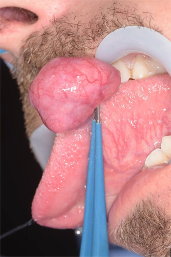
A 46-year-old man was urgently referred with a swelling on the left lateral border of the tongue. The swelling had been gradually enlarging over a period of one year, causing aesthetic and functional problems. His past medical history included asthma, which was well controlled through the use of inhalers. He smoked 20 cigarettes per day and denied consumption of any alcohol. Clinical examination revealed a swelling that was pedunculated and lobulated, with a maximum dimension of 2.5 cm (Figure 1). The overlying mucosa appeared thin, but not ulcerated. There were no complaints of paraesthesia or bleeding from the tongue, nor were there any palpable regional lymph nodes. A benign process was favoured, with differential diagnoses of fibro-epithelial polyp, granular cell tumour and lipoma. The mass was excised under general anaesthesia using carbon dioxide laser. Healing was uneventful and there has been no recurrence in one year.
Clinical presentation of case one.

Case two
A 20-year-old man was urgently referred with a swelling on the dorsum of the tongue, just to the left of the midline, which had been gradually enlarging over a period of 6 months. There was no history of trauma to the area in question. The patient was medically healthy, and did not consume tobacco and declared negligible alcohol consumption. On examination, there was a well-circumscribed polypoid growth with a sessile base 4 mm in maximum dimension. The overlying mucosa was normal and there was no lymphadenopathy. A provisional diagnosis of fibro-epithelial polyp was made. Excisional biopsy of the mass was performed under local anaesthesia. Healing was uneventful and there has been no recurrence in one year.
Case three
A 49-year-old woman was referred for a swelling on the dorsum of the tongue of 4 months’ duration. Although symptomless, she noticed it was increasing in size. The patient was medically fit and well, did not consume tobacco, and consumed alcohol within the recommended weekly limit. Clinical examination of the tongue revealed a firm, sessile polyp approximately 4 mm in diameter. The overlying mucosa was normal. Examination findings of the head and neck were otherwise unremarkable. A provisional diagnosis of fibro-epithelial polyp was made. Excisional biopsy of the mass was performed under local anaesthesia. Healing was uneventful and there has been no recurrence in 18 months.
Case four
A 46-year-old woman was referred for a swelling on the left side of her tongue of 1 year's duration. The patient recalled no traumatic episode prior to the development of the lump, which was symptomless and of constant size. The patient had a history of asthma and psoriatic arthritis. She was a non-smoker and consumed moderate amounts of alcohol. Clinical examination of the tongue revealed a defined nodule 7 mm in diameter on the lateral aspect of the left anterior tongue. The overlying mucosa was normal. Examination findings of the head and neck were otherwise unremarkable. A provisional diagnosis of a lipoma, fibroma or mucocoele was made. Excisional biopsy of the lesion was performed under local anaesthesia. Healing proceeded normally and there has been no recurrence in five years.
Histopathology and immunohistochemistry
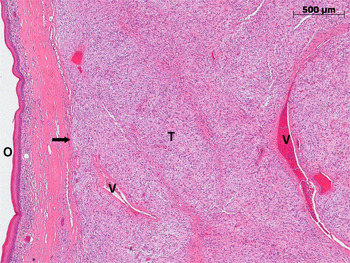
Microscopically, all cases involved a circumscribed tumour consisting of cytologically bland, densely packed spindle-shaped cells with minimal cytoplasm lying in a fibromyxoid stroma (Figure 2). Thin-walled blood vessels were present throughout and assumed a ‘staghorn’ appearance. Mitoses were scarce.
Low power view of solitary fibrous tumour of the tongue, showing densely packed spindle cells with prominent blood vessels (V). The tumour (T) has a defined margin (arrow). (H&E stain.) O = oral mucosal surface

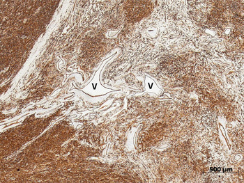
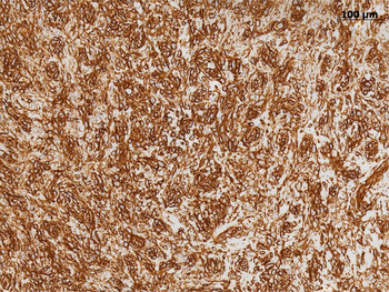
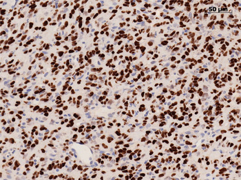
Immunohistochemistry showed strongly positive membranous and cytoplasmic expression of CD34 in all cases (Figures 3 and 4) and nuclear expression of STAT6 in two cases (cases one and four) (Figure 5). The cell proliferation index (as determined by nuclear expression of Ki67) was low (less than 5 per cent) in all four cases. These morphological and phenotypic features are consistent with solitary fibrous tumour.
Tumour is diffusely positive for CD34 (golden brown reaction product). (Immunoperoxidase stain.) V = ‘staghorn’ blood vessels

High power view showing the membranous staining pattern of CD34, which highlights the outline of the tumour cells. (Immunoperoxidase stain)

High power view showing the nuclear staining pattern of STAT6 in the tumour cells. (Immunoperoxidase stain)

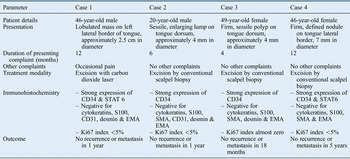
All tumours were treated as benign. Complete excision of the tumour was confirmed histologically in all cases. Table I summarises the details of the four cases.
Summary of clinical presentations, investigations, treatments and outcomes

CD = cluster of differentiation; EMA = epithelial membrane antigen; SMA = smooth muscle actin
Discussion
Solitary fibrous tumour was first described in 1992 by Klemperer and Rabin in the parietal or visceral pleura.Reference Klemperer and Rabin 1 However, the name betrays the fact that the tissue of origin is uncertain. Many reports have since shown it is not a tumour specific to this location.Reference Suster, Nascimento, Miettinen, Sickel and Moran 6 , Reference Guillou, Fletcher, Fletcher, Mandahl, Hogendoorn, Mertens and Bridge 8 , Reference Chan 10
Solitary fibrous tumour usually presents in the fifth decade of life, as was the case in three of the patients presented here, and represents less than 2 per cent of all soft tissue tumours.Reference Penel, Amela, Decanter, Robin and Marec-Berard 12 They are usually indolent and painless but if traumatised, may become symptomatic (as in case one).
Chan proposed a set of criteria to assist in histopathological diagnosis.Reference Chan 10 Specifically, solitary fibrous tumour is characterised by: short, spindled or ovoid cells, with scanty and poorly defined cytoplasm; alternating hypercellular and hypocellular foci, with sclerosis in the latter; circumscription; scanty mitotic figures (less than 4 mitoses per 10 high power fields); and intimate intertwining of collagen fibrils with spindle cells.Reference Chan 10 Nonetheless, in sections stained with haematoxylin and eosin (Figure 2), solitary fibrous tumour has a number of differential diagnoses,Reference Penel, Amela, Decanter, Robin and Marec-Berard 12 and immunohistochemistry for STAT6 and CD34 is necessary to clinch the diagnosis.Reference Chan 10 , Reference Matsuzaki, Iwamoto, Chiba, Saito, Yagisawa and Ichikawa 13
Of the 13 lingual solitary fibrous tumour cases already described in the literature, all were positive for CD34.Reference Piattelli, Fioroni and Rubini 2 – Reference Shnayder, Greenfield, Oweity and DeLacure 5 , Reference O'Regan, Vanguri, Allen, Eversole, Wright and Woo 7 , Reference Yoshida, Tsuta, Ohno, Yoshida, Narita and Kawai 11 , Reference Abrari and Bakshi 14 – Reference Yamashita, Satoh and Goto 19 However, our series is the first to demonstrate nuclear STAT6 reactivity in lingual solitary fibrous tumour (Figure 5). STAT6 is more specific for solitary fibrous tumour than CD34, and, although expressed in the majority of solitary fibrous tumours, STAT6 is not expressed by them all.Reference Han, Zhang, Yu, Han, Wang and Xu 20 It is currently unknown whether lack of STAT6 expression in solitary fibrous tumour has any prognostic significance.
Although benign, solitary fibrous tumour may be locally infiltrative, and either recur or, occasionally, metastasise. A review by Cox et al. showed that of 142 cases of solitary fibrous tumour in the head and neck region, 10 had atypical or malignant features, and recurrence was largely dictated by the presence of positive margins at the time of excision, rather than the microscopic grade of the tumour itself.Reference Cox, Daniels and Jordan 21
A high proliferation index is cited to be a risk factor in dictating aggressive tumour behaviour. Künzel et al. demonstrated that tumours could progress from a low to high proliferative index, and hence warranted long-term follow up.Reference Künzel, Hainz, Ziebart, Pitz, Ihler and Strieth 22
-
• Solitary fibrous tumours are of mesenchymal origin; several extra-pleural locations have been reported including the head and neck
-
• There are 13 reported cases of solitary fibrous tumour of the tongue in the literature
-
• All 13 cases were positive for CD34, but none reported nuclear STAT6 expression
-
• In our series of four cases involving the tongue, all were positive for CD34 and two demonstrated nuclear STAT6 expression
-
• Nuclear STAT6 expression is more specific than CD34 positivity in solitary fibrous tumour diagnosis
-
• Surgical excision is the ‘gold standard’ treatment; to date, there have been no recurrences following excision
There is only one report of a frankly malignant solitary fibrous tumour of the tongue.Reference Shnayder, Greenfield, Oweity and DeLacure 5 In addition to infiltration, this tumour showed severe nuclear atypia, a high mitotic count and areas of necrosis. However, neither this nor any of the previously reported cases of solitary fibrous tumour in the tongue have recurred or metastasised; all have successfully been treated with complete surgical excision. In all four of the cases reported here, there has been no recurrence or metastasis in at least one year.
Conclusion
Although rare, solitary fibrous tumour should be considered in the differential diagnosis of tongue swellings. All of our cases were initially diagnosed clinically as fibro-epithelial polyps. Solitary fibrous tumour is usually benign and can be treated with simple surgical excision. However, review is necessary, as solitary fibrous tumour has the potential to recur and on occasion metastasise.
Acknowledgements
The authors acknowledge the assistance of Messrs Mike Shelley, John Tighe and Brian Bisase (consultant oral and maxillofacial surgeons at Queen Victoria Hospital NHS Foundation Trust, East Grinstead) in reporting this case series. Consultant histopathologists Professor Phil Sloan (Royal Victoria Infirmary, Newcastle upon Tyne), Professor Archie Malcolm (Royal Shrewsbury Hospital) and Dr Lamios Munthali (Queen Victoria Hospital NHS Foundation Trust) confirmed the histological diagnoses.