INTRODUCTION
Information about the reproductive biology of an exploited cephalopod population is necessary both for further assessment and for setting management regulations. The macroscopic determination of the maturation stage by visual examination of gonads is a relatively rapid and inexpensive manner of determining the reproductive status (Sauer & Lipinski, Reference Sauer and Lipinski1990; Boyle & Rodhouse, Reference Boyle and Rodhouse2005). However, it is often difficult to set discrete categories and define discrete reference points based on macroscopic criteria because maturation progresses continuously. Besides, macroscopic analysis alone may lead to a misclassification of the maturity state of animals (Mangold, Reference Mangold and Boyle1987). On the other hand, histological analysis could provide more precise data about the changes occurring in gonads. It serves as a tool to validate the adopted macroscopic stages by linking categories of microscopic development (i.e. stages describing the degree of development of spermatogenesis or oogenesis) to macroscopic maturity stages, although it might be a time consuming and expensive task. In addition, the knowledge about the reproductive strategy of the cephalopod species would improve through histological observations (Sauer & Lipinski, Reference Sauer and Lipinski1990; Melo & Sauer, Reference Melo and Sauer1999; Gonçalves et al., Reference Gonçalves, Sendão and Borges2002; Díaz-Uribe et al., Reference Díaz-Uribe, Hernández-Herrera, Morales-Bojórquez, Martínez-Aguilar, Suárez-Higuera and Hernández-López2006).
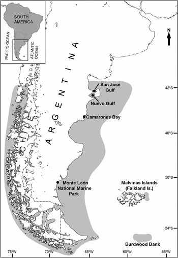
The Patagonian red octopus, Enteroctopus megalocyathus (Gould, 1852), is a relatively large octopus receiving growing attention regarding its fishing and its potential of culture in the southern tip of South America (Argentine and Chilean coasts) (Ortiz et al., Reference Ortiz, Ré and Márquez2006, Reference Ortiz, Ré, Márquez and Glembocki2011; Perez et al., Reference Pérez, López, Aguila and González2006; Farías et al., Reference Farías, Navarro, Cerna and Uriarte2011). Geographical distribution of this species, ranges from 41°S to 56°S in the south-west Atlantic Ocean, and from 42°S to 56°S in the south-eastern Pacific Ocean, with a bathymetric range of distribution from intertidal areas up 240 m depth (Ré, Reference Ré and Boschi1998; Osorio et al., Reference Osorio, Peña, Ramajo and Gracelon2006; Ibáñez et al., Reference Ibáñez, Camus and Rocha2009) (Figure 1). Along the Patagonian Atlantic coast, the species is harvested by artisanal fisheries and by sporting fishermen in intertidal and subtidal areas. Although the fisheries in this region have been operating at least since the 1960s, they are unregulated and official catches are not recorded (Cinti et al., Reference Cinti, Soria, Orensanz and Parma2003; Ortiz et al., Reference Ortiz, Ré, Márquez and Glembocki2011). On the southern Chilean coast, captures by diving approximate 500 tons per year, although at present in main fishing areas there exist regulations that forbid the fishing of this species (Ibáñez & Chong, Reference Ibáñez and Chong2008; Sernapesca, 2011).
Geographical distribution of Enteroctopus megalocyathus (in grey) according to Ré, (1998, Reference Ré and Boltovskoy2008) and Ibáñez et al. (Reference Ibáñez, Camus and Rocha2009). Dots indicate sampling areas along the Patagonian Atlantic coast.

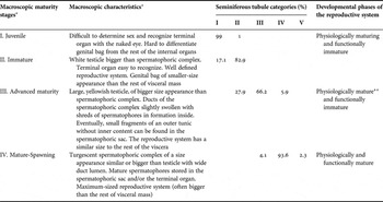
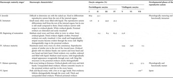
Sexual maturation of E. megalocyathus has recently been assessed on the northern Atlantic Patagonian coast (San José and Nuevo Gulfs) (Figure 1) (Ortiz et al., Reference Ortiz, Ré, Márquez and Glembocki2011). In that work, maturity stages were assigned taking into account the macroscopic (i.e. morphochromatic) characteristics of the reproductive system defined by scales developed for E. megalocyathus (Tables 1 & 2). Nevertheless, these macroscopic maturity scales remain to be validated. In addition, little is known about the gametogenesis of E. megalocyathus as well as many other aspects of its reproductive biology. The aims of this work were to define microscopic categories of development by histological analysis of the gonads of both sexes, and to validate the macroscopic scales of E. megalocyathus, in order to establish these scales as a tool that will allow for further maturity assessments.
Macroscopic maturity stages, frequency of seminiferous tubule categories and developmental phases resulting from the overall state of development and functioning of the reproductive system of Enteroctopus megalocyathus males.

*, from Ortiz et al. (Reference Ortiz, Ré, Márquez and Glembocki2011); **, some animals which presented spermatozoa in seminiferous tubules.
Macroscopic maturity stages, frequency of oocyte categories and developmental phases resulting from the overall state of development and functioning of the reproductive system of Enteroctopus megalocyathus females.

*, from Ortiz et al. (Reference Ortiz, Ré, Márquez and Glembocki2011); **, corresponding to only two oocytes found in one ovary.
MATERIALS AND METHODS
Samples were obtained from January 2006 to June 2007 by monthly diving surveys up to 25 m depth in San José Gulf and Nuevo Gulf. In the same period additional animals were collected in the intertidal areas of Camarones Bay and Monte Leon National Park (Figure 1). In all cases, animals were captured with a hook.
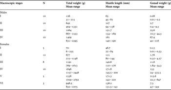
Total weight (TW), and dorsal mantle length (ML) were registered to the nearest 1 g and 1 mm respectively in 40 males and 43 females, which were assigned to a maturity stage (Tables 1 & 2). Besides, the weight of the ovary (OV), and the testis (TE) were recorded to the nearest 0.01 g (Table 3). Fresh gonads were fixed in Bouin's solution for 7 days and stored in ethanol 70% for histological examination. Tissue samples taken from the mid-area of testis and ovaries were then dehydrated through an alcohol series, cleared in xylol, and embedded in paraffin wax. Afterwards, sections 6 µm thick were cut with a rotary microtome and stained with eosin and haematoxylin.
Macroscopic maturity stages and morphometric and gravimetric characteristics of Enteroctopus megalocyathus histologically analysed.

Changes through the course of the seminiferous tubules development in males and of oocyte/follicular cell complexes development in females were characterized and classified into categories of microscopic development. Categories were counted along two random perpendicular 10 mm lines, running from the periphery towards the middle of the gonads. In addition, a fraction of oocytes in each category was measured under microscope using their largest diameter.
To assess the correspondence between macroscopic maturation stages and the level of seminiferous tubules and oocyte development, a histological maturity index (HMI) was calculated for each individual according to Sauer & Lipinski's (1990) equation:
 $${\rm HMI}=\displaystyle{{\sum\limits_{i=1}^s {\left({n_i\; x\; i} \right)} } \over {\sum\limits_{i=1}^s {n_i} }}$$
where i is a discrete number which represents each microscopic category, s is the maximum number of categories recognized for each sex, and n i the absolute frequency of seminiferous tubules or oocytes in the category i resulting from the sum of the counts made on each gonad. Kruskal–Wallis (KW) and t planned multiple comparison tests were performed to compare HMI between consecutive macroscopic maturity stages (Conover, Reference Conover1999). Statistical analyses were performed with GraphPad InStat and Excel packages.
RESULTS
Categories of microscopic development
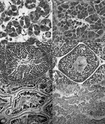
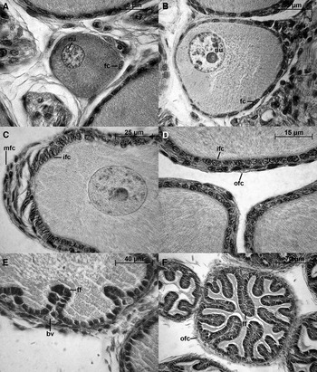
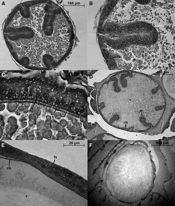
Five and seven microscopic developmental categories were recognized in gonads of males (Figure 2) and females (Figures 3 & 4) respectively.
Enteroctopus megalocyathus. Seminiferous tubule categories: (A) category I; (B) category II; (C) category III; (D) (early) and (E) (late), category IV; (F) category V. bv, blood vessel; ge, germinal epithelium; lu, central lumen; sc, spermatocytes; sg, spermatogonia; sph, spermatozoa heads; spt, spermatozoa tails; st, spermatids. Arrow head, an espermatogonia undergoing mitotic division.

Enteroctopus megalocyathus. Previtellogenic oocyte categories: (A) category I; (B) category II; (C & D) category III; (E & F) category IV. bv, blood vessel; fc, follicular cells; ff, follicular folds; ifc, inner layer of follicular cells; mfc, multiple layer of follicular cells; nu, nucleolus; ofc, outer layer of follicular cells; s, stalk side.

Enteroctopus megalocyathus. Vitellogenic oocyte categories: (A & B) category V; (C & D) category VI; (E & F) category VII. bv, blood vessel; ch, chorion; ff, follicular folds; fs, follicular syncytium; nfc, nucleus of the follicular syncytium; v, vitellus.

MALES
Seminiferous tubule category I: the tubule is poorly developed, has a low cell density and the central lumen is difficult to recognize. Cytologically, there are only rounded spermatogonia attached to the inner tubule wall (germinal epithelium) (Figure 2A) and occasionally a few early spermatocytes are present.
Seminiferous tubule category II: the tubule in this category is well defined and wider than that in category I. Central lumen is now recognized but there are no cells inside. At this stage, there is evidence of the cellular multiplication process. The spermatogonias predominate, and early and secondary spermatocytes appear, increasing cellular density. Smaller than spermatogonias and distinguished by their large nuclei, spermatocytes appear in groups that extend from the germinal epithelium towards the lumen (Figure 2B). No spermatids are present.
Seminiferous tubule category III: the tubule shows a well defined central lumen and a high cell density. In this stage the maturation process stands out and a decrease in cell size is evident from the germinal epithelium towards the central lumen (Figure 2C). There are spermatogonias, many early and secondary spermatocytes and small spermatids. A few spermatozoa are present in some of the samples partially occupying the central lumen, in which case they are noticeable by their acidophilic tails.
Seminiferous tubule category IV: the lumen is slightly wider and it is densely covered by the tails of spermatozoa. A great abundance of spermatids and numerous heads of spermatozoa arranged in groups are located in the periphery of the lumen. There are also many spermatocytes and very few spermatogonias (Figure 2D). Along with the discharge of spermatozoa from the tubule, cell density decreases and the central lumen increases in size. In advanced stages of this process, there seem to be no cells associated with the germinal epithelium (Figure 2E).
Seminiferous tubule category V: tubules are either empty or have very few sperm tails. Tubular limits defined in earlier stages by germinal epithelium are not easily recognized (Figure 2F).
FEMALES
Oocyte category I: the oocyte is spherical, it has a small average width of 35 µm (SD = 11.2, N = 60) (Figure 3A). The nucleus presents a prominent nucleoli and heterochromatic clumps. Follicle cells are scarce or absent. When present, they begin to differentiate themselves from the connective tissue overlaying the oocyte.
Oocyte category II: the oocyte has an ovoid shape and it is attached to the ovarian connective tissue by a short stalk. Ovoid follicle cells form a single-layer which spreads from the stalk side, and surround the oocyte, except in the area that will become the animal pole of the egg (opposite the side of the stalk). The mean width of the oocyte is 75 µm (SD = 17.7, N = 60) (Figure 3B).
Oocyte category III: follicle cells are distributed in two layers, except in the side opposite the stalk, where follicle flat cells have formed multiple layers. These cells become incorporated to the inner layer of follicle ovoid cells (Figure 3C). Follicular cells of the outer layer surrounding the oocyte appear dispersed (Figure 3D). In the nucleus of the oocyte a prominent nucleoli persists and the oocyte average width is 135 µm (SD = 21.1, N = 60).
Oocyte category IV: at the beginning of this stage, the inner layer of follicle cells starts to invade the oocyte as folds and they become cuboidal. Blood capillaries develop at the bases of the folds (Figure 3E). Proliferation of follicular epithelium continues. In the second phase, the folds extend deeper into the oocyte increasing their number. The outer layer is formed by scattered flat cells surrounding the oocyte. The inner layer of follicle cells become columnar but keep the separation between them (Figure 3F). The oocyte has an average width of 240 µm (SD = 68.8, N = 60).
Oocyte category V: the onset of vitellogenesis takes place (Figure 4A). The outer layer of follicle cells is not recognizable. Follicle cells of the inner layer have prominent nuclei and the separations between these cells disappear forming a follicular syncytium (Figure 4B). Oocytes width increases on average three times (mean = 607 µm, SD = 172, N = 60).
Oocyte category VI: droplets of chorionic material are secreted by follicle cells and fuse along the inner surface of the epithelium, beginning to form the chorion. The large nuclei of follicle cell epithelium are oriented to cytoplasm of the oocyte (Figure 4C). Folds of follicular syncytium begin to regress, their number decrease, and the oocyte width continues to increase (mean = 1550 µm, SD = 0.39, N = 60) by the active secretion of yolk droplets inside (Figure 4D).
Oocyte category VII: the follicle cell epithelium becomes thin (Figure 4E). The whole oocyte is packaged with yolk and the chorion is fully formed (Figure 4F). Detached from the egg string, the mature oocyte will be ready for ovulation. Oocyte width reaches the maximum (mean = 1980 µm, SD = 0.54, N = 28).
MICROSCOPIC CATEGORIES ALONG MACROSCOPIC MATURITY STAGES
In males, within each macroscopic stage of maturation and between consecutive stages, seminiferous tubules showed a regular and progressive pattern of development (Table 1). Spermatozoa were recorded from macroscopic stage III in seminiferous tubules categories III and IV. At the stage IV, almost all testes exhibited a central lumen of tubules filled with spermatozoa, except for one testis which presented partially spent tubules. Spent tubules (category V) were only observed in one male which exhibited a small testis (TE weight = 42 g) but still had spermatophores stored in the spermatophoric sac and terminal organ (Tables 1 & 3).
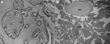
In females, the greatest overlap of oocyte categories was observed in macroscopic stages II and III. Previtellogenic oocytes were registered up to stage III, from which the first early vitellogenic oocytes (oocyte category V) appeared. Nearly mature and mature oocytes (categories VI and VII respectively) were observed from stage IV on, but mature oocytes reached their maximum frequency at macroscopic stage V (Table 2). In addition, at the latter stage, distinct postovulatory follicles begin to be observed indicating recent spawning. An increased number of postovulatory follicles, and a very few (only two mature oocytes in one ovary) or no oocytes inside ovaries were registered at macroscopic stage VI (Figure 5; Table 2).
Enteroctopus megalocyathus. Cross-section of an ovary at mature-spawning (A) and at spent stage (B). no, mature oocyte not evacuated; pof, postovulatory follicles; v, vitellus.

Validation of macroscopic maturity stages
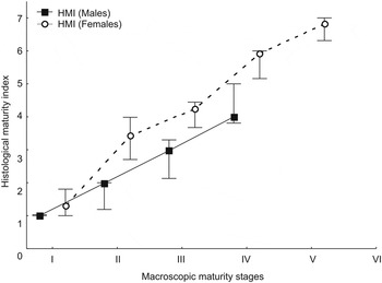
The HMI values of males and females increased and showed highly significant differences through macroscopic stages (KW = 36.1, P < 0.001 and KW = 30.6, P < 0.001 respectively) (Figure 6). In males, highly significant differences were found between all consecutive stages (I–II: t = 10, P < 0.01; II–III: t = 10, P < 0.01; III–IV: t = 9.5, P < 0.01). In females, significant differences were found between macroscopic maturity stages I–II (t = 8.8, P < 0.01), II–III (t = 9.2, P < 0.01), III–IV (t = 9.5, P < 0.01), and IV–V (t = 7, P < 0.05). Due to the fact that in macroscopic stage VI almost all oocytes have been expelled this stage was not used for the statistical comparison.
Median (± minimum–maximum) values of the histological maturity index (HMI) of males and females of Enteroctopus megalocyathus by macroscopic stages.

DISCUSSION
As for other cephalopod species, a comparison between spermatogenesis and oogenesis showed that the maturity process in males can be described in a lower number of categories than females. Besides, the development of seminiferous tubules and the oocyte/follicular cell complexes in Enteroctopus megalocyathus proceeds along schemes broadly similar to those described for other octopod species (Pujals, Reference Pujals1986; Coelho, Reference Coelho1990; Boyle & Chevis, Reference Boyle and Chevis1992; Gonçalves et al., Reference Gonçalves, Sendão and Borges2002; Rodríguez-Rúa et al., Reference Rodríguez-Rúa, Pozuelo, Prado, Gómez and Bruzón2005). In the case of females, the rapid increase of the area of the single and the double layers of follicular cells is often attributed to mitosis (Cowden, Reference Cowden1968; Boyle & Chevis, Reference Boyle and Chevis1992). However, it is remarkable that at oocyte category III, an active proliferation of follicular cells occurs at the opposite stalk side of the oocyte. These follicular cells migrate and eventually enclose the oocyte (Figure 3C). In addition, they also seem to contribute to increase the area of the follicular cell layer during the infolding process (i.e. at oocyte category IV) (Figure 3E, F). A similar pattern of follicular cells proliferation has been described for Eledone cirrhosa (Lamarck, 1798) (Boyle & Chevis, Reference Boyle and Chevis1992).
Histological analysis of gonads coupled with macroscopic observations would permit the definition of developmental phases during sexual maturation on the basis of the degree of development of sexual cells, and once they become mature, their transport and accumulation in different parts of the reproductive system (i.e. the overall state of development and functioning of the reproductive system) (Arkhipkin, Reference Arkhipkin1992; Sauer & Lipinski, Reference Sauer and Lipinski1990; Nigmatullin et al., Reference Nigmatullin, Sabirov and Zalygalian2003). In males at stages I and II, germinal cells were observed in early stages of differentiation (i.e. up to seminiferous tubule category II) and no spermatophores. These stages can be categorized into histologically (or physiologically) maturing and functionally immature phases. At stage III, TE weight increased sharply as a consequence of a continuous increase of the density and the diversity of the germinal cells. In addition, some animals at this stage exhibited a few spermatozoa in their testis and fragments of an outer tunic without inner content in the spermatophoric sac (Tables 1 & 3). The latter would be a process occurring before the animals reach functional maturation, prior to the formation of mature spermatophores (Lipinski & Underhill, Reference Lipinski and Underhill1995; Nigmatullin et al., Reference Nigmatullin, Sabirov and Zalygalian2003). Thus, at least a fraction of stage III males could be considered to be in a physiological maturity phase but still functionally immature. In contrast, stage IV males which presented the lumen of seminiferous tubules covered by spermatozoa (i.e. seminiferous tubule category IV) and mature spermatophores storage in the spermatophoric sac and/or the terminal organ could fall into a physiologically and functionally mature phase (Table 1). On the other hand, an analysis was conducted in stage IV males which exhibited signs of gonad exhaustion (i.e. seminiferous tubules partially spent or without sperm and reduced testis weight) (Table 1). Since these octopuses still had storage spermatophores, they could be in an active spawning condition, a fact that makes it difficult to have clear criteria to determine the spent stage in this sex. In females, the sharply OV weight increase at stage IV is primarily due to a size increase of the oocytes as a consequence of the vitellogenesis (Tables 2 & 3). As a proof that mating can occur in stage IV females, evaginated spermatophores were observed attached to their distal oviducts (Ortiz et al., Reference Ortiz, Ré, Márquez and Glembocki2011). Nevertheless, up to that stage, they should be considered to be in a functionally immature and physiologically maturing phase because most oocytes are still in a maturing developmental state (99.5% of oocyte categories V and VI). Conversely, stage V of females should be considered in a functionally and physiologically mature phase because mature oocytes are found inside ovary and distal oviducts (i.e. in a spawning condition) (Table 2). Thereafter, stage VI would not depend on the oocyte/follicular cells complexes development. At this stage, the morphological characteristics of the gonad define a clear set of criteria of the spent stage of this sex (Table 2) that is also used to recognize the spent stage in other octopus species (Pujals, Reference Pujals1986; Gonçalves, et al., Reference Gonçalves, Sendão and Borges2002; Leporati et al., Reference Leporati, Pecl and Semmens2008).
In both sexes of E. megalocyathus at the most advanced states of maturation, histological analysis reveals that animals do not have a stock of germinal cells that would permit a second spawning (Figures 2F & 5B). In addition, at macroscopic stage VI females undergo a decrease of TW, and ML (Table 3) (Ortiz et al., Reference Ortiz, Ré, Márquez and Glembocki2011). At spent stage, these microscopic and macroscopic features are conditions associated with senescence (Arnold & Williams-Arnold, Reference Arnold, Williams-Arnold, Giese and Pearse1977; Anderson et al., Reference Anderson, Wood and Byrne2002; Gonçalves et al., Reference Gonçalves, Sendão and Borges2002) expected to occur when E. megalocyathus approaches the end of its life span. On the other hand, although an overlapping of oocyte categories was observed through maturation, group-synchronous ovulation occurs in the ovary at mature-spawning stage. At that stage postovulatory follicles were seen alongside nearly mature and mature oocytes (Table 2; Figure 5A). These findings provide indirect evidence that E. megalocyathus has an egg lying period confined to a few days or weeks during which nearly mature oocytes become ready to be spawned. In addition, somatic growth does not seem to take place between spawning events (Table 3). Therefore, E. megalocyathus could be considered an intermittent terminal spawner (sensu Rocha et al., Reference Rocha, Guerra and González2001). Observations during spawning on captive E. megalocyathus could confirm this hypothesis.
The agreement between macroscopic and histological criteria confirms the validity of macroscopic maturity scales of both sexes. However, as in ichthyology, maturity staging by macroscopic examination could contain a subjective judgment (Gerritsen & McGrath, Reference Gerritsen and McGrath2006). This would be particularly relevant in the intermediate (‘maturing’) stages of the E. megalocyathus's scales that are defined by the relative size and colour of the different parts of the reproductive organs (Tables 1 & 2). These stages have been the most abundant within catches in fishing seasons and areas of the northern Atlantic Patagonian coast (Ortiz et al., Reference Ortiz, Ré, Márquez and Glembocki2011). Thus, assays investigating the variability among assessors in the assignment of macroscopic maturity stages of E. megalocyathus could be performed in order to check the accuracy of the data gathered in this process.
ACKNOWLEDGEMENTS
I express my gratitude to Dr María Edith Ré for carefully reading the early version of this manuscript. I thank Ms Norma Luján Bustos and Dr Alfredo Castro Vázquez for their valuable technical assistance and comments, Florencia Arroyabe for photograph processing, Erica Giarratano for corrections to my English, and Mr Miguel ‘Hormiga' Díaz, Mr Ricardo ‘Bebote' Vera, and Dr Alejandro Bortolus for their help with fieldwork. I also thank two anonymous referees and the assistant editor for their comments. This project was supported by Agencia Nacional de Promoción Científica y Tecnológica (PICT 2002 No. 12737). Financial support was also provided by research grant UEP Donación BIRF/GEF TF028272-AR and Subproyecto PNUD-GEF AB-54.