




No CrossRef data available.
Article contents
Echocardiographic considerations during deployment of the Helex Septal Occluder for closure of atrial septal defects
Published online by Cambridge University Press: 24 May 2005
Abstract
The Helex Septal Occluder is a new device used to close atrial septal defects via interventional catheterization. In order to study the role of echocardiography during its use, and to describe the morphologic variants of defects suitable for closure with this occluder, we evaluated all patients undergoing intended closure of an atrial septal defect with the Helex occluder. A combination of transthoracic, transesophageal, three-dimensional, and intracardiac echocardiography were used before, during, and after the procedure to characterize anatomy, assess candidacy for closure, guide the device during its deployment, and evaluate results. Among the 60 candidates included in the study, 11 were excluded because of transesophageal echocardiographic and/or catheterization data obtained in the laboratory. Attempts at closure were successful in 46 patients, and unsuccessful in 3. We successfully treated four types of defects. These were defects positioned centrally within the oval fossa with appreciable rims along the entire circumference of the defect, defects with deficient or absent segments of the rim, defects with aneurysm of the primary atrial septum, and defects with multiple fenestrations. Follow-up transthoracic echocardiograms taken at a median of 7 months demonstrated no residual defects in 21, trivial residual defects in 17, and small residual defects in 8 patients. In 20 patients, three-dimensional reconstructions were used to characterize the morphology of the defect and the position of the device. Because transesophageal echocardiography was often limited by acoustic interference from the device, intracardiac echocardiography was utilized in 3 cases to overcome this limitation.
Keywords
Information
- Type
- Original Article
- Information
- Copyright
- © 2003 Cambridge University Press
Multiple devices have been developed over the past 25 years for transcatheter closure of atrial septal defects in the catheterization laboratory.1–5 The Helex Septal Occluder (W.L. Gore and Associates, Flagstaff, AZ, USA) is a new device wound from a single strand of nickel-titanium (nitinol) wire and covered by an ultrathin membrane of microporous expanded polytetrafluoroethylene (Fig. 1).6, 7 When deployed, the nitinol wire forms two low profile, circular, flexible discs which straddle the atrial septum. Unlike other devices, the Helex is associated with a retrieval cord that allows for removal even after separation of the device from the delivery system. Deployment is performed under fluoroscopic and echocardiographic guidance. In this investigation, we have evaluated the role of echocardiography during use of this device, focusing specifically on the utility and limitations of transesophageal echocardiography, three-dimensional reconstruction, and intracardiac echocardiography during selection of patients and deployment of the device.

Figure 1. Photograph of a Helex Septal Occluder with its locking mechanism (arrow) (ePTFE = expanded polytetrafluoroethylene).
Materials and methods
Our object was to assess the eligibility for transcatheter closure of atrial septal defects within the oval fossa. This involved review of a prior trans-thoracic echocardiogram to evaluate the clinical significance of the defect with a qualitative assessment of right ventricular volume load, to assess of the size and location of the defect, to characterize of the primary septum and the surrounding structures, to search for additional atrial septal defects, and to exclude other intracardiac abnormalities that might require transcatheter or surgical intervention. We included all patients taken to the catheterization laboratory for possible treatment with the Helex occluder. All were intubated and received general anesthesia. Transesophageal echocardiography was performed using either a Phillips Sonos 5500 system (Andover, MA, USA) interfaced with a pediatric biplane or adult omniplane probe, or a General Electric Vivid 5 system (Milwaukee, WI, USA) interfaced with a pediatric or adult multiplane probe.
All transesophageal echocardiographic evaluations followed a strict comprehensive protocol similar to previously described methodology.8–10 Prior to assessment of the defect, all other intracardiac structures were carefully inspected. Special attention was given to each individual pulmonary vein, since partially anomalous pulmonary venous connection was considered a contraindication to use of the device. We evaluated the morphology of the defect,11–14 examining the atrial septum from multiple planes to look specifically for aneurysmal redundancy of the primary septum and/or additional fenestrations. The location of the defect(s) with respect to the superior and inferior caval veins, coronary sinus, tricuspid and mitral valves, and individual pulmonary veins was assessed to confirm eligibility for closure. Choosing the appropriate size for the device involved the following measurements:
- Width of the defect along the horizontal, vertical, and other available planes.
- Width of the atrial septum along the widest horizontal plane.
- Width of the antero-superior rim behind the ascending aorta as it coursed over the roof of the atrial septum (Fig. 2).

Figure 2. Measurement of the width of the antero-superior rim of the oval fossa behind the ascending aorta (Ao: ascending aorta; LA: left atrium; RA: right atrium).
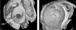
When possible, three-dimensional reconstruction of the defect and adjacent structures was performed using EchoPAC-3D, a General Electric three-dimensional reconstruction software associated with the Vivid 5 imaging system (Fig. 3a,b). Utility of this software was limited to the adult multiplane transesophageal probe, so reconstructions could be made only in those patients weighing more than 20 kg. Once the number of scanplanes from 0° to 180° was specified, the software mechanically rotated the transesophageal transducer and obtained cross-sectional echocardiographic data at each scanplane. Gating based on heart rate was performed using an electrocardiogram, and determined the time duration for gathering the entire echocardiographic data set. Because all patients were intubated for the procedure, apnea was induced during acquisition, and gating based on respiration was unnecessary. The data were then analyzed, and three-dimensional reconstruction was performed and displayed for review prior to selection of the device to be used for attempted closure.

Figure 3. This, and all subsequent three-dimensional reconstructions, are displayed from the left atrial vantage point such that the most anterior structure is the atrial septum before and after deployment. The right atrium is located directly behind the septum and the aorta is on the right side of the picture: (a) reconstruction of a circular central defect; (b) reconstruction after placement of a Helex device (Ao: aorta; ASD: atrial septal defect; SVC: superior caval vein).
Each patient then underwent catheterization to obtain physiologic and anatomic data pertaining to the septal defect. Other structural abnormalities were evaluated and addressed if possible. The stretched diameter of the defect was measured using a static sizing balloon technique (NuMed, Hopkinton, NY, USA). With the balloon expanded across the defect, complete occlusion was ascertained by color flow mapping. The diameter of the waist in the balloon at the site of indentation was then measured both fluoroscopically and echocardiographically. A ratio of 1.5 to 2 for the size of the device relative to the waist in the balloon was used to determine the appropriate device for each patient. The available sizes, measured as the diameter of the discs, were 15 mm, 20 mm, 25 mm, 30 mm, and 35 mm.
If a patient was not considered a candidate for closure because of the size of the defect, absence of the inferior or posterior rim, or other intracardiac abnormalities, an attempt was made to take the patient directly from the catheterization laboratory to the operating room for surgical intervention. In this “hybrid” approach, the patient would be subjected to general anesthesia only once during the hospitalization.
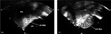
Deployment of the device followed previously described methodology.6, 7 Because the transesophageal transducer often interfered with fluoroscopic visualization of the conformation of the left atrial disc within the left atrial chamber, the transducer was usually pulled to a high esophageal position at this initial stage, and little echocardiographic data were available at this time. After conformation of the left atrial disc, the transducer was moved to a low esophageal position, and transesophageal echocardiography was utilized at several stages:
- After the conformed left atrial disc was pulled against the atrial septum (Fig. 4a).
- When the right atrial disc was conformed to complete occlusion of the defect.
- When the device was locked and separated from the delivery system.
- When the device was released from the retrieval cord.

Figure 4. (a) Conformation of the left atrial disc and retraction against the atrial septum; (b) conformation of the right atrial disc has resulted in distortion of the left atrial disc (LA: left atrium; RA: right atrium).
Prior to separation from the delivery system, the delivery system often caused tension on the device and the atrial septum. Echocardiography was used at this stage to assess resolution of this tension and of the associated distortion of the atrial septum. Inappropriate size or position of the device, significant residual defects, and impingement of the device on the mitral or tricuspid valve were indications for its removal.
If possible, three-dimensional reconstruction was used after deployment to evaluate the position of the device relative to the septum, and to identify and characterize residual defects if present (Fig. 3b). As previously described,15 intracardiac echocardiography was also attempted on several patients using the Acuson AcuNav diagnostic ultrasound catheter (Mountain View, CA, USA) interfaced with the Acuson Sequoia or Cypress imaging system. The AcuNav, a 10 French catheter, with a multiple frequency 5 to 10 MHz transducer at its tip, was placed in the right atrium to evaluate the atrial septum and adjacent structures, and data were obtained before deployment and after formation of the discs prior to separation from the delivery system.
After successful closure, transthoracic studies were obtained on the day after the procedure, and at the follow-up visits made at one month, six months, and one year. As in the transesophageal studies, particular attention was given to establishing the position of the device relative to the septum, residual leaks, flow from the superior and inferior caval veins into the right atrium and from the pulmonary veins into the left atrium, and the state of right ventricular volume loading.
Results
Between April 2000 and December 2001, 60 patients were taken to the catheterization laboratory for treatment with the Helex Septal Occluder. The median age was 5.7 years, with a range from 0.5 to 72 years, and a mean of 16.1 years. The median weight was 21 kg, with a range from 6.6 to 131 kg, and a mean of 39.2 kg. Transesophageal echocardiography was performed in all patients. Three-dimensional reconstruction was performed before and after successful deployment in 20, and intracardiac echocardiography was performed in 3. The time for three-dimensional reconstruction from the beginning of acquisition of data to the first display of a reconstruction was from 5 to 12 minutes.
Catheterization was needed twice in four of the 60 patients, in three because the defects were too large for devices available at the time. All three returned for successful closure when the 35 mm device became available. The transesophageal study for the fourth patient showed multiple fenestrations not amenable to closure with a single device. When compassionate permission was given, the defects were successfully closed using a 30 mm device together with a 15 mm device (Fig. 5).

Figure 5. Closure with two devices, showing slight overlap between the two devices on the rims of the oval fossa (RUPV: right upper pulmonary vein).
We excluded 11 of the 60 patients from attempted closure because of transesophageal echocardiographic and/or diagnostic catheterization findings in the catheterization laboratory (Table 1). The preceding transthoracic study did not diagnose partially anomalous connection of the right upper pulmonary vein into the superior caval vein in 3 patients, and had not identified defects confluent with the inferior caval vein (Fig. 6) in another 3 patients. Surgical repair was undertaken in 9 patients, four going directly from the catheterization laboratory to the operating room. A patient with unstable atrial fibrillation, and another with a defect confluent with the inferior caval vein, had not undergone surgical atrial defect closure at the time this paper was written.
Table 1. Patients excluded from closure with a Helex Septal Occluder.


Figure 6. This transthoracic subxiphoid short-axis image shows an atrial septal defect confluent with the inferior caval vein due to absence of the inferior rim of the oval fossa. Subxiphoid images were not obtained in the pre-intervention transthoracic study from an outside institution, and this image was obtained in the catheterization laboratory when confluence with the inferior caval vein was suspected subsequent to transesophageal echocardiography (ASD = atrial septal defect; IVC = inferior caval vein; LA: left atrium; SVC: superior caval vein).
We attempted closure in 49 patients, achieving success in 46 (94%), including the 4 patients who underwent 2 attempts and the 2 patients who required 2 devices to close multiple fenestrations. Among these 46 patients, the median size of the defect was 15 mm, with a range from 9 to 22 mm, and a mean of 16 mm. Successful closure was defined, using transesophageal echocardiography, fluoroscopy, and angiography, as an appropriate position of the device with, at most, a small residual defect along the margin of the device or elsewhere in the oval fossa. Most of the defects were eccentric in morphology (Fig. 7). Five patients had abnormal attachments of the primary septum within the left atrium, and 4 had abnormal attachments of the Eustachian valve to the primary septum.

Figure 7. Data on morphology for 46 successful closures: 9 central defects; 26 eccentric defects (22 with deficient or absent antero-superior rim behind the ascending aorta, 2 with deficient posterior rim, and 2 with deficient inferior rim); and 11 other defects with multiple fenestrations and/or aneurysm of the primary septum (5 with multiple fenestrations only, 2 with aneurysm only, and 4 with both). One patient with a deficient inferior rim also had multiple fenestrations and aneurysm of the primary septum.
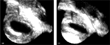
Attempts at closure were unsuccessful in 3 patients. In one patient, the defect was elliptical in shape and dynamic in size, as seen by three-dimensional echocardiography (Fig. 8). The defect acted like a “buttonhole”, allowing the formed discs to easily slip leftward or rightward across the defect. Each of the other 2 had a large defect with a relatively small oval fossa that could not accommodate the size of device necessary for successful closure. The abnormal position of the occluder on the septum, and the associated residual defects, resulted in elective removal of the device for these 2 patients.

Figure 8. Three-dimensional reconstruction of an elliptical defect during the cardiac cycle reveals dynamic changes in the size of the defect at different times in the cardiac cycle: (a) diastole; (b) systole.
For patients with successful closure, transesophageal evaluation immediately after implantation showed no residual defect in 21, a trivial residual defect less than 2 mm in diameter in 17, and a small residual defect from 2 to 3 mm in diameter in 8. Of the 21 patients without a residual defect on the transesophageal study, 7 had a trivial or small residual defect on the first subsequent transthoracic study one day after the procedure. Of the 17 patients with a trivial residual defect on the transesophageal study, 4 had a small residual defect on the first subsequent transthoracic study. At the time of the most recent follow-up transthoracic study, conducted at a median of 7 months after the procedure, with a range from 1 day to 18 months, 22 patients (48%) had no significant residual defect, 14 (30%) had a trivial residual defect, and 10 (22%) had a small residual defect. No patient had a residual defect greater than 3 mm in width. Furthermore, no patient had qualitative evidence for residual right ventricular volume overload.
Discussion
The most significant issue associated with echocardiographic evaluation of insertion of the Helex Septal Occluder involves the expanded polytetrafluoro-ethylene membrane bonded to the frame. The membrane contains microscopic air spaces that reflect ultrasound waves. As a result of acoustic interference from the membrane on the left atrial disc, it is often difficult to evaluate the shape and orientation of the right atrial disc, and to exclude residual defects along the margin of the device by transesophageal echocardiography (Fig. 9a). This may explain some of the discrepancies between results from the transesophageal study performed immediately following insertion and from the first subsequent transthoracic study. On several occasions, a transthoracic evaluation prior to locking the device or separation from the delivery system has been required to assure that the device is not impinging on adjacent structures. This points to the possible value of intracardiac echocardiography during deployment. Because the transducer on the tip of the catheter is situated in the right atrium, the conformed left atrial disc does not interfere with observation of the right atrial disc as it is conformed and pulled against the septum (Fig. 9b). In addition, residual left-to-right shunts along the margins of the device are more easily assessed. The air spaces within the membrane do fill with fluid in the days following implantation, so that acoustic interference is not an issue in follow-up transthoracic studies. Modification of the membrane to reduce acoustic interference is another possible solution, and is currently being tested (personal communication).

Figure 9. (a) Transesophageal echocardiogram showing acoustic interference created by microscopic air spaces in the left atrial disc precluding evaluation of the right atrial disc; (b) intracardiac echocardiography showing the right atrial disc clearly; the different degrees of acoustic interference created by the devices in these two images result from differences in image amplification or gain between the two studies (LA: left atrium, RA: right atrium).
Because of the spiral nature of the nitinol frame, acute changes in the orientation or shape of the device against the septum are frequently documented by transesophageal echocardiography. The process of conformation of the right atrial disc can distort the already formed left atrial disc, breaking its seal against the septum, until complete delivery is achieved and the apposed discs straddle the septum (Fig. 4b). Deployment of the locking mechanism involves tension on the central aspect of both discs, and retraction of the locking wire can also result in some distortion of either or both discs. Lastly, because the device does not exert direct pressure on the rims of the oval fossa to maintain its position, the orientation within the fossa can be physically manipulated along small distances by the delivery catheter following release of the device, but prior to removal of the retrieval cord. In patients with tiny residual defects along the antero-superior margin of the device, the delivery catheter has been successfully used to slide the entire device slightly anteriorly and/or superiorly until the residual defect is closed. The dynamic nature of the position of the device against the atrial septum may also explain some of the discrepancies between the immediate transesophageal findings after closure and the first subsequent transthoracic findings. The immediate transesophageal study in some patients has also revealed protrusion of part of the device into either atrium. Subsequent transthoracic studies have shown that these protrusions usually disappear as the device conforms to the shape of the oval fossa.
Three-dimensional reconstruction has been useful in delineating the static and dynamic shape of the oval fossa, as well as its spatial relationship to adjacent structures. It has been especially helpful in patients with a deficient antero-superior rim, wherein the exact orientation of the deficient fossa against the ascending aorta is more easily understood by viewing the frozen heart from multiple vantage points (Fig. 10a). Three-dimensional reconstruction for patients with multiple fenestrations has also provided useful information regarding the spatial relationship of all the defects. In patients with an aneurysmal primary septum, reconstruction has proven less valuable, probably because this requires dynamic assessment of the septum, and visualization from multiple vantage points is more difficult with dynamic images of the heart. Once a device has been deployed, three-dimensional reconstruction has shown how the left atrial disc conforms to planar and to non-planar atrial septal shapes (Fig. 10b). The mechanism for a residual leak can be assessed by differentiating between an inappropriate size of the device as opposed to incomplete seating of the disc against the septum, with some protrusion into the atrial chamber. The former requires placement of a different device, while the latter is usually acceptable, since the residual leak typically disappears as the disc conforms to the rims of the oval fossa over time.

Figure 10. (a) Three-dimensional reconstruction of a “teardrop”-shaped defect with a deficient antero-superior rim behind the ascending aorta; (b) reconstruction after placement of the device, with a residual leak along the antero-superior margin of the device (arrows) (Ao: ascending aorta).
Four types of defects within the oval fossa have been treated with the occluder:
- Central defects with an appreciable rim along the entire circumference of the defect.
- Defects with deficiency or absence of a segment of the rim.
- Defects associated with an aneurysm of the primary septum.
- Defects associated with multiple fenestrations.
Closure in patients with complete absence of a segment of the rim has been successful only in those where the deficient segment is positioned antero-superiorly behind the ascending aorta. In these cases, the antero-superior aspect of the left atrial and right atrial discs can straddle the ascending aorta, while the remaining portions of the discs straddle the available rims (Fig. 11). Complete absence of the inferior rim produces a defect that is confluent with the inferior caval vein. We did not find complete absence of the posterior rim. At all events, the findings can obviate the need for catheterization and attempted closure using a device. In patients with a large redundant primary septum, a small device may not be fully anchored to the static part of the atrial septum and may remain mobile and possibly unstable within the atrial chamber. Thus, in patients with an aneurysm of the primary septum, and/or multiple fenestrations, successful closure requires a device large enough to incorporate the mouth of the aneurysm and/or most or all of the fenestrations. We needed 2 devices to close all or most of the aggregate atrial defect in two patients. In one case, there is slight overlap of the 2 devices without disrupting the ability of either device to close its respective fenestration or group of fenestrations (Fig. 5). In patients with abnormal attachments of the primary septum or the Eustachian valve, the attachments may interfere with conformation of either disc. Careful conformation and situation of the discs against the atrial septum, without incorporating the attachments into the area of the discs, have resulted in at most only a small residual defect persisting in these patients.

Figure 11. After deployment of the device in a defect with an absent antero-superior rim, the antero-superior aspect of the right atrial and left atrial discs straddle the ascending aorta in order to close the defect (Ao: aorta).
Limited transthoracic windows in bigger children and adults can preclude complete assessment of the atrial septum and all the pulmonary veins. These findings were partially responsible for the 11 exclusions from attempts at closure using the device, including the 4 patients in whom the size of the defect was incorrectly assessed during the transthoracic study. Evaluation of the right upper pulmonary vein and inferior septal rim is usually only available in subxiphoid short-axis or right parasternal views (Fig. 6). If these views are unavailable, partially anomalous pulmonary venous connection, and defects confluent with the inferior caval vein, may not be diagnosed until a transesophageal study is performed. In addition, among all the patients who have undergone closure using the Helex device, just over half have a residual defect, albeit trivial or small. This number may not be entirely accurate because of the limited transthoracic windows. None of these patients, however, have residual right ventricular volume overload at a maximum follow-up of 18 months.
The unsuccessful attempts at closure occurred during the early stages of our study. The ability of interventionalists and echocardiographers to assess whether an atrial defect and atrial septum can accommodate a specific device size has improved with time and experience. Careful assessment of the width of the oval fossa, and strict adherence to a maximum ratio of the device to the waist in the balloon of 1.5 to 2, have resulted in no failures later in the study period.
The most commonly used devices for closure of defects within the oval fossa include the double-umbrella devices (CardioSEAL and Starflex, NMT, Boston, MA, USA), and the Amplatzer Septal Occluder (AGA, Golden Valley, MN, USA). Several studies have shown the utility of cross-sectional and three-dimensional echocardiography during their deployment.9–10, 14, 16–18 The double-umbrella devices consist of 2 sets of 4 arms supporting a Dacron patch located on both sides of the atrial septum. Although these devices are easily seen by transesophageal echocardiography, without the problems of acoustic interference, there is often ambiguity in determining the exact location of all 8 arms, with difficulty in excluding improper protrusion of an arm into the wrong atrium.16 The spiral frame of the Helex device alleviates the issue of improper placement of arms, though deployment can favor conformation of either disc by incomplete release and conformation of the opposing disc into its corresponding atrium. In other words, more than half of the spiral frame can be formed in one atrium. The Helex device, however, is difficult to visualize by transesophageal echocardiography secondary to acoustic interference. It does have the advantage of the potential for multiple deployments, and an abnormally positioned device can be pulled back into the delivery system and redeployed. Although bulkier in profile than the Helex device, the Amplatzer occluder is easily seen by transesophageal echocardiography without the problems of acoustic interference.10, 18 Because the waist of the device expands into the defect, there is less ambiguity in the location of the margins of the disc. Like the Helex device, the Amplatzer occluder also has the potential for multiple deployments.
In conclusion, the Helex Septal Occluder is a new and effective device with which to close atrial septal defects in the catheterization laboratory. Echocardiography is invaluable before, during, and after its deployment. A complete and careful transthoracic study is necessary to determine eligibility for the procedure. The transesophageal study prior to attempted closure, along with catheterization, are able to provide the data properly to choose the size of the device. As with other devices, transesophageal echocardiography also provides guidance during deployment. Transesophageal echocardiography during and after deployment, nonetheless, can be limited by acoustic interference from microscopic air spaces in the device. Intracardiac echocardiography, and/or modification of the membrane, may help overcome this limitation. Three-dimensional reconstruction has also helped to increase understanding of the morphology of the oval fossa prior to deployment, and has confirmed an appropriate position for the device after deployment.
Acknowledgments
Technical support has been provided by Brenda Ferrer, Linet Echavarria, Christina Pelczar, Ruby Whalen, and Nestor Ventura.
References
King TD, Thompson SL, Steiner C, Mills NL. Secundum atrial septal defect: non-operative closure during cardiac catheterization. JAMA 1976; 235: 2506–2509.Google Scholar
Rome JJ, Keane JF, Perry SB, Spevak PJ, Lock JE. Double-umbrella closure of atrial septal defects: initial clinical applications. Circulation 1990; 82: 751–758.Google Scholar
Lloyd TR, Rao PS, Beekman RH 3rd, Mendelsohn AM, Sideris EB. Atrial septal defect occlusion with a buttoned device (a multi-institutional U.S. trial). Am J Cardiol 1994; 73: 286–291.Google Scholar
Hausdorf G, Schneider M, Fink C, et al. Transcatheter closure of atrial septal defects within the oval fossa: medium-term results in children using the “ASDOS” technique. Cardiol Young 1998; 8: 462–471.Google Scholar
Masura J, Gavora P, Formanek A, Hijazi ZM. Transcatheter closure of secundum atrial septal defects using the new self-centering Amplatzer septal occluder: initial human experience. Cathet Cardiovasc Diagn 1997; 42: 388–393.Google Scholar
Zahn EM, Wilson N, Cutright W, Latson LA. Development and testing of the Helex septal occluder, a new expanded polytetra-fluoroethylene atrial septal defect occlusion system. Circulation 2001; 104: 711–716.Google Scholar
Latson LA, Zahn EM, Wilson N. Helex septal occluder for closure of atrial septal defects. Curr Interv Cardiol Rep 2000; 2: 268–273.Google Scholar
Seward JB, Khandheira BK, Oh JK, et al. Transesophageal echocardiography: technique, anatomic correlations, implementations, and clinical applications. Mayo Clin Proc 1988; 63: 649–680.Google Scholar
Rosenfeld HM, van der Velde ME, Sanders SP, et al. Echocardiographic predictors of candidacy for successful transcatheter atrial septal defect closure. Cath Cardiovasc Diag 1995; 34: 29–34.Google Scholar
Cooke JC, Gelman JS, Harper RV. Echocardiologists' role in the deployment of the Amplatzer atrial septal occluder device in adults. J Am Soc Echocardiogr 2001; 14: 588–594.Google Scholar
Sweeney LJ, Rosenquist GC. The normal anatomy of the atrial septum in the human heart. Am Heart J 1979; 98: 194–199.Google Scholar
Ferreira SM, Ho SY, Anderson RH. Morphological study of defects of the atrial septum within the oval fossa: implications for transcatheter closure of left-to-right shunt. Br Heart J 1992; 67: 316–320.Google Scholar
Podnar T, Martanovic P, Gavora P, Masura J. Morphological variations of secundum-type atrial septal defects: feasibility for percutaneous closure using Amplatzer septal occluders. Cathet Cardiovasc Intervent 2001; 53: 386–391.Google Scholar
Chan KC, Godman MJ. Morphological variations of fossa ovalis atrial septal defects (secundum): feasibility for transcutaneous closure with the clam-shell device. Br Heart J 1993; 69: 52–55.Google Scholar
Hijazi Z, Wang Z, Cao Q, Koenig P, Waight D, Lang R. Transcatheter closure of atrial septal defects and patent foramen ovale under intracardiac echocardiographic guidance: feasibility and comparison with transesophageal echocardiography. Cathet Cardiovasc Intervent 2001; 52: 194–199.Google Scholar
Maeno YV, Benson LN, Boutin C. Impact of dynamic 3D transoesophageal echocardiography in the assessment of atrial septal defects and occlusion by the double-umbrella device (CardioSEAL). Cardiol Young 1998; 8: 368–378.Google Scholar
Boutin C, Musewe NN, Smallhorn JF, et al. Echocardiographic follow-up of atrial septal defect after catheter closure by double-umbrella device. Circulation 1993; 88: 621–627.Google Scholar
Cao QL, Radtke W, Berger F, Zhu W, Hijazi ZM. Transcatheter closure of multiple atrial septal defects: initial results and value of two- and three-dimensional transesophageal echocardiography. Eur Heart J 2000; 21: 941–947.Google Scholar

Measurement of the width of the antero-superior rim of the oval fossa behind the ascending aorta (Ao: ascending aorta; LA: left atrium; RA: right atrium).
This, and all subsequent three-dimensional reconstructions, are displayed from the left atrial vantage point such that the most anterior structure is the atrial septum before and after deployment. The right atrium is located directly behind the septum and the aorta is on the right side of the picture: (a) reconstruction of a circular central defect; (b) reconstruction after placement of a Helex device (Ao: aorta; ASD: atrial septal defect; SVC: superior caval vein).
(a) Conformation of the left atrial disc and retraction against the atrial septum; (b) conformation of the right atrial disc has resulted in distortion of the left atrial disc (LA: left atrium; RA: right atrium).

Closure with two devices, showing slight overlap between the two devices on the rims of the oval fossa (RUPV: right upper pulmonary vein).
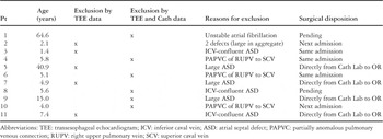
Table 1.

This transthoracic subxiphoid short-axis image shows an atrial septal defect confluent with the inferior caval vein due to absence of the inferior rim of the oval fossa. Subxiphoid images were not obtained in the pre-intervention transthoracic study from an outside institution, and this image was obtained in the catheterization laboratory when confluence with the inferior caval vein was suspected subsequent to transesophageal echocardiography (ASD = atrial septal defect; IVC = inferior caval vein; LA: left atrium; SVC: superior caval vein).

Data on morphology for 46 successful closures: 9 central defects; 26 eccentric defects (22 with deficient or absent antero-superior rim behind the ascending aorta, 2 with deficient posterior rim, and 2 with deficient inferior rim); and 11 other defects with multiple fenestrations and/or aneurysm of the primary septum (5 with multiple fenestrations only, 2 with aneurysm only, and 4 with both). One patient with a deficient inferior rim also had multiple fenestrations and aneurysm of the primary septum.
Three-dimensional reconstruction of an elliptical defect during the cardiac cycle reveals dynamic changes in the size of the defect at different times in the cardiac cycle: (a) diastole; (b) systole.
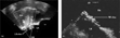
(a) Transesophageal echocardiogram showing acoustic interference created by microscopic air spaces in the left atrial disc precluding evaluation of the right atrial disc; (b) intracardiac echocardiography showing the right atrial disc clearly; the different degrees of acoustic interference created by the devices in these two images result from differences in image amplification or gain between the two studies (LA: left atrium, RA: right atrium).
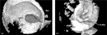
(a) Three-dimensional reconstruction of a “teardrop”-shaped defect with a deficient antero-superior rim behind the ascending aorta; (b) reconstruction after placement of the device, with a residual leak along the antero-superior margin of the device (arrows) (Ao: ascending aorta).

After deployment of the device in a defect with an absent antero-superior rim, the antero-superior aspect of the right atrial and left atrial discs straddle the ascending aorta in order to close the defect (Ao: aorta).
 You have
Access
- 8
- Cited by