INTRODUCTION
The earliest paper we found on toxoplasmosis in Portugal is that of Campos (Reference Campos1951), which was written in Portuguese. Many other papers are also in Portuguese and some important data are buried in unpublished dissertations and theses, not available to a wider audience. Here we review the epidemiology, prevalence and clinical manifestations of Toxoplasma gondii infection in humans and animals from Portugal. Topics for future research are also suggested in order to encourage international collaboration.
METHODS FOR PRESENT REVIEW
A review of the literature available until May 2014 was carried out by searching the PubMed database with reference combination of the following keywords: ‘(Toxoplasma gondii OR toxoplasmosis) AND Portugal’. Our initial search indicated 52 references, out of which 29 references on T. gondii infection or toxoplasmosis in humans and/or animals from Portugal were selected. Databases of universities, research institutes and scientific societies and the reference lists of all the retrieved articles were also searched for published reports deemed as relevant to this review. After this, three additional papers were retrieved from non-indexed but peer-reviewed Portuguese journals. A set of 32 papers is reviewed in the main document. Four abstracts from the proceedings of national conferences, another abstract from the proceedings of an international conference, four master's dissertations and two doctoral theses also accounted for the scientific literature used to prepare the present review. That information is provided as a supplementary file (Supplement 1 – in Online version only).
Detailed historical, serological, parasitological, clinical and additional information on T. gondii infections in humans and other animals are summarized in tables (Tables 1–4) throughout the review. Fortunately, most studies used the commercially available modified agglutination test (MAT) to detect T. gondii antibodies in human and animal sera. This is the same test as described by Desmonts and Remington (Reference Desmonts and Remington1980) and Dubey and Desmonts (Reference Dubey and Desmonts1987). It has been widely used to determine T. gondii seroprevalence in humans and animals (Dubey, Reference Dubey2010). This test assays only immunoglobulin (Ig) G antibodies, because the mercaptoethanol needed in the test destroys IgM and IgM-like antibodies. Cut-off values for MAT are listed wherever the authors provided the information. Data by other tests used is also provided.
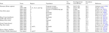
Seroprevalence of T. gondii infection in humans and domestic animals from Portugal

Ag, Algarve; At, Alentejo; C, Center; CMIA, chemiluminescence microparticle immunoassay; IFAT, indirect fluorescent antibody test; L, Lisbon; MAT, modified agglutination test; N, North; ND, not defined.
Antibody titres of cats from Portugal tested by the MAT

MAT, modified agglutination test.
a Catch and neuter programme in Lisbon.
b From individual owners.
c Veterinary clinics.
d Trapped by municipality.
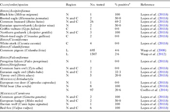
Seroprevalence of T. gondii infection in wild animals from Portugal sampled between 2008 and 2012

C: Center; N: North.
a Modified agglutination test (cut-off titre: 20).
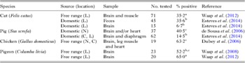
Detection of T. gondii in tissues of animals from Portugal

C: Center; L: Lisbon; N: North.
a In vitro isolation.
b PCR.
c Isolation by bioassay.
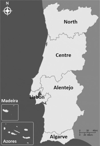
Portugal, officially the Portuguese Republic, is located in Southwestern Europe. The country is located in the western part of the Iberian Peninsula, where it is bordered to the north and east by Spain and to the south and west by the Atlantic Ocean. The Portuguese territory has a total area of 92 090 km2 and comprises a mainland and two insular autonomous regions: the archipelagos of Azores and Madeira, in the Northern Atlantic. Portugal has a population of around 10·8 million people, and joined the European Union in 1986. In the present work the country was divided according to the European Union Nomenclature of Units for Territorial Statistics (NUTS) II, namely North, Centre, Alentejo, Lisbon, Algarve, Azores and Madeira regions (Fig. 1).
Regions of Portugal according to the NUTS II. (adapted from Wikipedia).

TOXOPLASMOSIS IN HUMANS
Prevalence of T. gondii infection
Published information is limited to three surveys; two of them are more than 20 years old (Table 1) and reported high seroprevalence. Antunes et al. (Reference Antunes, Bacelar, Janz, Saraiva and Araújo1981) found a 0·8% yearly incidence of infection in pregnant women from Lisbon and suggested an incidence of congenital infection of around 0·3% per pregnancy.
In the study by Ângelo (Reference Ângelo1983) (Table 1), seroprevalence varied from 19·8% in people aged 8 months to 5 years old to much higher levels in the 6–15 years, 16–30 years, 31–45 years and older population (39·1, 55·1, 57·1 and 57·8%, respectively). The reasons for these increasing results are most likely associated with a cumulative exposure with age. Analysing the results obtained by geographical districts, it was found that the mean values of seroprevalence of human infection were higher in the North than in the South of the country (Alentejo), ranging from 56·4% in Viana do Castelo (North) to 33% in Évora (Alentejo).
In another report by Ângelo (Reference Ângelo2003), seroprevalence in the population varied from 35% in the South to 70% in the North; for women of childbearing age, these values were between 30 and 60%. There is no national data concerning the prevalence of infection in pregnant women or in newborns. Nevertheless, Ângelo (Reference Ângelo2003) suggested the figure of 11 cases of congenital infection per 10 000 live births, with an estimated number of 124 children congenitally infected each year.
We recently studied seroprevalence and associated risk factors of T. gondii in 401 women of childbearing age from the North of Portugal (Lopes et al. Reference Lopes, Dubey, Moutinho, Gargaté, Vilares, Rodrigues and Cardoso2012a ). Out of these, 343 women attending the gynaecology and obstetrics services of the public Hospital Centre of Trás-os-Montes e Alto Douro, in Vila Real, and 58 women attending private clinics participated in the study. Antibodies (IgG) to T. gondii were detected in 98 (24·4%) out of 401; two (2·0%) simultaneously had IgM. Almost 50% of the women studied by Lopes et al. (Reference Lopes, Dubey, Moutinho, Gargaté, Vilares, Rodrigues and Cardoso2012a ) did not have any knowledge regarding toxoplasmosis. Risk factors for T. gondii infection in decreasing order were: soil-related activities without gloves (odds ratio [OR] = 8·4), consumption of unwashed raw vegetables or fruit (OR = 7·6) and eating smoked or cured processed pork products (OR = 2·5). Consumption of raw or undercooked meat was not identified as a risk factor. Exposure to oocysts rather than tissue cysts in meat was the most important risk factor associated with T. gondii infection in women in this study.
There is no legislation in Portugal that requires screening for toxoplasmic infection in pregnant women; however, in 2000, guidelines about the prevention of toxoplasmosis were made available by the General Directorate of Health (Ângelo, Reference Ângelo2003).
Clinical toxoplasmosis
Congenital
To our knowledge no confirmed clinical case has been published on congenital toxoplasmosis in Portugal. Campos (Reference Campos1951) reported severe encephalitis associated with a progressive blindness in a 3·5-year-old child. The child died 3 years later and Toxoplasma-like tachyzoites were found in the brain at autopsy. It is not certain if the child was congenitally or postnatally infected.
Human immunodeficiency virus (HIV)
An HIV-seropositive patient with visceral leishmaniosis was further diagnosed with cerebral toxoplasmosis by the presence of IgG antibodies to T. gondii (dye and agglutination tests) and clinical and computer tomography (CT) scan features (Antunes et al. Reference Antunes, Carvalho, Tavares, Botas, Fortes, del Rio, Dutschmann, Costa, Abranches, Pereira, Paiva, Araújo and Baptista1987). The patient's condition improved after pyrimethamine and sulphadiazine therapy for toxoplasmosis and antimony for leishmaniosis. In the frame of a European multicentre clinical research programme, Antunes (Reference Antunes1991) reported 16 additional cases of toxoplasmic encephalitis in HIV patients.
A presumptive diagnosis of toxoplasmosis was made by Miranda et al. (Reference Miranda, Gomes, Guimarães, Nogueira, Abreu and Figueiredo1992) in 20 out of 95 HIV patients seen at the S. João Hospital, Oporto, between 1985 and 1991, based on symptoms (headache in 11, consciousness alterations in eight, vomiting in seven, fever in six, asthenia in five, and seizures in three), antibodies to T. gondii, CT, and favourable response to anti-T. gondii therapy (pyrimethamine and sulphadiazine in 16 patients and pyrimethamine with clindamycin in the remaining four). Diagnosis was confirmed in one patient by finding tachyzoites in brain biopsy. Two patients died after 3 to 4 weeks of therapy, but post-mortem diagnosis was not made. The serological study of T. gondii infection included detection of specific antibodies by enzyme-linked immunosorbent assay (ELISA).
In a retrospective study, Costa-Gomes et al. (Reference Costa-Gomes, Nunes, Pinto, Gouveia and Pais2012) provisionally diagnosed toxoplasmosis based on CT or magnetic resonance studies or both in 11 out of 196 HIV patients. Nothing was said of serologic tests use, if any. Similarly, in a paper evaluating the surgical techniques of obtaining biopsy of brainstem for diagnosis of various ailments, Gonçalves-Ferreira et al. (Reference Gonçalves-Ferreira, Herculano-Carvalho and Pimentel2003) mentioned two male HIV patients with focal infiltration in brainstem biopsied at a hospital in Lisbon: a 37-year old and a 45-year old (who also had concurrent Cytomegalovirus infection). No other details were given.
Although CT and serological examination are useful, definitive diagnosis of toxoplasmosis can only be made post-mortem or by biopsy examination. Other conditions, including lymphomas, can mimic toxoplasmosis, and the determination of Ig isotype and the magnitude of titre to T. gondii are not helpful in the differential diagnosis. In most HIV-infected patients clinical toxoplasmosis is a reactivation of chronic infection, and most of these patients have antibodies to T. gondii. Favourable response to anti-toxoplasmic therapy is helpful in ante-mortem diagnosis of toxoplasmosis.
Ocular
The only report on ocular toxoplasmosis we found is that of Palmares et al. (Reference Palmares, Coutinho and Castro-Correia1990). They reviewed case histories of 450 patients seen in a referral hospital in Northern Portugal. Toxoplasmosis-associated uveitis was diagnosed in 41 (9·1%) out of 450 patients seen from 1985 to 1989. The diagnosis criteria were the morphologic picture and seropositivity in any of the serological tests. The report is brief and there is no information regarding the serological tests used and the titres. Our concept of toxoplasmosis has changed during the last 25 years, when this information was presented at a symposium. For example, postnatally acquired toxoplasmosis is now increasingly recognized as cause of uveitis; previously most of it was considered due to congenital infection.
Organ transplant patients
Abecassis (Reference Abecassis1991) reported two cases of encephalitis by T. gondii among 60 patients who received bone marrow transplantations, between May 1987 and May 1991. The first case was a 24-year-old female patient bone marrow transplanted for severe aplastic anaemia, which had been diagnosed 4 years before. Six months after transplantation, fever with poor general condition, severe muscle atrophy and a convulsive episode arised. An axial CT scan revealed multiple ring lesions. The severe thrombocytopenia presented by the patient did not allow brain biopsy and the patient was treated with pyrimethamine and sulphadiazine without favourable evolution of lesions on CT. The patient died 3 weeks later; autopsy was not performed. To us, the diagnosis is at best suspicious. In the second case the patient was a 41-year-old woman, transplanted for chronic myeloid leukaemia in chronic phase, diagnosed 8 months before. Prior to bone marrow transplantation an evaluation had been done with both the recipient and the donor being seronegative for T. gondii infection by ELISA. However, there was a subsequent seroconversion of the recipient with the detection of IgG and IgM antibodies, reflecting a recent infection. Therapy with spiramycin was instituted. Approximately 3 weeks after bone marrow transplantation the patient developed unusual cardiac complications and fever. Cranial CT revealed extensive left ventricular intracerebral ring shape lesions. Given the patient's condition, a severe thrombocytopenia and the previous evidence of recent infection with T. gondii, therapy with sulphadiazine and pyrimethamine was initiated. A favourable development was observed with complete recovery without neurological sequels.
Da Cunha et al. (Reference da Cunha, Ferreira, Ramos, Martins, de Freitas, Borges, Côrte-Real, Mota, Meliço-Silvestre and Furtado1993a , Reference da Cunha, Ferreira, Ramos, Martins, de Freitas, Borges, Côrte-Real, Mota, Meliço-Silvestre and Linhares-Furtado b ) reported evidence of cerebral toxoplasmosis in a 42-year-old male patient after a successful renal transplantation in Portugal. The patient had headache and loss of consciousness; CT scan revealed focal lesion in brain. The patient had persistent serum antibodies to T. gondii and anti-Toxoplasma therapy (clindamycin and pyrimethamine) was initiated. When clinical and imaging improvement was observed, secondary prophylaxis was initiated with dapsone and pyrimethamine, with favourable evolution (da Cunha et al. Reference da Cunha, Ferreira, Ramos, Martins, de Freitas, Borges, Côrte-Real, Mota, Meliço-Silvestre and Furtado1993a , Reference da Cunha, Ferreira, Ramos, Martins, de Freitas, Borges, Côrte-Real, Mota, Meliço-Silvestre and Linhares-Furtado b ).
TOXOPLASMOSIS IN ANIMALS
Clinical
Virtually nothing is known of clinical toxoplasmosis in animals in Portugal.
Serological and parasitological prevalence
Data are summarized in Tables 1–4.
Cat
Seroprevalence in cats deserves special attention because of the epidemiological importance of cats. Of the four serological surveys in cats, three were from Lisbon (Tables 1 and 2). The fourth survey (Lopes et al. Reference Lopes, Cardoso and Rodrigues2008) was in domestic cats from Trás-os-Montes e Alto Douro (Northeastern Portugal); 161 cats were from Vila Real, and the remaining 46 cats were from other contiguous municipalities (Table 1). However, data provided were not further distinguished with respect to localities. All four studies with cats used the same MAT test, the cut-off titres (20–80) were different, and therefore data are not strictly comparable (Tables 1 and 2).
Serological prevalence varied from 20 to 44%; the highest being from stray cats from Lisbon. The stray cats were captured from different areas of Lisbon by the municipal services. It is noteworthy that a very high percentage of cats from Portugal had high titres (Table 2). In the study by Waap et al. (Reference Waap, Cardoso, Leitão, Nunes, Vilares, Gargaté, Meireles, Cortes and Ângelo2012), 68 (36·3%) out of 187 seropositive cats had MAT titres of 18 000 or higher reaching as much as 162 000. One of us has never encountered such high MAT titres from cats from other parts of the world (J. P. Dubey, personal observations). This result may be due to the T. gondii strains or very high environmental contamination leading to repeated infections in cats.
Risk factors such as age, gender, diet and lifestyle were assessed in these four surveys. Gender did not affect the seropositivity. Age was one of the main risk factors; seroprevalence increased with age. Age-related data were: 14·6% seropositivity in 2–11 months old vs 51·7% in 3- to 6-year-old cats (Lopes et al. Reference Lopes, Cardoso and Rodrigues2008); 20·6% in cats younger than 1 year vs 56·9% in cats older than 5 years (Waap et al. Reference Waap, Cardoso, Leitão, Nunes, Vilares, Gargaté, Meireles, Cortes and Ângelo2012); 7·7% in cats younger than 2 years vs 36·5% in cats older than 3 years (Esteves et al. Reference Esteves, Aguiar, Rosado, Costa, de Sousa, Antunes and Matos2014). These data indicate post-natal exposure of cats to T. gondii. There is no information on congenital transmission of T. gondii in cats in Portugal. Cats that were fed on diets including raw or undercooked viscera and/or meat also had a higher percentage (53·5%) of infection in comparison with animals receiving only commercial canned or dried food (22·9%; Lopes et al. Reference Lopes, Cardoso and Rodrigues2008). Feline contact was not shown to be a statistically significant risk factor for seropositivity in logistic regression analysis (Lopes et al. Reference Lopes, Cardoso and Rodrigues2008).
As of yet, viable T. gondii oocysts have not been demonstrated in cat feces in Portugal. Oocysts were not found by microscopic examination of feces of 231 cats from Lisbon (Duarte et al. Reference Duarte, Castro, Pereira da Fonseca, Almeida, Madeira de Carvalho, Meireles, Fazendeiro, Tavares and Vaz2010). Ferreira et al. (Reference Ferreira, Pereira-Baltasar, Parreira, Padre, Vilhena, Távora Tavira, Atouguia and Centeno-Lima2011) detected T. gondii–like oocysts in one out of 20 cats from private homes from region of Alentejo, but without bioassay a definitive diagnosis could not be made. Esteves et al. (Reference Esteves, Aguiar, Rosado, Costa, de Sousa, Antunes and Matos2014) reported the presence of T. gondii DNA in feces of 16 (35·5%) out of 45 cat stools from Lisbon. This is a very high rate of DNA positivity, but bioassay is necessary for a definitive diagnosis of T. gondii in feces. The demonstration of DNA should not be equated with the presence of viable T. gondii oocysts. T. gondii DNA was also detected in six (13·3%) out of 15 brains of cats.
Viable T. gondii was isolated from a relatively high number of seropositive cats (Table 4). Waap et al. (Reference Waap, Cardoso, Leitão, Nunes, Vilares, Gargaté, Meireles, Cortes and Ângelo2012) homogenized 1 g of brain from each of 56 seropositive cats, and isolated T. gondii from 15 of them in cell cultures seeded with this undigested brain homogenate. From another set of 15 cats they compared isolation of T. gondii in cell culture from brains (undigested 1 g) and pepsin digest of heart, and limb muscle (amount used not specified); T. gondii was isolated from muscles of 10 and brains of six cats.
This high rate of isolation (i.e. 15 [26·8%] out of 56) using 1 g of brain indicates high density of parasites in the brains of apparently healthy cats in Lisbon. These results are of interest because the success of isolation of T. gondii in cell cultures seeded by tissues of asymptomatic animals is generally low (Dubey, Reference Dubey2010).
Food animals as sources of infection
Poultry, pork, beef and mutton are the most important source of meat consumed by humans in Portugal. Little is known of the relative risk of T. gondii infection with respect to meat sold in grocery stores. Although T. gondii antibodies were found in 7·5% (Table 1) and 29·2% of cattle (Supplement 1, Table S1 – in Online version only), viable T. gondii has rarely been isolated from beef in other countries (Dubey, Reference Dubey2010). Thus, the role of beef in the epidemiology of T. gondii is uncertain.
In addition to data in Table 4, viable T. gondii was isolated from a bovine foetus (Canada et al. Reference Canada, Meireles, Rocha, da Costa, Erickson and Dubey2002). During the course of an investigation on Neospora caninum (a protozoan similar to T. gondii) bovine fetal tissues were bioassayed in mice. For this, homogenates of fetal brains were inoculated intraperitoneally into mice and protozoa harvested from the peritoneal cavity of infected mice were seeded on to cell cultures. One of these isolates proved to be T. gondii. The infected fetus was 5-month gestational age from a Holstein cow from the Azores (Canada et al. Reference Canada, Meireles, Rocha, da Costa, Erickson and Dubey2002). Confirmation of T. gondii as the causative agent of the abortion was not performed since the brains were not examined histologically.
Among the three surveys in pigs, seroprevalence varied from 7·1 to 15·6% (Table 1). Age and management were the two main factors concerning prevalence. Seroprevalence in feeder pigs (market age pigs) is important with respect to public health, because these pigs for human consumption are generally 6–10 months old at the time of slaughter. In this age group 5% out of 180 pigs from North of Portugal had antibodies to T. gondii; these pigs were from 14 farms (Lopes et al. Reference Lopes, Dubey, Neto, Rodrigues, Martins, Rodrigues and Cardoso2013a ). Similar results (i.e. 18 [6·8%] out of 264) were obtained from pigs from a slaughterhouse in Lisbon (Esteves et al. Reference Esteves, Aguiar, Rosado, Costa, de Sousa, Antunes and Matos2014). Seroprevalence were higher in older pigs, suggesting post-natal infection. As expected, pigs raised under extensive management had higher rate of T. gondii infection (Lopes et al. Reference Lopes, Dubey, Neto, Rodrigues, Martins, Rodrigues and Cardoso2013a ; Esteves et al. Reference Esteves, Aguiar, Rosado, Costa, de Sousa, Antunes and Matos2014).
No information was available concerning transplacental transmission of T. gondii, because only a few neonatal pigs were tested in both of these surveys. Transplacental transmission of T. gondii in pigs is uncommon (Dubey, Reference Dubey2010). De Sousa et al. (Reference de Sousa, Ajzenberg, Canada, Freire, da Costa, Dardé, Thulliez and Dubey2006) found seropositivity in 63 (15·6%) out of 333 pigs from the municipality of Vinhais, in Northeastern Portugal; these pigs were 1–4 years old and mainly used for traditional sausage production. Brains/hearts (50 g) of 37 of these seropositive pigs were bioassayed in mice (Table 4). Viable T. gondii strains (TgPiPr1-15) were isolated from 15 (40·5%) of these pigs (de Sousa et al. Reference de Sousa, Ajzenberg, Canada, Freire, da Costa, Dardé, Thulliez and Dubey2006). Toxoplasma gondii DNA was detected in 9 (14·5%: 7 [35·0%] out of 20 seropositive; 2 [4·8%] out of 42 seronegative) out of 62 brains and diaphragms of pigs sampled at a slaughterhouse in Lisbon (Esteves et al. Reference Esteves, Aguiar, Rosado, Costa, de Sousa, Antunes and Matos2014).
In addition to meat from domestic pigs, meat from wild boars may also be an important source of T. gondii infection in humans. Coelho et al. (Reference Coelho, Vieira-Pinto, Faria., Vale-Goncalves, Veloso, Paiva-Cardoso, Mesquita and Lopes2014) found T. gondii antibodies in 20 (20·6%) out of 97 wild boars from the North of Portugal. In Portugal there is some tradition of wild boar meat consumption. According to the official data, it is estimated that around 15 400 wild boars are hunted per year in Portugal. Additionally, this meat is widely used to make uncontrolled homemade sausages, prepared without any heat processing and, therefore, consumed raw, increasing the likelihood of T. gondii as a foodborne pathogen for humans (Coelho et al. Reference Coelho, Vieira-Pinto, Faria., Vale-Goncalves, Veloso, Paiva-Cardoso, Mesquita and Lopes2014). Additionally, infected viscera and carcasses left by hunters may pose a threat to scavenging susceptible animals.
Nothing is known of the prevalence of T. gondii in intensively raised chickens. Seroprevalence of T. gondii in free range chickens (small farms) is more indicative of soil contamination, because birds feed from the ground, than as a food source for the main population. Seroprevalence in 225 free range chickens from 18 farms was 20·8% using a MAT titre of 20 (Dubey et al. Reference Dubey, Vianna, Sousa, Canada, Meireles, Correia da Costa, Marcet, Lehmann, Dardé and Thulliez2006). Tissues (brain, leg muscle and heart) of 19 seropositive chickens were bioassayed in mice and viable T. gondii strains (TgCkPr1-16) were isolated from 16 of them (Table 4). Using the same MAT titre, seroprevalence was much lower (5·2%) in 96 field-bred chickens from different parts of Portugal (Supplement 1, Table S1 – in Online version only).
The ingestion of undercooked mutton may be a risk factor for T. gondii infection in humans, due to the high prevalence of T. gondii in sheep and goats in Portugal (Table 1). Sousa et al. (Reference Sousa, Thompson, Silva, Freire, Lopes, Correia da Costa, Castro, Carvalheira and Canada2009) found a 17·1% seroprevalence (Table 1) of T. gondii infection in 1467 sheep from 160 farms in Vinhais. They also determined that a MAT cut-off of 20 was optimal for serodiagnosis of T. gondii infection in sheep; 86·2% sensitivity, 89·1% specificity, and negative and positive predictive values of 90·3 and 84·5%, respectively were obtained at this titre. Age of 7 months or older was considered a risk factor for T. gondii infection in sheep studied by Lopes et al. (Reference Lopes, Dubey, Neto, Rodrigues, Martins, Rodrigues and Cardoso2013a ). We reported 50% or higher seropositivity in sheep older than 6 months, but only 34 sheep were included in this age group.
Contamination of environment with T. gondii
There are no specific data on the contamination of the soil and environment with T. gondii oocysts in Portugal. However, the high seroprevalence of T. gondii in cats (Tables 1 and 2) suggests that environment is likely to be contaminated, because cats that are seropositive have shed oocysts (Dubey, Reference Dubey2010). Although T. gondii oocysts are shed for only 1–2 weeks in the life of the cat, millions of oocysts can be shed and they can survive outdoors for months.
Pigeons were used as sentinels for T. gondii oocyst contamination in the city of Lisbon, because pigeons feed from the ground and they are herbivores (Waap et al. Reference Waap, Vilares, Rebelo, Gomes and Ângelo2008, Reference Waap, Cardoso, Leitão, Nunes, Vilares, Gargaté, Meireles, Cortes and Ângelo2012). Seroprevalence among pigeons from 33 sites ranged from 4·6 to 65%, suggesting a considerable contamination of several urban areas of the city with T. gondii oocysts. Nineteen out of these 33 sites were coincident with feeding sites from the study in 2008 (Waap et al. Reference Waap, Vilares, Rebelo, Gomes and Ângelo2008). Infection levels within the flocks ranged between 4·8 and 21·1%. The proportion of positive flocks rose from 36·8% (7/19) to 42·1% (8/19) at the same feeding sites regarding the previous study (Waap et al. Reference Waap, Vilares, Rebelo, Gomes and Ângelo2008). The proportion of seropositive pigeons varied depending on the trimester of the year when blood samples were collected, being higher in the first trimester (January–March) with 4·7% of seropositivity in pigeon flocks. Isolation of T. gondii from pigeons in this study (Table 4) using brain homogenates proved to be very efficient, with an overall isolation rate of 65% (13/20) and an isolation success of 100% (12/12) when considering pigeons with titres ≥180. This efficiency of isolation is noteworthy, because only 0·5 g of brain was used for isolation and cell cultures were seeded with undigested brain suspension.
The high seroprevalence of T. gondii in domestic and wild herbivores (Tables 1 and 3) also indicates that rural environment is also contaminated with oocysts. In 173 horses from the North of Portugal (Table 1), no statistical differences were found among the equine categories of gender (female, male and gelding), age (1·5–6, 7–12 and 13–30 years), type of housing (indoors and mixed/outdoors), ability (recreation, farming and sports) and clinical status (apparently healthy and sick) for both agents. These results indicate that horses in the North of Portugal are exposed to T. gondii and their tissues might harbour cysts. Although voluntary consumption of horse meat by people is not a common habit in Portugal, the risk of T. gondii transmission from horses to humans should not be neglected due to the recent contamination of beef with horse meat noticed in Europe (Lopes et al. Reference Lopes, Sousa, Dubey, Ribeiro, Silvestre, Cotovio, Schallig, Cardoso and Cordeiro-da-Silva2013b ).
Dogs have been used as sentinel animals for estimating T. gondii infection in the environment. Dogs are known to eat cat feces and roll over in cat feces. In the only report in dogs from Portugal, risk factors for T. gondii infection were age above 12 months (OR = 4·0), chance of eating birds or small mammals (OR = 4·0), housing exclusively outdoors (OR = 1·5), and diet including home-cooked food (OR = 3·0) or raw meat and viscera (OR = 7·7). Contact with other domestic animals was not identified as a risk factor. The major risk factor for T. gondii infection in dogs was the inclusion of raw meat or viscera in the diet (Lopes et al. Reference Lopes, Santos, Neto, Rodrigues, Kwok, Dubey and Cardoso2011a ).
Genetic characterization of T. gondii strains from Portugal
Only a small percentage of exposed adult humans or other animals develop clinical signs of disease. The severity of toxoplasmosis in immunocompetent hosts is a mix of host and parasitic factors (host variability, inoculum doses and parasite strain, among others). Recently, attention has been focused on the genetic variability among T. gondii isolates from apparently healthy and sick hosts. Severe cases of toxoplasmosis that have been reported in immunocompetent patients are considered to be due to infection with atypical T. gondii genotypes (Robert-Gangneux and Dardé, Reference Robert-Gangneux and Dardé2012). There is little information on genetic typing of T. gondii isolates circulating in Portugal. We are aware of only one characterization report of T. gondii from humans in Portugal (Supplement 1 – in Online version only). An indirect indication of genotypes circulating in humans is provided through serotyping methods (Sousa et al. Reference Sousa, Ajzenberg, Vilanova, Costa and Dardé2008). A serotyping based on detection of antibodies against GRA6 peptide found a majority of GRA6 Type II profile as in other European countries, but also a significantly higher prevalence of serotype I/III among Portuguese patients (15%) than among French patients (2%). These results might suggest a different epidemiological pattern of strains circulating in these two countries.
Information on genetic typing is summarized here. The quality of DNA is important for genetic typing and complete data can be obtained only from DNA extracted from large numbers of viable parasites, usually cell or mouse-cultured organisms. More limited information can be obtained on DNA extracted directly from tissues of asymptomatic animals. Humans become infected with T. gondii mostly by consumption of uncooked meat or the oocysts. Therefore, information on genotypes of isolates from animals, especially cats, is relevant to human infections.
Different methods have been used to type the isolates. Earlier information was obtained using polymerase chain reaction-restriction fragment length polymorphism (PCR-RFLP) with only SAG2 marker; out of the 12 analysed isolates from chickens, eight were Type II and four were Type III (Dubey et al. Reference Dubey, Vianna, Sousa, Canada, Meireles, Correia da Costa, Marcet, Lehmann, Dardé and Thulliez2006). Similar results were obtained with isolates from pigs. Using SAG2 and five microsatellite markers, 11 out of the 15 isolates from pigs were Type II and four isolates were Type III. These results were confirmed by using serotyping using the GRA6 and GRA7 derived peptides (Sousa et al. Reference Sousa, Canada, Correia da Costa and Dardé2010). Waap et al. (Reference Waap, Vilares, Rebelo, Gomes and Ângelo2008) characterized T. gondii DNA obtained directly from brain tissue from 12 out of 23 seropositive pigeons by five microsatellites; nine strains were Type II, two strains were Type III and one Type I.
Type II is the most prevalent T. gondii in Europe and North America (Su et al. Reference Su, Khan, Zhou, Majumdar, Ajzenberg, Dardé, Zhu, Ajioka, Rosenthal, Dubey and Sibley2012). Microsatellite and PCR-RFLP typing of the B1 strain from aborted bovine foetus (Canada et al. Reference Canada, Meireles, Rocha, da Costa, Erickson and Dubey2002) revealed a Type I strain, which is an unusual occurrence in the North of Portugal (A. P. Lopes, personal observations). This genotype is very rarely isolated in Europe, and its isolation on the Azores, may reflect a possible importation from the American continent.
Concluding remarks and future directions
Since congenital toxoplasmosis is of great concern to public health, with important medical, social and even economic consequences, and since infection acquired postnatally is seldom accompanied by clinical manifestations, knowledge on the epidemiology of infection in humans is of particular significance in the battle against this parasite. The knowledge of the main sources of infection for the human and animal populations in Portugal may allow the development of more effective measures for primary prevention of infection in the human groups at higher risk, including pregnant women, women in childbearing age and HIV patients, thus minimizing congenital and cerebral toxoplasmosis. There is a need for a population-based survey of T. gondii in humans in different regions and persons of different ethnicity.
Among other topics of interest, it would be important to deepen the study in wild carnivores, such as the European wild cat (Felis silvestris) and the Iberian lynx (Linx pardinus), in order to identify possible sources of infection that can take part in the food chain of these predators, such as birds and wild rabbits, and to verify the impact of this protozoal infection on the decline in the population of those endangered species. Furthermore, as Portugal produces livestock meat for internal consumption and for export, isolation of T. gondii from meat and further characterization of the isolates will be needed to better understand the risk that especially sheep infection may represent for the human health.
The information summarized here should be of help to veterinarians, physicians, public health workers and biologists.
SUPPLEMENTARY MATERIAL
To view supplementary material for this article, please visit http://dx.doi.org/10.1017/S0031182014001413.
ACKNOWLEDGEMENTS
The findings and conclusions in this report are those of the authors and do not necessarily represent the views of the U.S. Department of Agriculture.
FINANCIAL SUPPORT
This work was sponsored by the Foundation for Science and Technology (FCT), Ministry of Education and Science, Portugal, under the Project PEst-OE/AGR/UI0772/2014.