Introduction
In recent years, there have been considerable advances in the understanding of the systematics and phylogeny of the Schistosomatidae (e.g. Snyder & Loker, Reference Snyder and Loker2000; Lockyer et al., Reference Lockyer, Olson, Ostergaard, Rollinson, Johnston, Attwood, Southgate, Horak, Snyder, Le, Agatsuma, McManus, Carmichael, Naem and Littlewood2003). Species in the Schistosomatidae are found in all temperate and tropical regions of the world and are primarily associated with freshwater habitats (Rollinson & Southgate, Reference Rollinson, Southgate, Rollinson and Simpson1987). There are 13 recognized genera and approximately 100 species of schistosomes, the larval stages of which infect both pulmonate and caenogastropod freshwater snails, and adults that parasitize crocodiles, birds and mammals (Basch, Reference Basch1991; Platt & Brooks, Reference Platt and Brooks1997). Currently, two species of elephant schistosomes are known, Bivitellobilharzia nairi from the blood vessels of the liver parenchyma of Asian elephants and B. loxodontae (Vogel & Minning, Reference Vogel and Minning1940) in African elephants.
The first record of a schistosome parasite in elephants was made by Vogel & Minning (Reference Vogel and Minning1940), who encountered the infection in two Indian and two African elephants in captivity at the Hamburg Zoo, Germany, and erected the genus Bivitellobilharzia. Mudaliar & Ramanujachary (1945) found a parasite, designated as Schistosoma nairi, in the liver of a dead wild Indian elephant. Based on the presence of paired vitelline glands in females and the large number of testes in males, Dutt & Srivastava (Reference Dutt and Srivastava1955, Reference Dutt and Srivastava1961) reassigned this parasite to Bivitellobilharzia.
Although the morphological and surface topographical characteristics of mammal schistosomes have been well documented (Kruatrachue et al., Reference Kruatrachue, Riengrojpitak, Sahaphong and Upatham1982, Reference Kruatrachue, Riengrojpitak, Upatham and Sahaphong1983a, Reference Kruatrachue, Upatham, Sahaphong, Tongthong and Khunborivanb, Reference Kruatrachue, Upatham, Sahaphong, Tongthong and Khunborivan1987; Vongpayabal et al., Reference Vongpayabal, Sobhon, Upatham, Wanichanon, Mitranond, Tanphaichitr and Tumbel1982; Sobhon et al., Reference Sobhon, Upatham, Koonchornboon, Saitongdee, Khunborivan, Yuan, Vongpayabal, Ow-Yang and Greer1983, Reference Sobhon, Upatham and McLaren1984), there is little available information on the detailed morphology of B. nairi. The present study focuses on the morphological and scanning electron microscopy (SEM) topographic characteristics of B. nairi found in Sri Lankan captive elephants. Comparisons are made with species of Bivitellobilharzia and other schistosome species available in the laboratory.
Materials and methods
Parasitological procedures
Adult Bivitellobilharzia nairi were collected from a dead elephant near the Pinnawala Elephant Orphanage, Sri Lanka. The abdominal cavity was opened and all visceral organs were examined. The mesenteric veins and intestinal wall were examined for adult worms. The entire liver was collected into a cool box and then transported to the laboratory for further investigation. Portions of liver tissue were fixed in 10% buffered formalin (pH 7.0) for histological studies. An estimation of the parasite burden was made and adult worms from the portal vein and associated blood capillaries of the liver parenchyma were then collected, washed in saline and preserved in 99% ethanol. Voucher specimens were preserved and deposited in the Department of Veterinary Pathobiology, Faculty of Veterinary Medicine and Animal Science, University of Peradeniya, Sri Lanka.
Adult worms, fixed in ethanol, were stained with aceto-carmine as described by Fried & Manger (Reference Fried and Manger1992). Stained specimens were placed in clove oil until they were translucent, then mounted in Canada balsam. Measurements of body and organs were performed using an ocular micrometer. Mean values (ranges in parentheses) for 20 male and 20 female worms are given in the results.
Comparative morphology with other schistosomes
Comparative morphological studies were made using other schistosomes, i.e. S. bovis, S. japonicum, S. spindale, S. indicum, S. nasale, S. hippopotami, Orientobilharzia harinasutai and O. datti. All measurements were taken using an ocular micrometer, except for S. hippopotami, O. harinasutai and O. datti where published parameters were used due to unavailability of specimens (Dutt & Srivastava, Reference Dutt and Srivastava1952, Reference Dutt and Srivastava1961; Thurston, Reference Thurston1963). Furthermore, morphological features of Schistosomatium douthitti and Heterobilharzia americana, described by Farley (Reference Farley1971), were used for comparison.
Scanning electron microscopy
Ethanol-fixed adult male worms were washed with distilled water for 30 min, then fixed with 2.5% glutaraldehyde for 2 h, followed by a wash with 0.1 m sodium cacodylate buffer containing calcium acetate (pH 7.2) for 20 min. Thereafter, samples were prepared for SEM as described by McKinstry et al. (Reference McKinstry, Fairweather, Brennan and Forbes2003). Finally, specimens were examined on a Hitachi scanning electron microscope (SME) S-238N operating at 20–25 kV.
Results
Predilection site
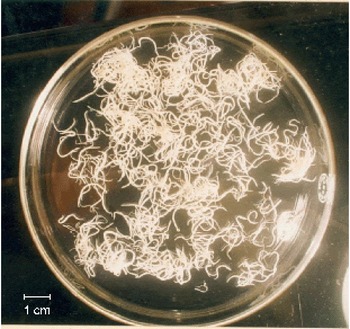
The mesenteric blood vessels and portal veins were examined for adult worms. The recovered adult worms were alive, in copula, and found only in the portal vein and small blood vessels of the liver parenchyma (fig. 1). The liver was enlarged, fibrotic, congested and with pinpoint red and whitish patches (not shown). The total number of worms recovered from a portion of the liver was 129,870. We suspect that this infection caused the death of the animal. However, there are no reports of B. nairi infection in wild elephants with which to compare this level of infection.
(colour online) Adult worms of B. nairi recovered from the liver.

Morphology under the light microscope
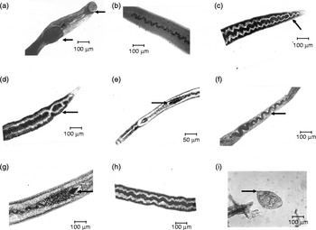
Male worms are shorter and thicker than females, their bodies attenuated at each end and widest in the mid-region. Body length is 12.04 mm (11.2–13.06 mm) and maximum width is 0.68 mm (0.59–0.71 mm) (1.28 mm when the margins of the gynaecophoric canal are opened out). Width at the level of the ventral sucker is 0.62 mm. The suckers are well developed: diameter of the oral sucker is 245 μm, and that of the ventral sucker is 372 μm (fig. 2a). The ventral sucker has a fimbriated margin. The body is constricted immediately posterior to the ventral sucker, narrowing to a width of 0.49 mm. The gynaecophoric canal extends from this constriction to the posterior end of the worm. The short oesophagus (1.07 mm), divides just anterior to the ventral sucker into two intestinal caeca, which follow a zigzag course to about 0.25 mm from the bladder and 0.46 mm from the posterior tip of the body, where they unite to form an extremely short common caecum (19 μm in length). The average number of testes is 57 (54–68). They are small and irregular in shape, and arranged in a zigzag fashion along the middle line of the body and each unit of the zigzag line is comprised of 2–4 testes (fig. 2b). The length of the testicular chain is 109 μm. The diameter of individual testicular follicles varies from 62 μm to 126 μm.
Male of B. nairi showing (a) the anterior end with an oval oral sucker and a larger ventral sucker and (b) mid-region with a chain of testes. Female B. nairi showing (c) posterior end with small caeca and (d) with caeca reunited; (e) anterior end and (f) anterior end with spiral-shaped ovary; (g) uterus with a single egg in the ootype; (h) two rows of vitelline glands one on either side of the body; and (i) an egg of B. nairi isolated from a fresh dung pat of the infected elephant.

Female worms are more slender and elongated than males, their bodies being 16.8 mm (14.1–19.5 mm) long and 0.31 mm wide at the widest point. The body is slender and delicate anterior to the ovary and considerably broader posterior to this (fig. 2e). The mouth is surrounded by an oral sucker measuring 98–118 μm in diameter. The ventral sucker is situated in a groove-like depression about 0.43 mm from the oral sucker. The alimentary canal is similar to that of the male, except that the oesophageal gland is less prominent, the caeca wider and the common caecum shorter. The posterior caecal union is close to the end of the body and is sometimes followed by rebranching and reunion of the caeca (fig. 2c and d). The ovary is extremely elongated, 980 μm in length and up to 67 μm in width, tubular and twisted into a loose spiral with 8–11 turns (fig. 2e and g). The oviduct originates posteriorly from the ovary, forms a loop around the anterior border of the vitelline glands and runs anteriorly dorsally in the body to the ootype, which typically contains a single immature egg (118 × 54 μm). The female genital opening is located just posterior to the ventral sucker (fig. 2g). A row of vitelline glands occurs on each side of the body, within the curves formed by the broad caeca (fig. 2h). Eggs are oval in shape and slightly asymmetrical (140–165 × 69–87 μm) and bear a straight, subterminal spine 9 μm long (fig. 2i).
Comparative morphology of Bivitellobilharzia nairi and species of Schistosoma
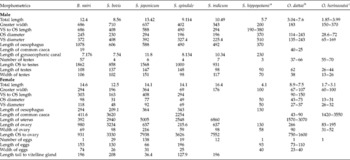
For comparative purposes, other species of schistosomes found commonly in mammals were mounted and measured (table 1). However, data for S. hippopotami, O. dattai, and O. harinasutai were taken from published papers (Dutt & Srivastava, Reference Dutt and Srivastava1952; Kruatrachu et al., Reference Kruatrachue, Bhaibulaya and Harinasuta1965) due to unavailability of specimens. Both male and female adult worms of B. nairi were comparable in length and width to other mammalian schistosomes except S. hippopotami, O. dattai and O. harinasutai (see table 1). Bivitellobilharzia nairi has many more testes (at least 54) than do Schistosoma species (3–8), but numbers that are comparable to those seen in H. americana, O. dattai and O. harinasutai, with 76, 52 and 62 testes, respectively. The salient character of male B. nairi is the small, posterior common caecum that is absent from other species, except members of the genus Austrobilharzia spp. (see Stunkard & Hinchcliffe, Reference Stunkard and Hinchcliffe1952) and Heterobilharzia (see Farley, Reference Farley1971). The female worm usually has a single egg in the uterus or ootype, as is also the case for S. hippopotami, O. dattai and O. harinasutai. However, B. nairi has vitellaria that spread toward the posterior end on either side of the caecal branches in a zigzag manner, a character not found in any other schistosomes.
Comparative morphometrics of B. nairi with other selected Schistosoma; all measurements are given in μm, except male and female body lengths (mm).

VS, ventral sucker; OS, oral sucker.
a Schistosoma hippopotami, Thurston (Reference Thurston1963).
b Orientobilharzia dattai, Dutt & Srivastava (Reference Dutt and Srivastava1952).
c Orientobilharzia harinasutai, Dutt & Srivastava (Reference Dutt and Srivastava1961).
Scanning electron microscopy
Male worms
Anterior-ventral surface
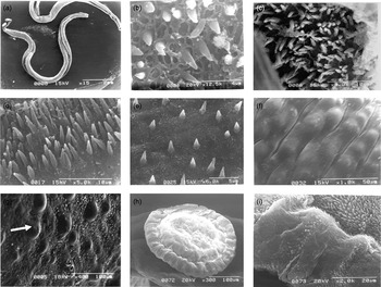
The ventral region between the oral sucker and the start of the gynaecophoric canal bears numerous elongated spines that are 2–2.5 μm in length. The surface of the tegument resembles a meshwork through which all spines emerge. Most of the papilla are somewhat conical and pointed in appearance, but a few of them have blunted ends (fig. 3b).
Scanning electron microscope (SEM) images of male B. nairi showing (a) anterior, middle and posterior parts of the gynaecophoric canal; (b) anterio-ventral surface of gynaecophoric canal; (c) middle-ventral surface of gynaecophoric canal; (d) middle-ventral surface of gynaecophoric canal; (e) posterio-ventral surface of gynaecophoric canal; (f) dorso-terminal end; (g) dorsal surface to show papillae (arrow); (h) ventral sucker; and (i) rim of the ventral sucker.

Mid-ventral surface
This is the widest portion of the body and contains most of the gynaecophoric canal, normally occupied by a female schistosome. It bears numerous long, sharp spines ranging from 3.5 to 4.0 μm in length. These spines are particularly dense within the gynaecophoric canal. Again, the surface of the tegument appeared as a meshwork through which the spines emerge (fig. 3c and d).
Posterior-ventral surface
This is the region posterior to the point where the hind body of the female emerges from the gynaecophoric canal. Spines, 2.5–3.0 μm in length, gradually decrease in number, becoming more scattered towards the posterior end and terminal part of the gynaecophoric canal (fig. 3e). No spines are seen on the tegument of the terminal end, but the surface here has a corrugated appearance (fig. 3f).
Dorsal and lateral surfaces
At low magnification, there appear to be no spines on the dorsal surface of the body. However, at higher magnification, the surface is roughly corrugated, and pitted, like a mesh (fig. 3g).
Discussion
Identification of schistosomes is based on morphological features and biometrical characteristics of eggs and cercariae, characteristics of the life cycle (prepatent periods, number of eggs produced, growth rate, host specificity) and, more recently, through DNA analysis (Agatsuma et al., Reference Agatsuma, Iwagami, Liu, Rajapakse, Mondal, Kitikoon, Ambu, Agatsuma, Blair and Higuchi2002, Reference Agatsuma, Rajapakse, Kuruwita, Iwagami and Rajapakse2004). The paucity of visible morphological features has left the taxonomy of many still in doubt. Bivitellobilharzia nairi is one of the members of the schistosome group that occurs in elephants in Sri Lanka. Here, we have provided for the first time a detailed morphological description and SEM topography of the parasite for comparison with other selected schistosomes in mammals and birds.
The morphological features of B. nairi described in this paper are very similar to those reported by Vogel & Minning (Reference Vogel and Minning1940), Yamguti (Reference Yamaguti1958), and Farley (Reference Farley1971). The measurements of internal organs and length of the worms, however, contradict the previous descriptions (Yamaguti, Reference Yamaguti1958; Farley, Reference Farley1971), which state that male and female worms are similar in size. The reasons for this difference are not known, but it may reflect an error in previous observations. It is important to stress that we were able to collect fresh specimens from the liver for detailed examination. Post-mortem examination of the infected elephant revealed that all adult worms occur in the small blood vessels and blood capillaries of the liver parenchyma, but not in the mesenteric vein as do other mammal intestinal schistosomes. The first report of a schistosome parasite in Asian elephants was by Mudaliar & Ramanujachary (Reference Mudaliar and Ramanujachary1945), using specimens which were collected several hours after the death of the elephant and, consequently, samples were poorly preserved. Therefore, there are some measurements from those previous studies that do not correspond with the results of the present paper.
Morphological comparisons revealed similarities and differences between B. nairi and other species of mammal schistosomes, particularly the length of both males and females, the shape of the egg and the site of infection. Bivitellobilharzia nairi is quite similar morphologically to other species of schistosomes, the eggs in the uterus being similar to those in S. hippopotami, O. dattai and O. harinasutai.
Conversely, some features, such as the number of testes in the male and the appearance of the vitelline glands in B. nairi, show distinct differences from other species of schistosomes. The number of testes in B. nairi is closer to that found in species of Heterobilharzia and Orientobilharzia (Farley, Reference Farley1971). In addition, B. nairi occurs inside blood vessels of the liver parenchyma, whereas other schistosomes reside in blood vessels outside the liver.
The tegument surface of numerous species has been described by different authors and used in some cases for a specific diagnosis (Kuntz et al., Reference Kuntz, Tulloch, Davidson and Huang1976, Reference Kuntz, Tulloch, Huang and Davidson1977, Reference Kuntz, Davidson, Huang and Tulloch1979; Hicks & Newman, Reference Hicks and Newman1977; Tulloch et al., Reference Tulloch, Kuntz, Davidson and Huang1977). In the present study, the topography of the tegument of B. nairi was examined using only male worms, since it has been suggested that SEM morphology of spines of male schistosomes is more distinct than that of females (Southgate et al., Reference Southgate, Rollinson and Vercruysse1986). Following a report by Senft et al. (Reference Senft, Philpott and Pelofsky1961) on the function and structure of the integument of adult S. mansoni, there have been many more published characterizations of adult Schistosoma via transmission and scanning electron microscopy (Morris & Threadgold, Reference Morris and Threadgold1968; Smith et al., Reference Smith, Reynolds and Von Lichtenberg1969; Miller et al., Reference Miller, Tulloch and Kuntz1972; Kuntz et al., Reference Kuntz, Tulloch, Davidson and Huang1976). We observed that the tegument of male B. nairi is smoother than in other schistosomes. Spines and papillae are present only on the ventral surface, particularly in association with the gynaecophoric canal. The inner surface of the canal is covered with long, sharp spines arranged irregularly. Spines are more dense in the middle part of the canal, but become smaller and more diffusely distributed posteriorly. Surface infrastructure and papillary organs are useful in the identification of infection site, as well as adaptations to the changes of living environments of the parasites (Robert et al., Reference Robert, George, David and Too-cheng1976, Reference Robert, George, Too-cheng and David1977). Unfortunately, there are no SEM studies of S. hippopotami with which to compare these data.
Our simultaneous study on the phylogenetic relationship of B. nairi with other mammal schistosomes (Agatsuma et al., Reference Agatsuma, Rajapakse, Kuruwita, Iwagami and Rajapakse2004) revealed evidence of a rather distant relationship between B. nairi and Schistosoma spp., but it clustered closely with Schistosomatium spp. (in the ITS2 gene), and with Heterobilharzia and Schistosomatium spp. (in the 28S RNA gene). However, maximum likelihood estimated trees from the combined data partitions of 18S, 28S and partial CO1 genes revealed that B. nairi consistently grouped basal to all Schistosoma spp. (Brant et al., Reference Brant, Morgan, Mkoji, Snyder, Rajapakse and Loker2006). In the present study, when we consider the number of testes in male worms, it is likely that B. nairi is more distant from the Schistosoma spp. and closer to species of the genera Heterobilharzia and Orientobilharzia. Hence, in this study, it is confirmed that elephant schistosomes are a unique genus within the Schistosomatidae.
Acknowledgements
We wish to express our thanks to the staff members of the Department of Pathobiology, Faculty of Veterinary Medicine and Animal Science, for collecting and preparing samples for this study. The authors would like to thank Professor David Blair, James Cook University, Australia for his invaluable comments on this paper. This work was supported by National Research Council of Sri Lanka Grant No.05-23.