Implications
Naringin is a type of citrus flavonoid that has shown antioxidant properties and a lowering effect on serum triacylglycerol and cholesterol levels in rabbits, rats and humans. However, according to the results observed in the fattening lambs of the present study, including naringin in the concentrate at a dose of 0.15% only improves the antioxidant status of the liver, whereas meat quality attributes do not seem to be affected.
Introduction
One of the main reasons for the deterioration of meat products during processing and storage is lipid peroxidation. Different synthetic antioxidants are being used as food additives to increase the shelf life of such products on the market, but the use of some of these is still controversial (e.g. butylated hydroxytoluene). A different approach for fresh meat is the inclusion of antioxidants in animal feedstuffs. For example, the efficacy of vitamin E in reducing lipoperoxidation in ruminant products has been extensively shown. However, it has been suggested that different antioxidants given alone or combined with vitamin E could be more efficient at enhancing the antioxidant status of animals than the use of large doses of vitamin E (Gladine et al., Reference Gladine, Rock, Morand, Bauchart and Durand2007; Gobert et al., Reference Gobert, Martin, Ferlay, Chilliard, Graulet, Pradel and Bauchart2009 and Reference Gobert, Gruffat, Habeanu, Parafita, Bauchart and Durand2010).
In this context, a special attention has been paid to the physiological effects promoted by the flavonoids, a type of polyphenol largely concentrated in citrus fruits. In human nutrition, the flavonoids have been suggested as being responsible for the prevention of chronic and degenerative diseases (Tripoli et al., Reference Tripoli, Guardia, Giammanco, Majo and Giammanco2007), but ruminant production might also benefit from these compounds. Thus, a supply of flavonoids with antioxidant properties could possibly be recommended for the shelf-life extension and colour stabilization of meat products (Luciano et al., Reference Luciano, Vasta, Monahan, López-Andrés, Biondi, Lanza and Priolo2011).
The inclusion of flavonoids in the diet of animals may also contribute towards obtaining healthier animal products for human consumption. On the one hand, the antioxidant properties of these compounds might help to prevent meat from cholesterol oxidation in the meat; the consumption of oxidized cholesterol seems to be more directly connected to the development of atherosclerosis and coronary heart disease than pure cholesterol (Hur et al., Reference Hur, Park and Joo2007). On the other hand, the enzymatic activity of some of the proteins related to lipid metabolism (e.g. acyl-CoA : diacylglycerol acyltransferase 1 (DGAT1), acyl-CoA : cholesterol acyltransferase (ACAT), hepatic 3-hydroxy-3-methylglutaryl coenzyme A (HMG-CoA) reductase) seems to be reduced by flavonoids (Casaschi et al., Reference Casaschi, Wang, Dang, Richards and Theriault2002; Jeon et al., Reference Jeon, Bok, Jang, Kim, Nam, Jeong, Park and Choi2002 and Reference Jeon, Park and Choi2004). However, little effort has been made to determine the effectiveness of flavonoids on the quality of ruminant products. In this sense, naringin, one type of grapefruit and citrus flavonoid with proven antioxidant properties (Jeon et al., Reference Jeon, Park and Choi2004) might be a good choice as it has been authorized as a feed additive in ruminant nutrition by the European Commission (Community Register of Feed Additives pursuant to Regulation (EC) No. 1831/2003).
Therefore, the aim of the present study was to examine the effects of naringin supplementation on biochemical parameters such as serum cholesterol (high density lipoprotein -HDL-, low density lipoprotein -LDL- and total cholesterol) and triglycerides (TAG) and their relationship with the chemical composition of liver and meat samples obtained from lambs fed a commercial feedstuff. In addition, the effect of naringin on the oxidative stability of meat (colour and lipoperoxidation) and liver (lipoperoxidation) samples was considered.
Material and methods
Genotyping of the flock
In order to partially avoid the genetic variation regarding intramuscular fat and other meat quality traits, all of the animals in the present study were selected on the basis of a single-nucleotide polymorphisms described for the leptin (LEP) gene. This mutation (A103G) has been described in intron 2 of the ovine LEP gene (Boucher et al., Reference Boucher, Palin, Castonguay, Gariepy and Pothier2006).
Animals of the flock were blood sampled by jugular venipuncture into Vacutainer tubes (10 ml) containing EDTA. The DNA was immediately extracted from a 200-μl aliquot of fresh whole blood using a QIAmp DNA Blood Mini Kit (Qiagen Ltd, Crawley, UK), and then stored at −20°C until use. The DNA was amplified by PCR using the primers described by Boucher et al. (Reference Boucher, Palin, Castonguay, Gariepy and Pothier2006) and then sequenced by the Automated Sequencer MEGABACE 500 (GE Healthcare, Chalfont St. Giles, UK).
Selected animals
Twenty Assaf lambs (initial age 13 to 15 weeks) were used in this experiment, all were heterozygous for the A103G polymorphism in the ovine LEP gene. The lambs were fed a commercial concentrate, barley straw and alfalfa hay until the trial began. The animals were dewormed with Ivomec (Merial Labs., Barcelona, Spain) and vaccinated against enterotoxaemia (Miloxan, Merial Labs).
After random stratification on the basis of body weight (BW; average BW, 24.8 ± 1.64 kg), the lambs were allocated into two groups (10 lambs per group) and housed individually. All handling practices followed the recommendations of the European Council Directive 86/609/EEC for the protection of animals used for experimental and other scientific purposes.
Diets and blood sampling method
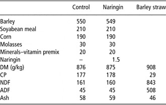
After 7 days of adaptation to the basal diet (barley straw and concentrate feed), all of the lambs were fed barley straw plus the concentrate feed alone (CONTROL group) or enriched with 0.15% of naringin (1.5 g/kg DM (NARINGIN group)) for 7 weeks. All of the ingredients for each concentrate (including naringin, which was obtained from Destilaciones Bordas Chinchurreta S.A., Seville, Spain) were mixed and ground in a mill and fed to the animals as dry meal. The concentrate and forage were supplied in separate feeding troughs at 0900 h every day, and fresh drinking water was always available. The ingredients and the chemical composition of both the concentrates and the barley straw are summarized in Table 1. The straw (200 g/day) and concentrate (30 g/kg BW per day) offered to each lamb were weighed daily.
Ingredients (g/kg) and chemical composition (g/kg DM) of the experimental concentrates and barley straw

DM=dry matter.
All of the animals were blood sampled by jugular venipuncture using Vacutainer tubes (10 ml) before administration of the experimental concentrate (day 0, week 0) and then each week until the final day of the experiment (day 49, week 7). Blood samples collected in tubes with no anticoagulant were allowed to clot for 30 min at room temperature and centrifuged at 2000 × g for 15 min at 4°C. Thereafter, serum was stored at −20°C until used for measuring the biochemical parameters.
Biochemical parameters of the serum samples
Total cholesterol, LDL, HDL and TAG concentrations in the serum samples were determined by an automated enzymatic colorimetric method with test kits from Roche Diagnostics (Madrid, Spain) on Cobas Integra 400 (Roche Diagnostic System, Madrid, Spain).
Slaughter procedure
Animals in the CONTROL and NARINGIN groups were weighed and slaughtered by exsanguination from the jugular vein, eviscerated and skinned. The hot carcass and the liver of each lamb were weighed, and liver samples were immediately frozen in liquid nitrogen and kept at −80°C. The carcasses were chilled at 4°C for 24 h, halved carefully and the longissimus thoracis et lumborum muscle was removed from the ribs of the left side of the carcass. The pH value of the longissimus thoracis muscle was determined in triplicate 24-h post mortem using a pH meter equipped with a penetrating glass electrode (pHmeter Metrohm® 704, Herisau, Switzerland).
Chemical parameters of the meat and liver samples
The longissimus thoracis samples were trimmed to eliminate connective tissue and intermuscular fat, then freeze-dried and homogenized in a food processor (Moulinex®, Bagnolet, France) in the same way as the liver samples. The fat content was determined in accordance with the methods described by the Association of Official Analytical Chemists (AOAC, 2003). The cholesterol content was determined by a colorimetric method using the Cholesterol/Cholesteryl Ester Quantitation Kit provided by Calbiochem (San Diego, CA, USA).
For polyphenol quantification, the freeze-dried samples (1.5 g) with 90 mg/kg of apigenin as the internal standard were extracted first with 20 ml of petroleum ether, with stirring, and taken to dryness at room temperature. Then, they were extracted using 150 ml of methanol in a Soxhlet extractor (B-811) (Buchi, Flawil, Switzerland) for 2 h under a nitrogen atmosphere. The methanolic extracts were taken to dryness at 40°C, under vacuum conditions in an evaporator system (Syncore Polyvap R-96, Buchi, Switzerland). The residue was re-dissolved in methanol and made up to 5 ml. The extracts were kept in vials at −80°C until analysis. For the high performance liquid chromatography (HPLC) analysis, a method adapted from Moñino et al. (Reference Moñino, Martínez, Sotomayor, Lafuente and Jordán2008) was performed on a reverse-phase ZORBAX SB-C18 column (4.6 × 250 mm, 5-μm pore size, Hewlett Packard, Palo Alto, CA, USA) using a guard column (ZORBAX SB-C18 4.6 × 125 mm, 5-μm pore size; Hewlett Packard) at room temperature. Extracts were passed through a 0.45-μm filter (Millipore SAS, Molsheim, France) and 30 μl was injected into a Hewlett Packard (Germany) system equipped with a G1311A quaternary pump and G1315A photodiode array UV/Vis detector. The mobile phase was acetonitrile (A) and acidified water containing 5% formic acid (B). The gradient was as follows: 0 min, 20%A; 15 min, 30%A; 25 min, 55%A; 30 min, 100%A; and held for 10 min before returning to the initial conditions. The flow rate was 1.0 ml/min and the wavelength of detection was set at 280 nm. Naringenin was identified by comparing the retention time and spectra of commercially available standard compounds. Linear regression models were used for quantification with standard dilution techniques and apigenin as the internal standard. The samples were run in triplicate.
Colour measurements of the meat samples
The L*a*b* system (l'Eclairage, Reference l'Eclairage1986) was used to determine the meat colour of the longissimus lumborum muscle using a chroma meter (Minolta® Croma Meter 2002, Langenhagen, Germany). For this purpose, each longissimus lumborum muscle (left side) was cut into 10 slices, each 2.5-cm thick. The slices corresponding to each animal were placed on Polyfoam trays, over-wrapped with an oxygen-permeable polyvinylchloride film and then stored under simulated retail display conditions (daily illumination and 4°C). For each display time (0, 1, 3, 5 and 9 days) two slices of fresh meat from each animal were measured for colour parameters at four different locations, and then averaged. After being measured for colour parameters the samples were frozen (−80°C) for use in trials related to the antioxidant status of the meat.
Antioxidant status of the meat and liver samples
The thiobarbituric acid-reactive substances procedure (TBARS) was performed on pre-thawed, raw meat and liver samples according to the following procedure (Maraschiello et al., Reference Maraschiello, Sárraga and García Regueiro1999). The acid hydrolysis of TEP (1,1,3,3-tetraethoxypropane) yielded malondialdehyde (MDA, the main product of lipid peroxidation), which was used to construct the standard curve. After thawing, the meat and liver samples were cut into 2.5 g pieces and homogenized for 30 s at 13 000 r.p.m. with 20 ml of distilled water using a T25 digital Ultraturrax (IKA, Staufen, Germany) provided with a 18G dispersal tool. Then, 5 ml of 25% trichloroacetic acid was added, centrifuged, filtered and 3.5 ml was transferred to a 10-ml screw-cap tube with 1.5-ml thiobarbituric acid (0.6%). The samples were heated at 70°C for 30 min and, after being cooled on ice for 10 min, the absorbance was measured at 532 nm. The results were expressed as μg MDA/g for meat or liver.
Cholesterol oxidation products (COPs), also called oxysterols, were determined in meat samples after 3 and 9 days on display according to the method proposed by Grau et al. (Reference Grau, Codony, Grimpa, Baucells and Guardiola2001). Briefly, meat samples were thawed, cooked under a grill at 180°C, while controlling the core temperature, and then cooled at 4°C for 30 min. Lipids were extracted according to the method of Folch et al. (Reference Folch, Lees and Sloane Stanley1957) from 3 g of meat using a mixture of chloroform : methanol (2 : 1, v/v). The internal standard used was 19-hydroxycholesterol (19-HC). Ten milliliters of 1.5 methanolic potassium hydroxide was then added and the mixture was kept in an orbital shaker for 20 h at room temperature under an N2 atmosphere and darkness to complete the cold saponification. The unsaponificable material was extracted three times with diethyl ether in a separating funnel, and then purified by solid phase extraction according to the procedure described by Guardiola et al. (Reference Guardiola, Codony, Rafecas and Boatella1995), using Sep-Pak Vac 6 cc (1 g) silica cartridges (Ref. WAT036910; Waters Corporation, Milford, USA). The COPs were derivatized (Sylon BTZ and Anhydrous Pyridine, Supelco, Bellefonte, PA, USA) to trimethylsilyl esters before gas chromatographic analysis on an Agilent 7890 Series gas chromatograph (Agilent Technologies, Santa Clara, CA, USA) provided with a flame ionization detector (FID), by splitless injection into a VF-5ms CP8947 capillary column (50 m × 250 μm × 0.25 μm, Varian, Palo Alto, CA, USA). The chromatographic conditions were as follows: injection volume 1 μl; initial oven temperature 75°C, increased to 250°C at 30°C/min, to 290°C at 8°C/min and to 292°C at 0.05°C/min; the injector and detector temperatures were 250°C and 280°C, respectively. Helium was used as the carrier gas at a flow rate of 1 ml/min. The oxysterols 7α-hydroxycholesterol (7α-HC), 7β-hydroxycholesterol (7β-HC), 5,6α-epoxycholesterol (α-CE), 5,6β-epoxycholesterol (β-CE), cholestanetriol (CT), 25-hydroxycholesterol (25-HC) and 7-ketocholesterol (7-KC) were identified by comparing their retention times with those of authentic standards (Steraloids Inc., Newport, RI, USA) and quantified using the internal standard. All determinations were performed in duplicate.
Statistical analyses
Data of serum total cholesterol, LDL, HDL and TAG concentrations were analysed as repeated measures using the MIXED procedure of SAS (SAS Institute Inc., Cary, NC, USA). The model included the fixed effects of treatment, day, the treatment-by-day interaction, the random effect of lamb nested within treatment and the residual error. The values observed on day 0 were used as the covariates. Data of feed intake, slaughter and physico-chemical and antioxidant parameters of the meat and livers were subjected to one-way ANOVA using the GLM procedure of SAS.
Results
There was no significant difference between the NARINGIN and the CONTROL groups in concentrate or barley straw intake (P > 0.05, overall mean values of 723 ± 21.1 and 154 ± 17.8 g/animal per day, respectively), thus reaching the established feed intake. As expected, the gain in BW was not affected by dietary treatment (173 ± 7.2 g/animal per day).
Biochemical parameters of the serum samples
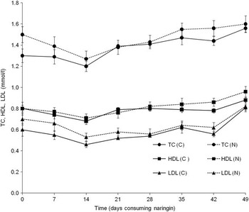
Figure 1 summarizes the changes in total cholesterol, HDL and LDL levels in the serum from lambs in response to the dietary treatments. As can be seen, there were no significant differences (P > 0.05) in the average values of total cholesterol, HDL and LDL levels in the sera between the NARINGIN group and the CONTROL group (Figure 1).
Evolution of total cholesterol (TC), HDL and LDL concentrations (mmol/l) in serum samples of CONTROL (C) and NARINGIN (N) lambs.

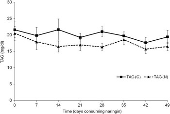
The mean values for the TAG levels in the CONTROL and NARINGIN groups can be observed in Figure 2. The NARINGIN lambs showed lower TAG levels (P = 0.087) compared with the CONTROL group (Figure 2).
Evolution of triacylglicerol (TAG) concentration (mg/dl) in serum samples of CONTROL (C) and NARINGIN (N) lambs.

Slaughter, weight gain and liver weight
Mean BW (33.3 ± 0.57 kg) and carcass weight (15.9 ± 0.34 kg) were similar in both groups. The liver weight (average values in CONTROL and NARINGIN groups of 578 g and 584 g, respectively, P > 0.10) was not significantly affected by the dietary treatment of the NARINGIN lambs.
Physical and chemical parameters of meat and liver samples
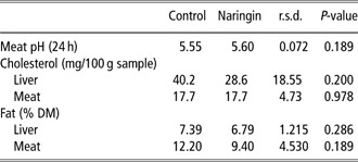
No significant differences (P > 0.05) were observed in the fat or cholesterol contents of both liver and meat samples (Table 2). Only numerical differences (i.e. not statistically significant), with lower values in the fat content of the NARINGIN group, were detected (Table 2).
Physical and chemical parameters of liver and meat samples

DM = dry matter.
The accumulation of naringenin (aglycone fraction of naringin) in the liver of the NARINGIN group (22.5 g/kg) was remarkable, whereas it was not detectable in the liver of the CONTROL lambs. This compound could not be detected in any of the groups at the meat level.
Colour measurements of the meat samples
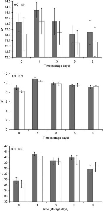
The evolution of meat colour parameters (L*, a*, b*) during storage at 4°C is represented in Figure 3. These parameters were not significantly affected by the dietary treatment.
Evolution of meat colour parameters under refrigerated storage (4°C) for CONTROL (C) and NARINGIN (N) lambs.

Antioxidant status of the meat and liver samples
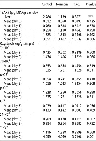
The mean values of the TBARS and oxysterols are summarized in Table 3. The TBARS levels in the liver samples were significantly lower in the NARINGIN group (P < 0.001; Table 3), whereas no significant differences were observed between the groups in the meat samples.
Antioxidant status (TBARS) of liver and meat samples and oxysterols content
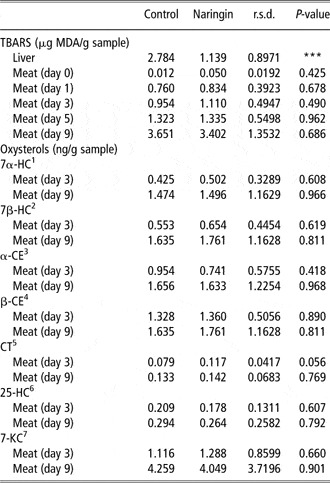
TBARS = thiobarbituric acid-reactive substances procedure; MDA=malondialdehyde; 7α-HC = 7α-hydroxycholesterol; 7β-HC = 7β-hydroxycholesterol; α-CE = 5,6α-epoxycholesterol; β-CE = 5,6β-epoxycholesterol; CT = cholestanetriol; 25-HC = 25-hydroxycholesterol; 7-KC = 7-ketocholesterol. ***P < 0.001.
Regarding the oxysterol contents in the cooked meat samples, the total COP values were significantly higher (P < 0.001) after 6 days of refrigerated storage in both CONTROL and NARINGIN lambs (Table 3). However, naringin supplementation showed no significant effect when both groups were compared on the same storage day; similar values (i.e. not significantly different, P > 0.05) were detected for all of the oxysterols studied (Table 3).
Discussion
Biochemical parameters of the serum samples
The lack of significant differences (P > 0.05) in the average values of total cholesterol, HDL and LDL levels of the serum from lambs in the NARINGIN group when compared with lambs in the CONTROL group is similar to the results reported by Gobert et al. (Reference Gobert, Martin, Ferlay, Chilliard, Graulet, Pradel and Bauchart2009), who found no differences in plasma cholesterol and TAG contents in dairy cows when being fed linseed supplemented with plant extracts rich in polyphenols and vitamin E. However, these results seem to differ from the results described in hypercholesterolaemic subjects who consumed naringin (Kim et al., Reference Kim, Oh, Park, Lee, Seo and Choi2004 and Reference Kim, Kim, Lee, Jeon, Do, Kwon, Cho, Kim, Jeong, Park, Ha and Choi2006; Baba et al., Reference Baba, Natsume, Yasuda, Nakamura, Tamura, Osakabe, Kanegae and Kondo2007), where a reduction in total cholesterol and LDL cholesterol levels plus an increase in HDL cholesterol was observed. This effect was attributed to an inhibitory effect of naringin on HMG-CoA reductase and ACAT, a rate-limiting enzyme of the cholesterol biosynthetic pathway and a cholesterol esterifying enzyme-regulating intracellular cholesterol homeostasis, respectively (Jung et al., Reference Jung, Kim, Lee, Lee, Kim, Park, Kim, Jeong and Choi2003). An increase in the excretion of faecal sterols was also described in subjects who consumed naringin (Kim et al., Reference Kim, Oh, Park, Lee, Seo and Choi2004). However, as stated beforehand all of these effects of naringin have been described in hypercholesterolaemic subjects, whereas no changes have been found in normocholesterolaemic cases. This is in agreement with the results observed in the present study, where the lack of significant differences (CONTROL v. NARINGIN) in serum total cholesterol, HDL and LDL levels in the serum samples could be explained as a result of the normocholesterolaemic status of the lambs that consumed feedstuff of a vegetable origin.
In contrast with previous studies (Gobert et al., Reference Gobert, Martin, Ferlay, Chilliard, Graulet, Pradel and Bauchart2009), the trend to significance (P = 0.087) found for the difference in TAG levels between both groups is noteworthy (Figure 2). The naringin-lowering effect on serum TAG levels might be related to an inhibitory effect of flavonoids on DGAT1 activity, one of the main enzymes involved in TAG synthesis in the liver (Casaschi et al., Reference Casaschi, Wang, Dang, Richards and Theriault2002; Jeon et al., Reference Jeon, Park and Choi2004). In fact, these compounds were suggested for used in the clinical treatment of hypertriglyceridaemia (Casaschi et al., Reference Casaschi, Wang, Dang, Richards and Theriault2002).
Slaughter, weight gain and liver weight
The lack of significant differences in the liver weight between CONTROL and NARINGIN lambs may appear to be contradictory when compared to the results previously published for hypercholesterolaemic rabbits (Jeon et al., Reference Jeon, Bok, Jang, Kim, Nam, Jeong, Park and Choi2002 and Reference Jeon, Park and Choi2004; Kim et al., Reference Kim, Oh, Park, Lee, Seo and Choi2004). However, liver hypertrophy is usually induced by hypercholesterolaemia, so naringin might have helped to maintain normal liver weights in those hypercholesterolaemic rabbits probably due to the downregulation exerted by this flavonoid on the expression of hepatic enzymes involved in cholesterol synthesis (HMG-CoA reductase). On the contrary, no effect was observed for naringin on the liver weight of rats fed a cholesterol-free diet (Seo et al., Reference Seo, Jeong, Lee, Park, Jung, Kim and Choi2003). In other words, the lack of a significant difference in liver weight between our lamb groups (CONTROL v. NARINGIN), which is in agreement with the biochemical parameters found in the serum samples, was probably a consequence of the normocholesterolaemic status of these animals that consumed a feedstuff of a purely vegetable origin.
Physical and chemical parameters of the meat and liver samples
Despite the tendency to significance observed in serum TAG levels when both groups were compared, no significant differences (P > 0.05) were detected in the fat contents of the liver or meat samples. Furthermore, no significant differences were observed in the cholesterol contents of liver or meat samples either. All animal cells require cholesterol as it is an essential structural component of the membrane (van Meer et al., Reference Van Meer, Voelker and Feigenson2008), so lowering cholesterol in body tissues such as meat seems to be difficult in animals raised under the same feeding system (Madruga et al., Reference Madruga, Medeiros, de Sousa, de Cunha, Pereira Filho and Queiroga2009). Consequently, research should be focused on protecting cholesterol from oxidation as a way of improving meat quality (Rankin and Pike, Reference Rankin and Pike1993). Therefore, in the present study, the content of COPs (oxysterols) in meat samples was measured, as discussed below.
Again, it must be stressed that naringenin (aglycone fraction of naringin) was found to accumulate in the liver of the NARINGIN group (22.5 g/kg), whereas it was not detectable in the liver of CONTROL lambs. In fact, the glycosidic flavanone naringin is hydrolysed by ruminal microorganisms in order to allow absorption of the aglycone fraction (naringenin) by the animal (Gladine et al., Reference Gladine, Rock, Morand, Bauchart and Durand2007). After absorption, naringenin is transported to the liver, where it forms glucuronide and/or sulphate or methyl conjugates before being normally excreted through the urinary and faecal systems (Spencer et al., Reference Spencer, Abd El Mohsen, Minihane and Mathers2007). The metabolism of naringenin in the liver might have promoted a higher concentration of this compound in this organ, whereas not enough naringenin would have reached the muscles (where it was not detected). It is tempting to suggest that this polyphenolic compound might be recognized by the organism as a strange substance to be eliminated, which would be in agreement with the accumulation of this compound in the liver but not in the muscle (Robles-Sardin et al., Reference Robles-Sardin, Bolaños-Villar, González-Aguilar and de la Rosa2009).
Colour measurements of the meat samples
Meat colour is affected by the oxidation of myoglobin and the consequent accumulation of metmyoglobin, which is responsible for meat browning. However, the evolution of meat colour parameters during storage at 4°C was not significantly affected by the dietary treatment. Luciano et al. (Reference Luciano, Vasta, Monahan, López-Andrés, Biondi, Lanza and Priolo2011) reported that the consumption of tannins could improve the colour stability of lamb meat, although the doses of polyphenols used by these authors were higher than in the present experiment. Moreover, longissimus lumborum has been described as a muscle with pronounced colour stability compared with highly oxidative muscles with elevated concentrations of myoglobin and higher oxygen consumption rates, such as m. gluteus medius (Young and West, Reference Young and West2001). However, the lack of a naringenin accumulation at this level might also explain the finding of no significant differences in meat colour parameters between the dietary treatments.
In any case, it must be noted that previous studies have suggested that lower levels of haemoglobin and myoglobin in lambs fed tannins (different polyphenolic compounds) might have been responsible for significant lightness differences in meat samples (Priolo et al., Reference Priolo, Waghorn, Lanza, Biondi and Pennisi2000). In addition, Gnanamani et al. (Reference Gnanamani, Sudha, Deepa, Sudha, Deivanai and Sadulla2008) suggested a chelating effect of naringenin (the aglycone fraction of naringin) on the ferrous ion needed to form the central core of both haemoglobin and myoglobin. Both reports are in agreement with the lower red blood cell counts detected in the NARINGIN group when compared with the CONTROL group (López-Campos et al., Reference López-Campos, Bodas, Prieto, Giráldez, Pérez and Andrés2010). Regarding the effects on meat colour, only numerical differences (∼3.7%), with lower values in the redness parameter (a*) of the NARINGIN group, were observed from the beginning of the display time (day 0) until the end of this period (day 9; see Figure 3). However, these differences (CONTROL v. NARINGIN) were not statistically significant at any of the display times.
Antioxidant status of meat and liver samples
Although in the present study the use of naringin did not seem to significantly affect the antioxidant status of the meat, previous studies carried out on finishing cows and lambs showed an improvement in this parameter when plant extracts rich in polyphenols were included in the diet (Moñino et al., Reference Moñino, Martínez, Sotomayor, Lafuente and Jordán2008; Gobert et al., Reference Gobert, Gruffat, Habeanu, Parafita, Bauchart and Durand2010; Luciano et al., Reference Luciano, Vasta, Monahan, López-Andrés, Biondi, Lanza and Priolo2011). This effect was related to the presence of polyphenols in muscle (Luciano et al., Reference Luciano, Vasta, Monahan, López-Andrés, Biondi, Lanza and Priolo2011). Our finding, however, is in agreement with the colour measurements and the lack of naringenin accumulation at the meat level. Only a similar increase in TBARS values of raw meat samples was observed in both groups (CONTROL and NARINGIN) as a consequence of higher lipoperoxidation at advancing displaying times.
Conversely, the TBARS levels in liver samples were significantly lower in the NARINGIN group, probably due to naringenin accumulation in this organ. In fact, this polyphenol acts as a superoxide scavenger due to its hydrogen-donating ability and as a hydrogen peroxide counteragent, while also maintaining a vitamin E-sparing effect (Jeon et al., Reference Jeon, Bok, Jang, Kim, Nam, Jeong, Park and Choi2002). Moreover, an indirect effect of naringenin in the liver was previously described, as it promotes an increase in hepatic antioxidant enzyme activities such as superoxide dismutase, catalase and glutathione reductase activities (Kim et al., Reference Kim, Oh, Park, Lee, Seo and Choi2004). Additional increases in glutathione peroxidase activity and glutathione levels in the livers of ethanol-treated rats fed low levels of naringin have also been observed previously (Seo et al., Reference Seo, Jeong, Lee, Park, Jung, Kim and Choi2003). However, contrasting results regarding the antioxidant properties of naringin can be found in the literature revised, which seem to be explained by a dose-dependent effect of the flavonoids (Ostrowska et al., Reference Ostrowska, Gabler, Sterling, Tatham, Jones, Eagling, Jois and Dunshea2007). For example, the low doses of naringin (0.5 g/kg diet) offered to rabbits fed a high cholesterol diet by Jeon et al. (Reference Jeon, Bok, Jang, Kim, Nam, Jeong, Park and Choi2002) showed no reduction of hepatic TBARS data, whereas in the present study the TBARS values in liver samples were significantly lowered when the 1.5 g/kg naringin diet was offered to the lambs.
Regarding the oxysterol contents in the meat samples, these compounds can be absorbed through the intestinal tract into the blood stream, thus increasing the susceptibility of the consumer to coronary heart disease. As the main source of oxysterols in meats is heat processing, these substances were determined in the cooked meat samples. After 9 days of storage at 4°C, the total COP values were two or three times higher than they were on day 3. Similar data were described previously by Rey et al. (Reference Rey, Kerry, Lynch, Lopez-Bote, Buckley and Morrissey2001), who found that the COPs in pork significantly increased during refrigerated storage.
It has been suggested that hydroperoxides of polyunsaturated fatty acids formed during lipid oxidation might be necessary for the initiation of cholesterol oxidation, so cholesterol oxidation might be synergistically increased by unsaturated fat (Smith, Reference Smith1987). Indeed, several studies have suggested that cholesterol oxidation is higher in cooked meat from pigs fed n-3-enriched diets, which may lead to more undesirable health effects than potential benefits (Rey et al., Reference Rey, Kerry, Lynch, Lopez-Bote, Buckley and Morrissey2001). Vitamin E administered to animals has been shown to be effective in reducing COP contents in cooked meat from pigs (Rey et al., Reference Rey, Kerry, Lynch, Lopez-Bote, Buckley and Morrissey2001; Eder et al., Reference Eder, Müller, Kluge, Hirche and Brandsch2005) and chickens (Grau et al., Reference Grau, Codony, Grimpa, Baucells and Guardiola2001). Regrettably, the NARINGIN lambs of the present study showed similar values (i.e. not significantly different, P > 0.05) for all of the oxysterols studied when compared with the CONTROL group. This is in agreement with the data discussed previously for the TBARS and colour parameters, and might again be explained again by the lack of naringenin accumulation in the muscle.
Independent of naringin administration, the low levels of CT and 25-HC found in the cooked meat samples (CONTROL or NARINGIN groups) at both times of refrigerated storage must be highlighted. This may have been a consequence of the high oxidative stability of longissimus muscle, possibly enhanced by the biohydrogenation process at the rumen level. However, this fact is particularly important for consumers who are concerned with healthier meat products, as both oxysterols have been described as being the most atherogenic ones, responsible for acute injury to the endothelium (Taylor et al., Reference Taylor, Peng, Werthessen, Tham and Lee1979; Peng et al., Reference Peng, Bruce Taylor, Hill and Morin1985).
Conclusion
The antioxidant status of the liver of lambs is improved when the animals are fed naringin at a rate of 0.15%, but the quality characteristics of the meat (oxidative stability, colour parameters) of longissimus do not seem to be significantly affected. The low levels of the most atherogenic oxysterols, independent of the inclusion of naringin in the diet of the lambs, were remarkable, and might have been due to the high oxidative stability of the chosen muscle. Future experiments focusing on the effect of antioxidant compounds on meat quality traits should compare the effect of these compounds on muscles with different levels of oxidative stability.
Acknowledgements
Financial support received from ‘Consejería de Educación de la Junta de Castilla y León’ (Project GR158) and CSIC ‘Intramural’ Project 200840I116 is gratefully acknowledged. Raúl Bodas and Nuria Prieto have a JAE-Doc contract from the CSIC under the programme ‘Junta para la Ampliación de Estudios’.