Introduction
It is well documented that bovine, ovine and swine oocytes exhibit a slow maturation progress in vivo and transition to GVBD phase demands de novo synthesis of several proteins. Proteins that pre-exist in the oocyte can support oocyte meiosis only until the chromatin condensation phase that starts 6 h after in vitro maturation through the activation of maturation/metaphase promoting factor (MPF) (Sirard et al., Reference Sirard, Florman, Leibfried-Rutledge, Barnes, Sims and First1989; Fissore et al., Reference Fissore, He and Vande Woude1996). Activation of MPF is either spontaneous or takes place under the influence of exogenous compounds such as growth factors or hormones, present in the IVM medium. MPF activation brings about phosphorylation of many protein kinases expressed in the oocyte and in cumulus cells that are involved in the control of oocyte maturation via different signalling pathways. Among these, mitogen-activated protein kinases (MAPK) ERK1/ERK2 (alias MAPK3 and MAPK1) and Jun N-terminal kinases JNK1/2, MAP kinase p38 (alias MAPK14) and AKT (alias protein kinase B) are included (Vigneron et al., Reference Vigneron, Perreau, Dupont, Uzbekova, Prigent and Mermillod2004; Salhab et al., Reference Salhab, Tosca, Cabau, Papillier, Perreau, Dupont, Mermillod and Uzbekova2011).
The MAPK superfamily consists of serine threonine protein kinases; their activation regulates many protein targets in the cytoplasm and the nucleus affecting cell proliferation, nuclear membrane formation, chromatin condensation, microtubular reorganization and the mode of expression of various genes (Torner et al., Reference Torner, Kubelka, Heleil, Tomek, Aim, Kuzmina and Guiard2001; Bai et al., Reference Bai, Zhao, Cao, Wen, Li and Meng2012).
ERK1/2 (isoforms ERK1 with 44 kDa and ERK2 42 kDa) are members of the MAPK superfamily, being expressed in immature mammalian oocytes. Phosphorylation of ERK1/2 starts around the GVBD stage of meiosis, but the exact time of activation differs among species. In murine and ovine oocytes ERK is phosphorylated after the GVBD, while in swine and bovine oocytes this occurs at the GVBD stage (Torner et al., Reference Torner, Kubelka, Heleil, Tomek, Aim, Kuzmina and Guiard2001; Bai et al., Reference Bai, Zhao, Cao, Wen, Li and Meng2012).
In bovine oocytes Fissore and co-workers (Reference Fissore, He and Vande Woude1996) studying the kinetics of H1 histone kinase and MAP kinase, found that the activation of both kinases occurs simultaneously; their activation is low during the first 6 h of maturation, and then it increases steadily until the 15th hour of maturation. In agreement with the previous study, other researchers found that ERK1/2 kinase become phosphorylated at 6 to 8 h and stays in this active form for 24 h (Verlhac et al., Reference Verlhac, Kubiak, Clarke and Maro1994; Torner et al., Reference Torner, Kubelka, Heleil, Tomek, Aim, Kuzmina and Guiard2001). At the same time other kinases are activated, independently of the MPF and MAP kinase activation, acting through different molecular pathways also controlling the maturation process and the de novo protein synthesis. Among these the PI3/Akt/PKB seems to be the most important. Akt (protein kinase B, PKB) is abundant in murine and bovine oocytes (Vigneron et al., Reference Vigneron, Perreau, Dupont, Uzbekova, Prigent and Mermillod2004; Tomek & Smiljakovic, Reference Tomek and Smiljakovic2005) and its activation is regulated by the phosphorylation at Thr 308 and Ser 473 (Tomek & Smiljakovic, Reference Tomek and Smiljakovic2005). The same authors reported that at GV and MII stages Akt activity is basal, while it peaks around GVBD and MI stages. Akt can phosphorylate proteins at membrane, cytosolic and nuclear levels acting as anti-apoptotic, antioxidant and glucose utilization factor.
Cumulus cells possess an essential role for oocyte growth, meiotic resumption and cytoplasmic maturation by protecting the oocyte from oxidative stress and providing energy (Salhab et al., Reference Salhab, Dhorne-Pollet, Auclair, Guyader-Joly, Brisard, Dalbies-Tran, Dupont, Ponsart, Mermillod and Uzbekova2013). Similar to the oocyte, molecular pathways are activated in cumulus cells during maturation (Salhab et al., Reference Salhab, Tosca, Cabau, Papillier, Perreau, Dupont, Mermillod and Uzbekova2011). In bovine, mice and porcine cumulus cells, ERK is constantly expressed throughout the maturation period. ERK inhibition during bovine IVM resulted in decreased ERK1/2 phosphorylation and subsequent reduced cumulus expansion and arrested oocytes in the pro-metaphase-I stage (Tosca et al., Reference Tosca, Uzbekova, Chabrolle and Dupont2007).
In parallel, regulation of cumulus cell function, survival, antioxidant and anti-apoptotic protection depend on the activation of the PI3-AKT pathway. When the Akt pathway in cumulus cells was inhibited, most of the in vitro matured bovine oocytes were arrested at the MI stage (Tomek & Smiljakovic, Reference Tomek and Smiljakovic2005).
Ghrelin is a peptide hormone with its main action on controlling growth hormone secretion. However, a body of empirical evidence suggests that ghrelin has a multifaceted role contributing to metabolism regulation, appetite and food intake (Veldhuis & Bowers, Reference Veldhuis and Bowers2010). In the female reproduction, ghrelin acts centrally regulating GnRH–LH–FSH secretion possessing a rather suppressive role on gonadotropin hormone secretion, and inhibiting in vitro bovine embryo development (Dovolou et al., Reference Dovolou, Chadio, Messinis, Rekkas, Deligiannis, Kalogiannis and Amiridis2013, Reference Dovolou, Periquestam, Messinism, Tsiligianni, Dafopoulos, Gutierrez-Adan and Amiridis2014b; Chouzouris et al., Reference Chouzouris, Dovolou, Dafopoulos, Georgoulias, Vasileiou, Fthenakis, Anifandis and Amiridis2016). At the molecular level, ghrelin exhibits strong antioxidant, anti-apoptotic and mitogenic actions, and appears to favour glucose utilization in various cell types such as osteoblasts and preadipocytes; these actions are exerted mainly through the activation of PI3/AKT and the MAP/ERK1/2 pathways (Delhanty et al., Reference Delhanty, van der Eerden, van der Velde, Gauna, Pols, Jahr, Chiba, van der Lely and van Leeuwen2006; Granata et al., Reference Granata, Settanni, Biancone, Trovato, Nano, Bertuzzi, Destefanis, Annunziata, Martinetti, Catapano, Che, Isgaard, Papotti, Ghigo and Muccioli2007).
Our knowledge on the effects of ghrelin on oocyte maturation is very limited as a very small number of published reports is currently available. In women undergoing IVF, serum and follicular fluid ghrelin concentrations correlate negatively with embryo cleavage rates and embryo viability at day 3 (Li et al., Reference Li, Ferin, Sauer and Lobo2011). In in vitro porcine oocyte maturation, ghrelin decreased the oocyte maturation rate for cumulus cell-enclosed oocytes, while no effect was shown in denuded oocytes (Suzuki et al., Reference Suzuki, Sasaki, Shimizu, Matsuzaki, Hashizume and Kuwayama2010). In contrast, ghrelin promoted in vitro maturation of ovine oocytes via the ERK1/2 pathway (Bai et al., Reference Bai, Zhao, Cao, Wen, Li and Meng2012). In bovine in vitro oocyte maturation, Popelkova et al. (2006) found that ghrelin, at a concentration of 5 ng ml–1, did not improve the maturation rate although an increased expression of ERK-MAP kinase was measured.
We have previously shown that inclusion of 800 pg ml–1 of acylated bovine ghrelin into in vitro maturation medium significantly accelerated bovine oocyte maturation (Dovolou et al., Reference Dovolou, Messinis, Periquesta, Dafopoulos, Gutierrez-Adan and Amiridis2014a). In vitro maturation for 18 h, in the presence of ghrelin, was adequate for competent maturation; this was evidenced by nuclear staining, detection of overexpression of the COX2, SOD2 and GPX1 genes in oocytes, the GREM1 and COX2 genes in cumulus cells, and eventually by elevated blastocysts formation rates. In contrast, 24 h maturation, in the presence of ghrelin resulted in aging of the oocytes and was manifested by the overexpansion of cumulus cells, overexpression of CCNB1 and GADPH in oocytes, CCNB1, SLC2A1 and LDHA in cumulus cells, and finally by a severe reduction in blastocyst formation rate (Dovolou et al., Reference Dovolou, Messinis, Periquesta, Dafopoulos, Gutierrez-Adan and Amiridis2014a).
The mechanisms through which ghrelin leads to acceleration of oocyte maturation is not known, hence, in the present study we aimed to investigate for the first time the possible molecular pathways through which ghrelin exerts its actions. To this end, we studied the phosphorylation of two important kinases in bovine oocytes and cumulus cells: Akt1, the second step in PI3 pathway that regulates antioxidant protection and apoptosis in cells, and ERK1/2, which is part of the MAPK pathway that regulates cell proliferation, gene expression and nuclear function in cells.
Materials and methods
Unless differently stated, all chemicals were purchased from Sigma Chemical Company (Poole, UK).
COCs collection and in vitro embryo production (IVP)
The techniques for in vitro embryo production have been previously described (Dovolou et al., Reference Dovolou, Clemente, Amiridis, Messinis, Kallitsaris, Gutierrez-Adan and Rizos2011). Briefly, immature cumulus–oocyte complexes (COCs) were collected from abattoir ovaries, by aspirating follicles having diameters from 2 to 8 mm. In total, 872 COCs were used in three replicates (632 COCs for IVP, 120 COCs for nuclear maturation assessment, and 120 COCs for protein analysis). COCs were matured in groups of 50 in TCM-199 supplemented with 10% (v/v) fetal calf serum (FCS), and 10 ng ml–1 epidermal growth factor (EGF), at 39°C under an atmosphere of 5% CO2 in air, with maximum humidity, with 800 pg ml–1 (Ghr18 n = 170 and Ghr24 n = 160) or without ghrelin (C18 n = 150 and C24 n = 152) for 18 or 24h. Matured oocytes were co-cultured for 24 h with swim-up separated frozen/thawed bull spermatozoa, and presumptive zygotes were cultured for 8 days in H-SOF enriched with 5% FCS in 25 μl droplets containing 25 embryos under oil at 39°C under an atmosphere of 5% CO2 and 5% oxygen tension with maximum humidity.
Evaluation of nuclear maturation
To access nuclear maturation, 30 oocytes (10 at each replicate) from each group were completely denuded by vortexing, and then were placed on a glass slide overlaid with a coverslip that was supported by four droplets of a vaseline:paraffin mixture (40:1). After slight compression, oocytes were fixed for 48 h in acetic acid:methanol fixative (1:3), subsequently, stained with aceto-orcein (1% orcein in 45% acetic acid) and finally examined under phase-contrast microscopy. An oocyte was considered mature if it displayed a chromatin configuration corresponding to telophase I, or metaphase II.
For ERK1/2 and Akt1 pathways assessment, 30 COCs from each group were removed from the maturation medium after 0, 6, 10, 18 and 24 h of maturation. COCs washed three times in phosphate-buffered saline (PBS) and then the oocytes were denuded mechanically, snap frozen in liquid nitrogen and stored at −80°C. The cumulus cells surrounding the aforementioned oocytes were also collected and snap frozen for protein analysis.
Phosphorylated protein assays
Whole-cell extracts from oocytes or cumulus cells were prepared by lysis on ice using the following buffer: 10 mM Tris–HCl (pH 7.5), 100 mM NaCl, 1 mM EDTA, 1% (v/v) Triton X-100, 0.5% (w/v) sodium deoxycholate containing protease inhibitors cocktail (1:100, Sigma, Germany) and 1 mM sodium orthovanadate. Analyte-specific bead kits and required reagents were obtained from EMD Millipore (Darmstadt, Germany), and assays were performed as recommended by the manufacturer. Phospho-Akt1/PKBalpha (Ser473) and phospho-ERK/MAP Kinase 1/3 (Thr185/Tyr187) beadmates (EMD Millipore, Darmstadt, Germany) were used together. Protein expression was measured on a Luminex 200 instrument (Luminex Corporation). The data output gives the mean fluorescence intensity (mFI) as a measure of protein abundance. The ratio of phosphoprotein mFI to total protein in treated or control oocytes, or cumulus cells was calculated. Values represent the mean value of three independent experiments ± standard error of the means.
Statistical analysis
The nuclear maturation, cleavage, and blastocyst formation rates were analyzed by chi-squared test. The ratio of phosphoprotein mFI to total protein in treated or control oocytes and cumulus cells was calculated and results were analyzed by t-test. In all cases significance was set at P < 0.05.
Results and Discussion
Nuclear staining and in vitro embryo production
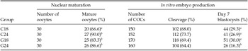
Data on oocyte nuclear maturation and on in vitro embryo production are presented in Table 1. At 18 h, the percentage of matured oocytes in the Ghr group tended to be higher compared with that of the C18 group (83.3% vs. 66.6%, P = 0.08).
Data on nuclear maturation and in vitro embryo production in 4 groups of COCs that were matured for 18 or 24 h in the absence (groups C) or in the presence (groups G) of ghrelin

a,b Within columns, different superscripts indicate significant differences (P < 0.05).
Our results imply that in the presence of ghrelin oocyte maturation was accelerated. The data on embryo yield revealed that blastocyst formation rate was the lowest in Ghr24, while no differences were detected between the remaining three groups. These data confirm our previous findings that the presence of ghrelin during in vitro maturation affects in vitro embryo development rates (Dovolou et al., Reference Dovolou, Messinis, Periquesta, Dafopoulos, Gutierrez-Adan and Amiridis2014a). It has to be noted that this experimental part was carried out in the present study in order to be used as an internal control for the ensuing study on protein analysis.
Akt1 in oocytes
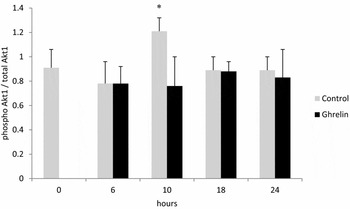
Data on Akt1 phosphorylation in control and ghrelin-treated oocytes are given in Fig. 1.
Akt1 expression in oocytes collected from COCs that were matured for 0, 6, 10, 18 or 24 h in standard IVM medium (Control), or in IVM medium supplemented with 800 pg ml–1 bovine acylated ghrelin. Bars of the same pair marked with an asterisk differ significantly (P < 0.05).

Oocytes matured in the presence of ghrelin expressed significantly lower (59.2%) Akt1 phosphorylation rate at 10 h compared with the respective controls. Akt phosphorylation in oocytes is constant with a peak at 10th hour of maturation (Salhab et al., 2011). In our experiment, control oocytes followed this pattern, while, in ghrelin-treated oocytes Akt expression was lower and similar to those detected at the ensuing stages of maturation. We infer that since ghrelin accelerates maturation, oocytes passed the 10th hour stage earlier and the peak of Akt1 activation was not picked up by our sampling protocol.
ERK1/2 in oocytes
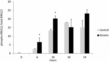
The results on ERK1/2 phosphorylation in ghrelin-treated and control oocytes are summarized in Fig. 2.
ERK1/2 expression in bovine oocytes that were matured for 0, 6, 10, 18 or 24 h in standard IVM medium (Control), or in IVM medium supplemented with 800 pg ml–1 bovine acylated ghrelin. Bars of the same pair marked with an asterisk differ significantly (P < 0.05).

At 6 and 10 h, ERK1/2 phosphorylation was increased in ghrelin-treated oocytes compared with the respective controls (264.9% and 52.5%, respectively), while no differences were detected between groups at 18 and 24 h.
Immature oocytes are arrested in GV stage and meiosis starts with the dissolution of the nuclear membrane, which in vivo is brought about by the LH surge, while in vitro it seems to be initiated after the removal of inhibitors contained in the follicular fluid. GVBD starts after 6–8 h in IVM with the activation of MPF and MAP kinases and consequently with protein synthesis and protein phosphorylation. Activation of MPF and MAP kinases starts at 6 h and increases several fold until 15 h of maturation and stays in this active form for 24 h (Torner et al., Reference Torner, Kubelka, Heleil, Tomek, Aim, Kuzmina and Guiard2001; Verlhac et al., Reference Verlhac, Kubiak, Clarke and Maro1994). In our previous experiment, at 18 h of in vitro maturation, we detected increased expression of cyclin B in ghrelin-treated oocytes that is indicative for increased activity of MPF (Dovolou et al. Reference Dovolou, Messinis, Periquesta, Dafopoulos, Gutierrez-Adan and Amiridis2014a); this result could explain the accelerated maturation rate of these oocytes. The increased ERK1/2 phosphorylation at 6 and 10 h found in the present work may promote increased protein synthesis resulting in earlier GVBD and a sooner pass to metaphase I. The determining role of protein synthesis in oocyte maturation process, has been previously shown (Sirard et al., Reference Sirard, Florman, Leibfried-Rutledge, Barnes, Sims and First1989). In this work inhibition of protein synthesis after the 6th hour of maturation, completely arrested the oocytes at the GVBD stage, while, the use of inhibitors after the 9th hour allowed 33% of the oocyte to pass the metaphase I. Increased phosphorylation of ERK with the use of Mos shortens by approximately 6 h the required time for oocytes to reach MII (18 instead of 24 h in IVM) (Fissore et al., Reference Fissore, He and Vande Woude1996).
Similar results were obtained from the present study with the use of ghrelin. Increased ERK1/2 phosphorylation in oocytes, beyond accelerated maturation, possibly contributed to increased blastocyst formation rates after 18 h of oocyte maturation.
Akt1 in cumulus cells
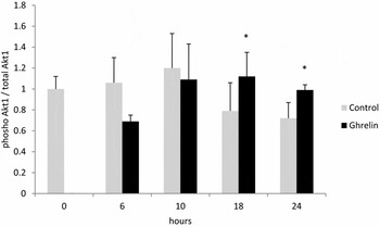
Data on Akt1 in cumulus cells are presented in Fig. 3.
Akt1 expression in bovine cumulus cells that were matured for 0, 6, 10, 18 or 24 h in standard IVM medium (Control), or in IVM medium supplemented with 800 pg ml–1 bovine acylated ghrelin. Bars of the same pair marked with asterisk differ significantly (P < 0.05).

At 18 and 24 h ghrelin caused significant increase in phosphorylation of Akt1 in cumulus cells compared with the respective controls (42.0% and 37.5%, respectively). Proper oocyte maturation is tightly linked with unhampered cumulus cells–oocyte communication, as cumulus cells provide nutrients, and factors for antioxidant protection to the oocyte. In our previous work we have shown increased expression of COX2 in cumulus cells exposed to ghrelin for 18 h (Dovolou et al., Reference Dovolou, Messinis, Periquesta, Dafopoulos, Gutierrez-Adan and Amiridis2014a). Elevated COX2 expression induces prostaglandin production, modifications of intercellular junctional complexes between oocyte and cumulus cells, and at the same time leads to increased phosphorylation of the PI3-Akt pathway (Nuttinck et al., Reference Nuttinck, Reinaud, Tricoire, Vigneron, Peynot, Mialot, Mermillod and Charpigny2002). These findings may explain the increased activity of Akt1 in ghrelin-treated cumulus cells at 18 and 24 h of maturation. In vitro matured oocytes exhibit higher Akt phosphorylation rates compared with in vivo matured ones (Salhab et al., Reference Salhab, Dhorne-Pollet, Auclair, Guyader-Joly, Brisard, Dalbies-Tran, Dupont, Ponsart, Mermillod and Uzbekova2013). With the progression of COC maturation, metabolism increases steadily that inevitably promotes oxidative stress (Sutton et al., Reference Sutton, Cetica, Beconi, Kind, Gilchrist and Thompson2003). In our previous study (Dovolou et al. Reference Dovolou, Messinis, Periquesta, Dafopoulos, Gutierrez-Adan and Amiridis2014a) we have shown increased activity of GPX1, SOD2 in oocytes implying increased anti-apoptotic, antioxidant protection and increased glucose utilization (upregulation of LDHA, SLC2A1, GAPDH) in cumulus cells. The strong anti-apoptotic and antioxidant actions of ghrelin have been shown in many cell types and these actions are mediated through the Akt and ERK1/2 pathways. For example, in rat neuronal cells cultured under oxygen and glucose deprivation ghrelin prevented apoptosis that was associated with increased activity of Akt1 and ERK1/2 kinases and with elevated levels of the anti-apoptotic Bcl-2 proteins family (Chung et al., Reference Chung, Seo, Moon and Park2008). The activation of the same pathways is linked to the proliferative actions of ghrelin in human osteoblasts, and to the mitogenic, anti-apoptotic and elevated glucose transport in adipocytes and in pancreatic β-cells (Kim et al., Reference Kim, Yoon, Jang, Park, Shin, Park, Ryu, Park, Park, Lee, Kim, Lee, Kim and Park2004; Delhanty et al., Reference Delhanty, van der Eerden, van der Velde, Gauna, Pols, Jahr, Chiba, van der Lely and van Leeuwen2006; Granata et al., Reference Granata, Settanni, Biancone, Trovato, Nano, Bertuzzi, Destefanis, Annunziata, Martinetti, Catapano, Che, Isgaard, Papotti, Ghigo and Muccioli2007).
The continuing increased activity of Akt1 in the ghrelin group in the present work it may imply that ghrelin exerts its anti-apoptotic, antioxidant protection and glucose utilization in cumulus cells through this pathway, which might be the same with the one described for other cell types.
ERK1/2 in cumulus cells
No differences were detected in ERK1/2 phosphorylation among groups (6 h 1.02 ± 0.81 vs. 0.68 ± 0.19; 10 h 0.31 ± 0.16 vs. 0.37 ± 0.12; 18 h 0.17 ± 0.05 vs. 0.23 ± 0.14; 24 h 0.14 ± 0.06 vs. 0.24 ± 0.15 for control versus ghrelin-treated cumulus cells respectively). ERK expression is constant during IVM in cumulus cells while inhibition of phosphorylation with UO126 beside the reduced cumulus expansion, arrests the oocyte to pre-metaphase I stage (Tosca et al., Reference Tosca, Uzbekova, Chabrolle and Dupont2007).
Here, we provide supportive data for our previous observation that ghrelin accelerates in vitro maturation of bovine oocytes. Our initial hypothesis, which was based on the pattern of various gene expression, is now attested by the results of the activation pattern of Akt1 and ERK1/2 in cumulus cells and in oocytes. The most plausible scenario includes that ghrelin acts directly on the oocyte through the early activation of the ERK1/2 pathway, while activation of Akt1 in cumulus cells supports the maturational process of the oocyte possibly through antioxidant protection and provision of nutrients.
Statement of interest
None