Introduction
Nasal obstruction due to inferior turbinate hypertrophy is a common problem. Hypertrophy of the turbinate is caused by either allergic or non-allergic chronic rhinitis, and is due to glandular submucosal hypertrophy, vascular congestion and collagen deposition. Nasal obstruction due to turbinate hypertrophy significantly impairs patients' quality of life.
Over the past century or more, a variety of surgical methods have been developed to reduce the volume of the inferior turbinates, creating a more patent nasal airway. These include ‘standard’ turbinectomy (excision using scissors), a variety of methods for submucosal resection of the turbinate, vidian neurectomy, electrocautery (either monopolar, using a spinal needle, or bipolar), cryotherapy, laser cautery (using several different types of laser), radiofrequency reduction (e.g. coblation) and endoscopic procedures (including the use of a debrider).
Surgical methods are associated with varying degrees of morbidity, and the outcome of the various procedures described is variable.
Material and methods
We performed an endoscopic, assisted, modified turbinoplasty technique with mucosal flaps on adult and adolescent patients who complained of nasal obstruction and were diagnosed with hypertrophied inferior turbinates. The procedure was carried out with or without other endoscopic procedures for the treatment of concomitant sino-nasal pathology, such as deviated nasal septum and chronic sinusitis. We operated only on patients who failed to respond to medical therapy. All patients underwent evaluation, including a thorough history of prior medical or surgical treatment, physical examination including anterior rhinoscopy, nasal endoscopy, and a computed tomography scan when chronic sinusitis was suspected.
Surgical technique
The procedure was performed under either local anaesthesia alone or combined local and general anaesthesia. Local anaesthesia was delivered via nasal neurosurgical cotton gauze (American Surgical company, Lynn, MA, USA) soaked with lidocaine and 2 per cent adrenaline 1:10 000 solution and packed around the inferior turbinate, the inferior meatus and the nasal cavity. After extraction of the soaked cotton gauze, the inferior turbinates were infiltrated with an intra-thecal 22 gauge needle delivering lidocaine 2 per cent and 1:100 000 adrenaline solution, for additional anaesthesia and haemostasis.
A standard 0° sinus endoscope was used in all turbinoplasty procedures.
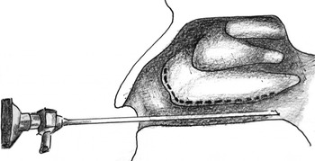
A longitudinal incision was made running inferiorly from the caudal end of the inferior turbinate up to the anterior portion (Figure 1), using a sickle knife, and completed with micro-scissors if necessary. The medial mucoperiosteal layer of the turbinate was elevated from the bony part of the turbinate in an antero-posterior direction, and from the inferior to the superior border of the turbinate, using a Freer elevator and the tip of the suction tube. In cases in which the inferior incision failed to completely release the mucoperiosteal flap, we additionally used turbinate micro-scissors.
Diagram showing the initial longitudinal incision (dotted line) running inferiorly from the caudal end of the inferior turbinate up to the anterior portion.

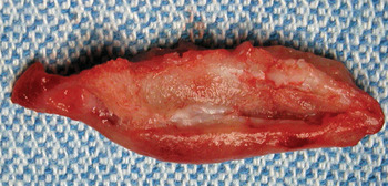
After elevation of the flap, the turbinate bone was denuded on its medial surface (Figure 2). Micro-scissors were introduced perpendicular to the dissected inferior turbinate and progressively excised an adequate volume of the turbinate, working in an antero-posterior direction. After partial excision of the turbinate bone with its attached lateral mucosa, the posterior end of the turbinate was cauterised to avoid late bleeding.
Clinical photograph showing the excised portion of the inferior turbinate; note that the medial aspect of the bone is denuded.

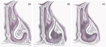
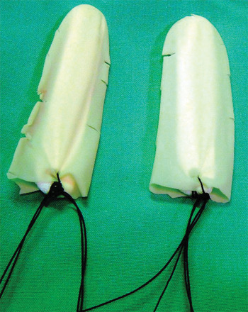
The previously elevated medial mucosal flap was then laterally rotated and repositioned to cover the remaining denuded turbinate bone and mucosal stump (Figure 3). This mucosal flap was secured in place by the introduction of a standard 8-cm nasal tampon (Merocel, Mystic, Connecticut, USA), which had previously been inserted into and sutured to a disposable glove finger, the glove finger fenestrated with scissors (Figure 4) and the whole unit smeared with synthomycin 5 per cent ophthalmic ointment. The tampon was inserted after the flap had been retracted laterally by an elongated nasal speculum. The tampon was removed after 24 hours.
Diagram of the right nose, coronal plane, showing the three consecutive surgical steps: (a) incision (dotted line); (b) mucosal flap creation and partial turbinate excision; and (c) mucosal flap repositioning over the turbinate stump.

Prepared nasal tampons: inserted into and sutured to a disposable glove finger which has been fenestrated with scissors.

This technique has been used since February 2002 for more than 400 patients undergoing inferior turbinectomy. All patients were instructed to perform nasal irrigations with saline solution for a period of up to two weeks after removal of packing. Patients were followed up for between six months and eight years, in either community or hospital-based out-patient clinics. Although we have not formally researched the effectiveness of this treatment compared with standard turbinectomy, we have observed excellent results regarding nasal obstruction, with fewer side effects and complications than the standard procedure. The great majority of our patients have reported improved nasal breathing; however, it is difficult to assess in all patients what part of their improvement is due to other concomitant procedures such as septoplasty and, occasionally, endoscopic sinus surgery.
Commonly reported complications of turbinectomy include bleeding, crusting, synechiae and empty nose symptoms.
Following the above-mentioned procedure, we did not encounter immediate post-operative bleeding requiring additional intervention. Occasionally, after tamponade removal there was a short and minimal bleeding episode, which was always controlled by local pressure. We did not encounter any late bleeding (i.e. over 24 hours after surgery), in contrast to standard inferior turbinectomy, following which there is occasionally severe bleeding from the posterior turbinate requiring haemostasis
We encountered some nasal crusting between one to two weeks after surgery. This was treated with nasal irrigation.
Rarely, small synechiae were encountered, following surgery incorporating septoplasty in addition to partial inferior turbinectomy. These synechiae were between the residual inferior turbinate and the septum. They were usually asymptomatic and easily treated.
Empty nose symptoms, such as dryness and post-nasal drip, are occasionally encountered following other turbinectomy techniques. No such symptoms were seen following our procedure.
Discussion
The primary goal of performing surgery on hypertrophied inferior nasal turbinates is to maximise the nasal airway for as extended a period as possible, while minimising complications such as excessive nasal drying, crusting, haemorrhage and pain. The advantages, disadvantages, complications and controversies of each form of treatment have been reviewed and discussed by many authors. Wight et al. Reference Wight, Jones and Beckingham1 reported that anterior trimming of the inferior turbinates cannot be recommended as a form of treatment, as the initial improvement in nasal airflow does not persist in the long term. Their conclusion, together with our observation of some unsatisfactory results of the non-endoscopic technique (caused by hypertrophy of the posterior part of the inferior turbinate), led us to develop our endoscopic technique.
The technique we describe is similar to that previously reported by Kawai et al. Reference Kawai, Kim, Okuyama and Yoshida2 but uses endoscopic instrumentation, which allows an improved view, greater precision, access to the inferior turbinate tail, and accurate mucosal flap handling and positioning.
In our experience, the healing process is simpler than in cases in which bone and mucosa are left denuded, with secondary crusting and inflammation. Removal of the tampon, prepared as described, is usually painless and bloodless.
Hol and HuizingReference Hol and Huizing3 reviewed 13 surgical treatments for inferior turbinate pathology, including electrocautery, chemocautery, cryosurgery, (subtotal) turbinectomy, laser surgery and radiofrequency. They concluded that these methods may have destructive effects on mucosal and sub-mucosal physiology, and that, judging from this perspective, they should not be used.
Jackson and KochReference Jackson and Koch4 stated that although most procedures are technically easy to perform, there is variable long-term success and significant risks, including necrosis of conchal bone, eschar formation and haemorrhage.
Passali et al. Reference Passali, Passali, Damiani, Passali and Bellussi5 reported long-term results in 382 patients randomly assigned to receive electrocautery, cryotherapy, laser cautery, submucosal resection or turbinectomy. In this study, improvement in nasal airflow following resection of the inferior turbinate was accompanied by a clinically significant loss of humidification, decreased efficiency of mucociliary transport and reduced secretory immunoglobulin A defence activities. Objective tests indicated that submucosal resection resulted in the greatest increase in airflow and nasal respiratory function with the lowest risk of long-term complications.
Wexler et al. Reference Wexler, Berger, Derowe and Ophir6 studied the long-term histological effects of inferior turbinate laser surgery, and found near-total elimination of the seromucinous gland in the region of laser treatment, with a connective tissue regenerative response, together with marked reduction in venous sinusoids.
Elwany and Abdel-Moneim,Reference Elwany and Abdel-Moneim7 conducted an electron microscopy study following carbon dioxide laser turbinectomy, and observed a decreased number and activity of seromucinous glands, fibrosis of the connective tissue stroma, and a reduction in the number and congestion of cavernous blood spaces.
Sapci et al. Reference Sapci, Sahin, Karavus and Akbulut8 concluded that although laser ablation of the turbinate was effective in improving nasal obstruction, it significantly disturbed mucociliary function.
Berger et al. Reference Berger, Ophir, Pitaro and Landsberg9 studied histopathological changes after coblation of the inferior turbinates, and found significant fibrosis, glandular and venous sinusoid depletion, and partial epithelial shedding.
Our technique combines the advantages of the submucosal and standard resection techniques. It consists of removal of hypertrophied bone and mucosa, while avoiding the undesirable depletion of important submucosal histological structures, and the harmful effects of leaving raw surfaces, by using the mucosal flap to cover the stump.
Our method usually enables resection of approximately half the turbinate volume, including both mucosa and bone. Performing this procedure endoscopically enables improved precision, better visualisation during mucosal flap incision and dissection, and precise placement of the mucosal flap and tampon. It also allows bipolar cauterisation of the inferior turbinate tail, as it enables the surgeon to approach the remote area of the tail, which often constitutes both the last obstacle to air passage and a source of secondary failure (in cases in which non-endoscopic surgery is used). Our method can reduce a large volume of the turbinate while preserving the mucosal continuity and submucosa by covering the raw surface with a mucosal flap. We believe that our method is rarely associated with post-operative side effects and complications such as dryness, infection, bleeding and pain.
Conclusion
Every reported surgical treatment for hypertrophied inferior turbinates has been noted to have advantages and drawbacks.
We believe that our endoscopic, assisted, modified turbinoplasty technique provides effective control of airway obstruction without overly interfering with the physiology of the turbinates. Furthermore, by leaving the mucosal lining continuity and submucosa intact, our technique reduces the incidence of undesirable side effects and complications associated with alternative procedures. We recommend this technique in view of its described hypothetical advantages, although we acknowledge the greater surgical skill and instrumentation required.
Additional studies are required in order to compare this technique to previously described procedures with regards to operating time, adverse effects and post-operative surgical success.