Introduction
Hormones are regulatory molecules that send signals to distant target tissues and transduce environmental and genetic signals via hormone receptors to affect gene transcription in target tissues, thereby exerting multiple phenotypic effects (Finch and Rose Reference Finch and Rose1995; Sinervo and Svensson Reference Sinervo and Svensson1998; Flatt et al. Reference Flatt, Tu and Tatar2005). Juvenile hormone (JH) and molting hormone (MH) are two important hormones in insects with major effects on various aspects of development and life history (Flatt et al. Reference Flatt, Tu and Tatar2005). For example, JH titres may affect flight behaviours or flight-capable morph by the photophase in Gryllus firmus (Scudder) (Orthoptera: Gryllidae) (Zhao and Zera Reference Zhao and Zera2004). In particular, a change in the JH and MH titres is a critical aspect of the hormonal signalling mechanism that determines variations in life history strategies in many insects (Riddiford Reference Riddiford1980; Tawfik et al. Reference Tawfik, Tanaka and Tanaka2002a; Flatt et al. Reference Flatt, Tu and Tatar2005). Despite the diversity in JH and MH secretions, the dominant JH and MH in most of the species examined include sesquiterpenoid JH with 16 carbon atoms (JH III) and 20-hydroxyecdysone (20E), which are the most widespread and biologically active hormones (Flatt et al. Reference Flatt, Tu and Tatar2005).
Juvenile hormone and/or MH regulation of diapause, which is induced by environmental conditions, such as photoperiod and temperature, is well understood (Christiansen-Weniger and Hardie Reference Christiansen-Weniger and Hardie1999; Munyiri and Ishikawa Reference Munyiri and Ishikawa2004; Singtripop et al. Reference Singtripop, Manaboon, Tatun, Kaneko and Sakurai2008). According to the positioning of diapause in an insect's life cycle, JH acts as a regulator in two distinct phases, i.e., in the larva and in the adult stage (Munyiri and Ishikawa Reference Munyiri and Ishikawa2004). Diapause in other developmental stages is generally under the control of ecdysteroids (Hartfelder Reference Hartfelder2000). A low ecdysteroid titre due to the inactivation of the prothoracic glands is considered the cause of the failure of cell differentiation and consequent developmental arrest in diapausing insects (Munyiri and Ishikawa Reference Munyiri and Ishikawa2004).
Diapause is programmed well in advance of its onset, which implies that there is a prediapause period that can be used to prepare the insect for the special conditions that lie ahead. Hence, it is common for diapause-destined insects to have a somewhat prolonged period of prediapause development, attain a larger size, and accumulate additional reserves (Denlinger et al. Reference Denlinger, Yocum and Rinehart2011). In many insects, diapause is influenced by the environmental conditions that the maternal generation experienced, as well as the diapausing generation experienced (Mousseau and Dingle Reference Mousseau and Dingle1991a, Reference Mousseau and Dingle1991b). The maternal influence that operates in controlling embryonic diapause in Bombyx mori (Linnaeus) (Lepidoptera: Bombycidae) has been well defined. In the case of bivoltine races of the silkworm, B. mori, environmental stimuli (temperature and daylight) received during the mother's embryonic development determines whether embryonic development in the next generation is arrested or continues without interruption (Katagiri et al. Reference Katagiri, Ando and Yamashita1998). 20E in the nondiapausing eggs of B. mori was observed to arise from hydrolysis of maternal conjugated ecdysteroids (Hoffmann and Lagueux Reference Hoffmann and Lagueux1985; Sonobe et al. Reference Sonobe, Tokushige, Makka, Tsutsumi, Hara and Fujimoto1999); in contrast, the metabolic process does not function in diapausing eggs (Horike and Sonobe Reference Horike and Sonobe1999; Sonobe et al. Reference Sonobe, Tokushige, Makka, Tsutsumi, Hara and Fujimoto1999), which signifies that the difference in 20E production in yolk cells determines the insect's developmental fate (Yamada and Sonobe Reference Yamada and Sonobe2003; Yamada et al. Reference Yamada, Yamahama and Sonobe2005). In some social insects, such as Apis mellifera (Linnaeus) (Hymenoptera: Apidae) (Yamazaki et al. Reference Yamazaki, Kiuchi, Takeuchi and Kubo2011) and Bombus terrestris (Linnaeus) (Hymenoptera: Apidae) (Geva et al. Reference Geva, Hartfelder and Bloch2005), a close association between the ovary status (volume of ovary or length of oocyte) and the ovarian ecdysteroid content exists; this association is strongly related to the social environment and is consistent with the premise that ovaries are the main source of ecdysteroids, just like in females of other insects (Nijhout Reference Nijhout1994). Hence, studying hormonal changes has been considered to be widely effective in understanding the maternal information transmission and diapause induction.
Cotesia vestalis (Haliday) (Hymenoptera: Braconidae), a solitary larval endoparasitoid, is one of the most important biological control agents of the diamondback moth Plutella xylostella (Linnaeus) (Lepidoptera: Plutellidae). When subjected to temperatures below 17 °C and <13 hours light per day, C. vestalis may be induced to enter facultative diapause in the prepupal stage (Alvi and Momoi Reference Alvi and Momoi1994), a transition state from larva to pupa. The second and third larval instars as a whole were sensitive stage for diapause induction (Ahmed Reference Ahmed2007). Like in a number of parasitic Hymenoptera, higher Diptera, and some Lepidoptera the mother decides the diapause fate of her progeny (Denlinger et al. Reference Denlinger, Yocum and Rinehart2011), in C. vestalis, all the maternal developmental stages had significantly effects on progeny diapause induction, especially two consecutive developmental life stages (i.e., either egg–larval and cocoon stages or cocoon and adult stages) (Ahmed Reference Ahmed2007). When parasitoids from Jilin, China were reared at 15 °C and a 11:13-hour light:dark photoperiod, the induction of diapause depended on maternal photoperiod. More than 90% of prepupae in the progeny entered diapause when the maternal generation was reared at 25 °C and a photoperiod of 8:16 hours light:dark, whereas this rate was 80.31% when the maternal generation was reared at the same temperature with a longer light phase (12:12 hours light:dark) and only 21% of progeny entered diapause when the maternal generation was reared at the same temperature under a 16:8-hour light:dark photoperiod (Ahmed et al. Reference Ahmed, Shi, Guo, Zou, Hao and Pang2007). This may be particularly significant when the mother has access to environmental cues not readily accessible to her progeny and the mother is not simply exerting some direct effect on her progeny, but initiating an effect expressed at a much later stage of development (Denlinger et al. Reference Denlinger, Yocum and Rinehart2011). In spite of the intriguing nature of these maternal effects, very little is known about the mechanisms that regulate these transgenerational effects. Hormones are well-known contributors to diapause regulation (Denlinger et al. Reference Denlinger, Yocum and Rinehart2011), so it is presumed that hormones may be as signals transmitted to the offspring.
Hormonal effects can be manipulated by administering hormone agonists, antagonists, or by removing endocrine organs (Kiuchi et al. Reference Kiuchi, Yasui, Hayasaka and Kamimura2003). However, these experiments are indirect procedures and may not necessarily affect the hormone physiologically (Islam et al. Reference Islam, Nankaku, Marui, Shahjahan, Kong and Yamanaka2005). Therefore in this study, the 20E and JH III contents in samples from cultures reared at 25 °C under 8:16-hour light:dark, 12:12-hour light:dark, and 16:8-hour light:dark photoperiods were examined directly.
Several methods may be used to measure hormones, such as nuclear magnetic resonance spectroscopy, radioimmunoassay (RIA), gas chromatography/mass spectrometry (GC/MS), and high-performance liquid chromatography (HPLC), and each method has its advantages and weaknesses. Gas chromatography/mass spectrometry and RIA, although very sensitive and specific, use specialised and costly equipment that often requires extensive derivatisation of the epoxide group or antisera (Bede et al. Reference Bede, Goodman and Tobe2000). High-performance liquid chromatography is an effective, precise, and convenient method of measuring hormones by comparison with the retention times of references (Sonobe et al. Reference Sonobe, Tokushige, Makka, Tsutsumi, Hara and Fujimoto1999; Gaudry et al. Reference Gaudry, Blais, Maria and Dauphin-Villemant2006; Zhang et al. Reference Zhang, Luo and Jiang2008; Lapenna and Dinan Reference Lapenna and Dinan2009). Some authors have concluded that no significant differences in hormone titres were observed in a comparison of HPLC with other methods (Eizaguirre et al. Reference Eizaguirre, Prats, Abellana, Lopez, Llovera and Canela1998; Bede et al. Reference Bede, Goodman and Tobe2000). Thus, in this study, HPLC was used to comparatively analyse differences in hormones under different photoperiods.
Materials and methods
Insect culture and sample preparation
Plutella xylostella was collected from cruciferous vegetable fields in the eastern suburbs of Hangzhou, Zhejiang Province, China (30°14′N, 120°10′E), in September 2009. The insect culture was maintained in the laboratory at 25 ± 1 °C, under 60–80% relative humidity (RH) and a 12:12-hour light:dark photoperiod, and then reared on potted cabbage (Brassica oleracea var. capitata (Linnaeus) (Brassicaceae) cultivar Jingfeng No. 1). Cotesia vestalis was collected from cabbage fields in the suburbs of Changchun, Jilin Province, China (43°52′N, 126°33′E), in August 2009. The stock population was maintained in the laboratory with the third instar larvae of P. xylostella at 25 ± 1 °C, 60–80% RH, and 12:12-hour light:dark photoperiod. Under such a condition, no individuals enter diapause. The populations of P. xylostella and C. vestalis had been reared for four to six generations in the laboratory prior to the experiments.
Parasitised P. xylostella larvae were obtained according to the method described by Ahmed et al. (Reference Ahmed, Shi, Guo, Zou, Hao and Pang2007), divided into three groups, and then reared on cabbage leaves held in plastic containers under three photoperiods of 8:16 hours light:dark, 12:12 hours light:dark, and 16:8 hours light:dark at 25 °C with ∼70% RH. All the results discussed were time-dependent events that occurred during a standard life cycle under culture conditions by anatomic identification. Newly formed cocoons were collected. Some of the cocoons were dissected to remove their silk shells, and groups of 10 prepupae without silk shells were used as nondiapausing prepupa samples G0 nondiapausing prepupae). The remaining collected cocoons were reared under the same conditions as that in the larvae to obtain adults. The newly emerged adults were allowed to mate, and groups of 10 mated adult females were used as adult samples (G0-mated females). The abdomens of other groups of 10 mated adult females were dissected and used as abdomen samples. The remaining mated females were used to parasitise third instar P. xylostella larvae. Groups of 200 eggs from each set of two-day-old mated females (G1 eggs before oviposition) and newly parasitised P. xylostella larvae (G1 eggs after oviposition) were collected as egg samples by dissecting mated females and the host larvae in cold saline. In order to remove possible contamination of female wasp or host larvae JH/20E, the collected eggs were rinsed five times in cells of 96-well cell culture plate by adding and removing the cold saline.
The parasitised P. xylostella larvae by adults reared at 25 °C under 8:16 hours light:dark and ∼70% RH were bred in a programmable incubator at 13 °C and 70% RH with 8:16-hour light:dark photoperiod to obtain diapauseing prepupae (G1 diapausing prepupae) (Ahmed et al. Reference Ahmed, Shi, Guo, Zou, Hao and Pang2007). Groups of 10 diapausing prepupae in each set were used as diapausing prepupa samples after the silk shells were removed.
Each prepupa or adult sample was weighed to the nearest 0.0001 g using a Sartorius electronic balance (Sartorius, Goettingen, Germany). All samples were stored at −86 °C before use in the analytical assay.
Size determination
Insects were dissected under a Leica MZ 16A stereomicroscope (Leica Microsystems, Wetzlar, Germany). Photographs of the samples were taken and observed under a Leica DM IRB fluorescence microscope (Leica Microsystems) and Nikon Stereoscopic Microscope Stand SMZ1500 (Nikon Corporation, Tokyo, Japan). Lengths and widths were measured from the microscope images using IM1000 V1.20 (Leica Microsystems AG, Heerbrugg, Switzerland) image acquisition and Photoshop software (Adobe Systems Incorporated, San Jose, California, United States of America) that were calibrated for each magnification with an image scale. Average data were acquired through multiple measurements. The volume of sample can be calculated using the following formula: V = π/4 × L × W 2 (V, volume; L, length; W, width) (Sato Reference Sato1980).
Preparation of standard solutions
Standard 20E (purity ⩾ 93%) and JH III (purity = 93%) were purchased from Sigma Chemical Company (St. Louis, Missouri, United States of America). All solvents and chemicals used for 20E and JH III content determination were of HPLC grade. Standard stock solutions of 20E and JH III were separately prepared in methanol at a concentration of 10 ng/μL. These stock solutions were diluted to six different concentrations to establish 20E and JH III calibration curves with the HPLC conditions as described in the sections “20E extraction and quantification” and “JH III extraction and quantification”. Lower diluted solutions were used to estimate the limit of detection (LOD), which is the lowest concentration level resulting in a peak area of three times the baseline noise, S/N > 3, and the limit of quantification (LOQ), which is the lowest concentration level resulting in a peak area of 10 times the baseline noise, S/N > 10.
20E extraction and quantification
20E was extracted as described by Gaudry et al. (Reference Gaudry, Blais, Maria and Dauphin-Villemant2006) with some modifications. Samples were homogenised in 1 mL 75% methanol and then centrifuged at 14 000 g for 10 minutes. Pellets were reextracted three times in the same method and supernatants resulting from each extraction were pooled. After the methanol was evaporated, the residues were partitioned three times using chloroform/double distilled water (1:1, vol/vol) to remove lipids. Aqueous phases were collected and pooled in an Eppendorf tube. After the water in the extract was evaporated at 80 °C, the extracts were dissolved in 200 μL methanol. About 20 μL of the dissolution mixture was injected into a Zorbax SB-C18 column (I.D. 5 μm diameter, 4.6 × 250 mm) on a Waters 600 controller (Waters, Milford, Massacusetts, United States of America) using methanol:distilled water (45:55, vol/vol) as the mobile phase with 0.4 mL/min flow rate and 248 nm detector wavelength. The retention time was identified by running the external standard (20E) at each series of the analyses. The 20E content in the extract was calculated by comparing the peak area with the external standard (20E). The MH content was expressed as the amount (in ng) of 20E equivalents per milligram of fresh weight (FW) or per 100 eggs (Gaudry et al. Reference Gaudry, Blais, Maria and Dauphin-Villemant2006). Each treatment was performed in triplicate.
Juvenile hormone III extraction and quantification
Juvenile hormone was extracted as described by Jiang et al. (Reference Jiang, Huang and Luo2011) with some modifications. Samples were homogenized in 1 mL of a mixture of methanol:ethyl ether (1:1, vol/vol). The mixture was sonicated for one to three minutes, extracted using 2 mL of hexane for another one minute, and then centrifuged at 11 180 g for 10 minutes at 4 °C. The hexane phase was collected, and the remainder was reextracted five times. Hexane extracts were combined and dried under a stream of N2, and then dissolved in 200 μL 80% methanol in water before analysis. Juvenile hormone III contents were determined by HPLC using the Waters 600 system equipped with a Zorbax SB-C18 column, a Waters 717 plus autosampler, and a Waters 2487 UV detector (Waters, Milford, Massachusetts, United States of America). Juvenile hormone was eluted with 80% methanol in water at a flow rate of 0.5 mL/minute with the ultraviolet light detector monitored at 218 nm. Juvenile hormone samples were identified by the retention time of the standard JH III, and quantified by comparing the peak area with the external quantitative standard (JH III) (Zhang et al. Reference Zhang, Luo and Jiang2008; Tian et al. Reference Tian, Guo, Wang, Liu, Jiang and Cao2010). The JH content was expressed as amount (in ng) of JH III equivalents per mg FW or per 100 eggs. Each treatment containing three samples was analysed three times using HPLC, and the average was set as the JH content of that treatment.
Recovery rates of authentic standards
Recovery experiments were performed to check the accuracy of the methods. 100 μL of the 10 ng/μL standard 20E and standard JH III were spiked into blank sample matrix to extracted and quantified with the same method used in extraction and quantification for 20E or for JH III.
Data analysis
All data pertaining to JH III and 20E are expressed as 100% recovery to allow comparison of different experiments (Bylemans et al. Reference Bylemans, Borovsky, Ujvary and De Loof1998). Data in this study were obtained from three independent determinations and are presented as mean ± SE. Statistical analysis was performed using the analysis of variance (ANOVA) and Fisher's least significant difference (LSD) multiple comparison tests using SAS 9.1 software (SAS Institute Incorporated, Cary, North Carolina, United States of America). P < 0.05 was set as the statistical significance criterion. Differences between preoviposition and postoviposition eggs, prepupae and mated females, as well as diapausing and nondiapausing prepupae, reared under the same photoperiod were evaluated by the Student's t-test with 95% confidence interval.
Results
Volume
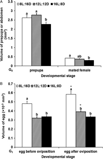
The volume of G1 diapausing prepupae was significantly larger than that of G0 nondiapausing prepupae (Table 1, Fig. 1A). The volumes of G0 prepupae under eight- and 12-hour light were significantly larger than those under 16-hour light (F = 5.04, P = 0.0085). No significant difference existed between volumes under eight- and 12-hour light. The volume of mated female abdomen increased as the light period decreased (F = 5.98, P = 0.0037).
Data of G1 diapausing and G0 nondiapausing prepupae in Cotesia vestalis.

Note: The values are the mean ± SE of three replicates. Each replicate contains 10 diapausing prepupae.
FW, fresh weight; JH, juvenile hormone.
Sizes of G0 prepupae, abdomens in mated females and G1 eggs (before and after oviposition) at 25 °C under three photoperiods (8:16, 12:12, and 16:8 hours light:dark) in Cotesia vestalis. (A) Volumes of G0 prepupae and abdomens in mated females and (B) volumes of eggs before and after oviposition. Values are expressed as mean ± SE (mm3) derived from triplicate experiments. Each replicate contains 10 prepupae, 10 mated females, or 10 eggs. Lowercase letters represent significant differences among three different photoperiods at the same developmental stage (Fisher's LSD, P < 0.05). *Indicates significant differences between the preoviposition and postoviposition eggs reared under the same photoperiod (Student's t-test, t = 0.05).

The volumes of G1 eggs before (F = 20.05, P < 0.0001) and after oviposition (F = 40.65, P < 0.0001) under eight-hour light were significantly larger than those under 12- and 16-hour light. No significant difference existed between volumes under 12- and 16-hour light. Compared with the volume of eggs before oviposition, egg volumes after oviposition increased significantly under eight- and 12-hour light. However, the egg volumes did not increase under 16-hour light (Fig. 1B).
HPLC method validation
Yang et al. (Reference Yang, Dai, Han and Wang1997) reported that, during the hormonal extraction process, the extractive solution was purified by thin layer chromatography, but the effect was not evident. The direct use of crude extracts appears to be most suitable in terms of low hormone titres in samples. The samples are more active and the controls cause fewer malformations because less processing is required (Eizaguirre et al. Reference Eizaguirre, Prats, Abellana, Lopez, Llovera and Canela1998).
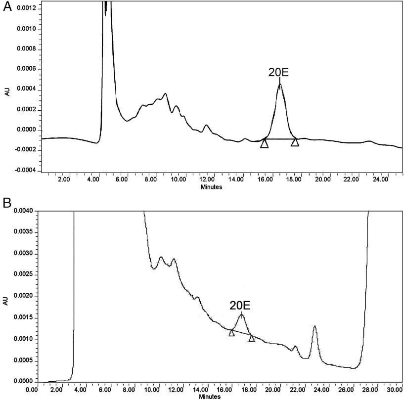
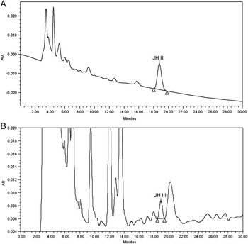
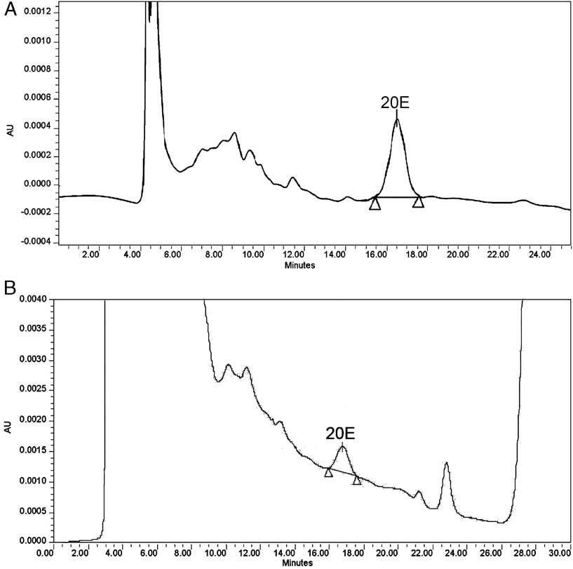
Retention times of standard 20E and standard JH III were 16.496 minutes (Fig. 2A) and 18.753 minutes (Fig. 3A), respectively. 20E and JH III in the samples were identified based on the retention times, which were similar to the references (Figs. 2B and 3B).
High-performance liquid chromatography results of the crude reaction mixture of 20E in an analytical Zorbax SB-C18 column subjected to a mobile phase (methanol:distilled water = 45:55, vol/vol) at a flow rate of 0.4 mL/minute. The effluent was monitored at λ = 248 nm. (A) 10 ng/μL authentic 20E standard and (B) G0 prepupae reared under 16:8-hour light:dark photoperiod.
High-performance liquid chromatography results of the crude reaction mixture of JH III in an analytical Zorbax SB-C18 column subjected to a mobile phase (methanol:distilled water = 80:20, vol/vol), at a flow rate of 0.5 mL/minute. The effluent was monitored at λ = 218 nm. (A) 10 ng/μL authentic JH III standard and (B) G0 prepupae reared under 16:8-hour light:dark photoperiod.

The calibration curves of the standard solutions of known amounts of the 20E and JH III authentic standards were linear with the range of 0.1 ng/μL–10 ng/μL (R 2 > 0.9991). The LOD of 20E and JH III was estimated to be 0.03 ng/μL and 0.04 ng/μL, respectively. The LOQ of 20E and JH III was 0.1 ng/μL. The recovery rates of standard 20E and standard JH III were higher than 87.71% and 89.51%, respectively.
HPLC-identified characteristics of the standard 20E and standard JH III were distinct, and the obtained retention times and peak areas were stable. The recovery rates were high, and the concentrations of 20E and JH III from samples were above the LOQ. Hence, the HPLC conditions used in our study are reliable.
20E and JH III contents
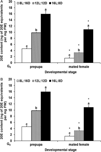
The 20E content was significantly lower in G1 diapausing prepupae than that in G0 nondiapausing prepupae (Table 1, Fig. 4A). 20E contents were reduced at both the G0 nondiapausing prepupal (F = 120.76, P < 0.0001) and adult stages (F = 144.75, P < 0.0001) as the light period was shortened and were significantly lower in G0-mated females than that in G0 prepupae (Fig. 4A). In G1 eggs (both before and after oviposition), 20E contents were lowest under the eight-hour short-light condition. Compared with 20E contents in eggs before oviposition, the content in eggs after oviposition increased significantly under the eight-hour short-light and 16-hour long-light conditions. However, the content did not increase under the 12-hour light condition (Fig. 4B).
20E contents of G0 prepupae, mated females and G1 eggs (before and after oviposition) at 25 °C under three photoperiods (8:16, 12:12, and 16:8-hour light:dark photoperiod) in Cotesia vestalis. (A) 20E contents of G0 prepupae and mated females and (B) 20E contents of G1 eggs before and after oviposition. Values are expressed as mean ± SE (ng of 20E equivalents/mg fresh weight or per 100 eggs) derived from triplicate experiments (using an average of 10 prepupae, 10 mated females, or 200 eggs for each homogenate). Lowercase letters represent significant differences among three different photoperiods at the same developmental stage (Fisher's LSD, P < 0.05). *Indicates significant differences between the prepupae and mated females and between the preoviposition and postoviposition eggs reared under the same photoperiod (Student's t-test, t = 0.05).

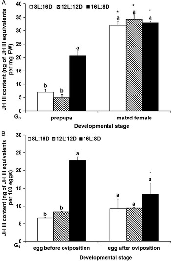
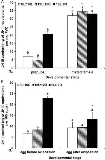
No significant difference was detected between JH III contents in G0 nondiapausing prepupae reared under eight hours of short-light and G1 diapause prepupae (Table 1, Fig. 5A). The JH III content in G0 nondiapausing prepupae reared under 16 hours of long light was significantly higher than that reared under eight hours of short light or 12 hours of light (F = 33.91, P = 0.0005). Moreover, the JH III contents in the G0-mated females reared under all three photoperiods showed no significant differences among each other but were significantly higher than those in G0 prepupae (Fig. 5A). In G1 eggs before oviposition, JH III contents were significantly lower under eight hours or 12 hours of light than those under 16 hours of light (F = 243.46, P < 0.0001). However, after oviposition, no significant difference was detected in JH III contents among eggs reared under the three photoperiods. The JH III content in eggs after oviposition decreased significantly under 16 hours of light periods, but those under eight hours and 12 hours of light did not, compared with the content in eggs before oviposition (Fig. 5B).
Juvenile hormone III contents of G0 prepupae, mated females, and G1 eggs (before and after oviposition) at 25 °C under three photoperiods (8:16, 12:12, and 16:8-hour light:dark photoperiod) in Cotesia vestalis. (A) Juvenile hormone III (JH) contents of G0 prepupae and mated females and (B) JH III contents of G1 eggs before and after oviposition. Values are expressed as mean ± SE (ng of JH III equivalents/mg fresh weight or per 100 eggs) derived from triplicate experiments (using an average of 10 prepupae, 10 mated females, or 200 eggs for each homogenate). Lowercase letters represent significant differences among three different photoperiods at the same developmental stage (Fisher's LSD, P < 0.05). *Indicates significant differences between the prepupae and mated females and between the preoviposition and postoviposition eggs reared under the same photoperiod (Student's t-test, t = 0.05).
Discussion
Diapause is programmed by environmental cues well before the onset of arrest, and diapause-destined insects often alter their physiology during the prediapause preparatory period. Changes in body size and the accumulation of energy reserves are among the most conspicuous alterations that occur during the prediapause period (Denlinger et al. Reference Denlinger, Yocum and Rinehart2005). Increased body mass is typically correlated with increased nutrient reserves, and commonly observed in diapause-destined insects; therefore, larger body sizes in diapausing individuals are generally considered to be adaptive because of their greater reserves (Hahn and Denlinger Reference Hahn and Denlinger2007, Reference Hahn and Denlinger2011). In Calliphora vicina (Robineau-Desvoidy) (Diptera: Calliphoridae), fewer small individuals entered diapause than large individuals (60% versus 95%) (Saunders Reference Saunders1997). In the present study, the G1 diapausing prepupae were larger than G0 nondiapausing prepupae, and the G0 nondiapausing prepupae were significantly larger under short photoperiods than under long photoperiods, supported by our previous study showing that the larger insects own more reserves. Because growth does not occur during the adult stage, thus the size of the adult reflects the mass of the larva at the end of larval development (Nijhout Reference Nijhout2003). Similar to prepupal stage, the size of mated females in C. vestalis was also larger under short light than under long light. Affected by maternal generation, the trends of G1 egg volumes before and after oviposition were similar to those at adult stage. It implies that C. vestalis initiates to accumulate reserves during prediapause preparatory period, such as glycogen (the primary carbohydrate reserve) and triglyceride accumulation, suggesting that the expression of alternative developmental tactics is influenced by environmental condition through the alteration of the body size and internal nutritional state (Hahn and Denlinger Reference Hahn and Denlinger2007).
The distinctive differences, such as reserves and body sizes, between diapause-destined and nondiapause-destined individuals were most likely regulated by the endocrine system (Denlinger et al. Reference Denlinger, Yocum and Rinehart2011). Juvenile hormone and ecdysteroid are key endocrine signals in insect development (Riddiford et al. Reference Riddiford, Hiruma, Zhou and Nelson2003), and can be influenced by photoperiod and temperature (Eizaguirre et al. Reference Eizaguirre, Prats, Abellana, Lopez, Llovera and Canela1998; Tawfik et al. Reference Tawfik, Tanaka and Tanaka2002b; Munyiri and Ishikawa Reference Munyiri and Ishikawa2004; Zhao and Zera Reference Zhao and Zera2004). Hormones not only regulate various physiological metabolisms, but also transmit environmental and genetic signals via hormone receptors to affect gene transcription in target tissues, thereby exerting multiple phenotypic effects (Flatt et al. Reference Flatt, Tu and Tatar2005; Marchal et al. Reference Marchal, Vandersmissen, Badisco, Van de Velde, Verlinden and Iga2010). Previous studies have implicated 20E signalling in the fat body of Drosophila melanogaster (Meigen) (Diptera: Drosophilidae) was a key component in size control (Mirth and Riddiford Reference Mirth and Riddiford2007). Rharrabe et al. (Reference Rharrabe, Bouayad and Sayah2009) found that 20E induced a decrease in glycogen, lipid contents, and larval weight in Plodia interpunctella (Hübner) (Lepidoptera: Pyralidae).
The 20E content was significantly lower in G1 diapausing prepupae than that in G0 nondiapausing prepupae (Table 1, Fig. 4A), and within the nondiapausing prepupae of C. vestalis, 20E contents decreased in individuals with decrease of photoperiods. Similarly in Antheraea mylitta (Drury) (Lepidoptera: Saturniidae), a relatively high 20E level was observed in nondiapause-destined generation larvae under long light on the last day of the fifth instar and at gut purge compared with the same stages of diapause-destined generation larvae under short light (Kumar et al. Reference Kumar, Subrahmanyam, Sharan, Mishra, Singh and Suryanarayana2008). Perhaps under long light, pupation needs more ecdysteroid to be initiated (Pérez-Hedo et al. Reference Pérez-Hedo, Goodman, Schafellner, Martini, Sehnal and Eizaguirre2011), and under short light the bigger individuals having more reserves affect adversely the accumulation of 20E (Marchal et al. Reference Marchal, Vandersmissen, Badisco, Van de Velde, Verlinden and Iga2010).
Adults integrate and respond to environmental information through subtle changes in ecdysone content (Schwedes and Carney Reference Schwedes and Carney2012). Under the three photoperiods, the variation trend of 20E content in mated females of C. vestalis was similar to that in prepupae, increasing with prolonged photoperiod, while the volumes of G0 female abdomen and G1 egg under eight-hour light were significantly larger than those under 16-hour light. This difference in ecdysteroid production among the three photoperiods might be due to the difference of volume of organ or the amount of substrate stored and available in these organs (Yamazaki et al. Reference Yamazaki, Kiuchi, Takeuchi and Kubo2011). A significant study by Colombani et al. (Reference Colombani, Bianchini, Layalle, Pondeville, Dauphin-Villemant and Antoniewski2005) demonstrated that disruption of 20E signalling in the fat body of Drosophila results in larger animals at pupariation. In B. terrestris, queens heading colonies have large active ovaries with high ecdysteroid content, whereas virgin gynes and mated queens before and after diapause have undeveloped ovaries with low ecdysteroid content (Geva et al. Reference Geva, Hartfelder and Bloch2005). Geva et al. (Reference Geva, Hartfelder and Bloch2005) considered the volumes as the indices of ovarian status. In fact it is well known that ecdysteroids are secreted by the follicle cells of the ovaries (Lafont et al. Reference Lafont, Dauphin-Villemant, Warren and Rees2005), and the follicle cell diameter increases more or less in parallel with the increase in oocyte length (Pascual et al. Reference Pascual, Cerdá, Benito, Tomás, Piulachs and Bellés1992). However, in overwintering Polistes gallicus (Linnaeus) (Hymenoptera: Vespidae) females, high ecdysteroid titres were detected, when their ovaries contained only small follicles (Strambi et al. Reference Strambi, Strambi and De Reggi1977). This suggests that the relationship between the oocyte sizes and ecdysteroid titres is different in different insect species. The contents of 20E in mated females were lower than those in prepupae in the present study. This result is consistent to the reports on D. melanogaster and Spodoptera littoralis (Boisduval) (Lepidoptera: Noctuidae), where 20E in adult was also at lower levels than in earlier developmental stages (Polanska et al. Reference Polanska, Maksimiuk-Ramirez, Ciuk, Kotwica and Bebas2009; Schwedes and Carney Reference Schwedes and Carney2012). The lower 20E level in adult was probably due to that prepupal stage is a transition state from larva to adult and that the prothoracic glands in the adult stage is degenerating (Tawfik et al. Reference Tawfik, Vedrova, Li, Sehnal and Obeng-Ofori1997).
Ecdysteroids secreted by the follicle cells of the ovaries in adult females are converted to conjugates, and linked to the yolk proteins, then incorporated into eggs (Lafont et al. Reference Lafont, Dauphin-Villemant, Warren and Rees2005; Denlinger et al. Reference Denlinger, Yocum and Rinehart2011). The conjugated ecdysteroids, present in the yolk, are hydrolysed by phosphatases during the embryonic development (Hagedorn Reference Hagedorn1985; Martini et al. Reference Martini, Chieco, Dindo and Baronio2011). The maternal effect on diapause of progeny is transmitted prior to the onset of embryogenesis, probably during oogenesis (Henrich and Denlinger Reference Henrich and Denlinger1982; Denlinger et al. Reference Denlinger, Yocum and Rinehart2011). In eggs of C. vestalis, 20E was also lowest in short-day conditions. These results are consistent with the results obtained by Tawfik et al. (Reference Tawfik, Tanaka and Tanaka2002a, Reference Tawfik, Tanaka and Tanaka2002b), who found that 20E contents in Locusta migratoria (Linnaeus) (Orthoptera: Acrididae) were significantly lower in the ovaries from short-day females than those from long-day females and in newly laid eggs by short-day females than by long-day females. Tawfik et al. (Reference Tawfik, Tanaka and Tanaka2002a, Reference Tawfik, Tanaka and Tanaka2002b) considered that the embryonic diapause is mediated by maternally produced hormone or hormones (ecdysteroids), and these hormones are produced depending on the environmental conditions experienced by the female parents and transmitted into their eggs to determine whether the offspring will enter diapause or develop to hatch. Because a change in the ecdysteroid titres is well known to be a critical aspect of hormonal signalling mechanism in many insects (Riddiford Reference Riddiford1980), the discrepant accumulation of 20E under different photoperiods might be a signal transmitted from ovary to eggs in progeny to influence the development of progeny. After oviposition, the contents of 20E in eggs under eight and 16 hours were higher compared with before oviposition, which were possibly due to de novo synthesis or/and dephosphorylation of maternal 20E 22-phosphate (Sonobe et al. Reference Sonobe, Tokushige, Makka, Tsutsumi, Hara and Fujimoto1999). The surge in the level of 20E probably relates to ecdysteroids participating in controlling early events of embryonic development (Makka et al. Reference Makka, Seino, Tomita, Fujiwara and Sonobe2002), such as gastrulation, germ band retraction, head involution, cuticle formation (Hoffmann and Lagueux Reference Hoffmann and Lagueux1985), and resistance to immunity of host (Genta et al. Reference Genta, Souza, Garcia and Azambuja2010; Schwedes and Carney Reference Schwedes and Carney2012).
In the present study, the JH III content in G1 diapausing prepupae did not significantly differ from that in G0 nondiapausing prepupae reared at the same photoperiod (8:16 hours light:dark). In Nasonia vitripennis (Walker) (Hymenoptera: Pteromalidae), no significant difference was observed between the JH contents in diapausing and nondiapausing larvae (De Loof et al. Reference De Loof, van Loon and Vanderroost1979). In Mamestra brassicae (Linnaeus) (Lepidoptera: Noctuidae), diapausing pupae had lower JH contents than nondiapausing pupae (Endo et al. Reference Endo, Fujimoto, Kondo, Yamanaka, Watanabe and Kong1997). In contrast, in Sesamia nonagrioides (Lefebvre) (Lepidoptera: Noctuidae) (Eizaguirre et al. Reference Eizaguirre, Schafellner, Lopez and Sehnal2005, Reference Eizaguirre, Lopez, Schafellner and Sehnal2007) and Psacothea hilaris (Pascoe) (Coleoptera: Cerambycidae) (Munyiri and Ishikawa Reference Munyiri and Ishikawa2004), diapausing larvae had higher JH contents than nondiapausing larvae. In S. nonagrioides and P. hilaris, larvae undergo stationary molts during diapauses, and JH titres remain elevated, thus confirming that molting can be stationary rather than progressive (Denlinger Reference Denlinger2002). Hence, the JH content differentiation between diapausing and nondiapausing individuals depends on the insect species and the environment experienced by the nondiapausing individuals. Juvenile hormone is not an active player in many forms of insect diapause (Denlinger et al. Reference Denlinger, Yocum and Rinehart2011).
Similar to the trend of 20E, JH III titres in nondiapausing prepupae under long photoperiod (16:8 hours light:dark) were higher than those under short photoperiodic (8:16 hours light:dark, 12:12 hours light:dark) conditions. However, the content was significantly higher than that in prepupae reared under 12 hours of light, and lower than that under 16 hours of light. Obviously, the differences in JH contents in G0 nondiapausing prepupae were caused by the different photoperiods since the temperature used was consistent. This result was consistent with the report on the pea aphids, Acyrthosiphon pisum (Harris) (Homoptera: Aphididae). They had lower JH III titres when reared under short-day lengths than those reared under long-day lengths (Ishikawa et al. Reference Ishikawa, Ogawa, Gotoh, Walsh, Tagu and Brisson2012). Ishikawa et al. (Reference Ishikawa, Ogawa, Gotoh, Walsh, Tagu and Brisson2012) suggested that JH esterase was a good candidate as a critical factor regulating JH titres in response to the decrease of photoperiod. Ferenz (Reference Ferenz1981) examined the effect of photoperiod on corpus allatum (CA) activity in vitro and showed that in Pterostichus nigrita (Paykull) (Coleoptera: Carabidae), short-day conditions were correlated with low levels of CA activity resulting in low JH III titres. Hodkova et al. (Reference Hodkova, Okuda and Wagner2001) thought that CA activity was stimulated by long light, via the brain, thereby secreting JH.
In fifth instar larvae of P. hilaris (Pascoe) (Coleoptera: Cerambycidae), the contents of JH III and 20E under long day were more than under short day, and the content of JH III was lower than that of 20E. The developmental profile showed 20E titres in the seventh instar larvae remained at lower levels than those in the fifth instar prediapause larvae, and the content of JH III was higher than 20E (Munyiri and Ishikawa Reference Munyiri and Ishikawa2004). In our study, the contents of JH III in prepupae were higher than those of 20E under 8:16-hour light:dark and 16:8-hour light:dark photoperiods, and inversely it was lower than that of 20E under 12:12-hour light:dark photoperiod, suggesting that although 20E and JH coregulate the developmental progresses (Suzuki et al. Reference Suzuki, Sakurai and Iwami2011), relative superiority or inferiority is influenced by photoperiod, developmental stage, and insect species.
Juvenile hormone III titres of C. vestalis-mated females were higher than those in prepupae, although no differences were observed among three photoperiods. The higher JH level in the adult was probably because JH plays a major role in the control of sexual maturation and reproduction of insects (Bendena et al. Reference Bendena, Zhang, Burtenshaw and Tobe2011; Wijayaratne et al. Reference Wijayaratne, Fields and Arthur2012). A decrease in JH production in the CA induces cessation of reproduction, specifically, the arrest of vitellogenesis and regression of the ovaries (Wasielewski et al. Reference Wasielewski, Wojciechowicz, Giejdasz and Krishnan2011b). Cotesia vestalis adult stage does not enter diapause so the normal capacity of reproduction needs to be preserved regardless of photoperiods.
Eggs before oviposition contained less JH under short-day (8:16-hour light:dark, 12:12-hour light:dark photoperiod) than under long-day (16:8-hour light:dark photoperiod) conditions. Perhaps, the differential accumulation of JH III, which is involved in differential synthesis of vitellogenic proteins (Richard et al. Reference Richard, Jones, Barbarito, Cerula, Detweiler and Fisher2001) and absorption (Stoffolano et al. Reference Stoffolano, Li, Zou and Yin1992), influences the size of oocytes (Wasielewski et al. Reference Wasielewski, Giejdasz, Wojciechowicz and Skrzypski2011a) and performs an important nutritional role during embryogenesis, acting as a source of amino acids, lipids, and carbohydrates for the developing embryo (Masuda and Oliveira Reference Masuda and Oliveira1985) to cooperate with 20E to complete information transmission. Short photoperiods had no effect on JH titres of eggs before and after oviposition, but long photoperiods decreased JH titres in eggs after oviposition; thus, egg JH levels after oviposition were similar under all photoperiodic regimes. Owing to significantly increased 20E in eggs after oviposition under long light, decreased JH III titres might be used to resist the immunity of host, suggesting that the eggs reared under long light were weaker at responding to stress. The contents of 20E both in eggs before and after oviposition were higher than those of JH III, suggesting that 20E plays more important role compared with JH III during embryonic development.
When the photoperiodism module produces an output signal that ultimately results in diapause, the hormones involved in diapause must be synthesised, secreted, and degraded at the appropriate rates and times. Hence, the hormonal control of diapause can be considered as a second module between photoperiodism and diapause itself (Emerson et al. Reference Emerson, Bradshaw and Holzapfel2009). In G1 diapausing prepupae of C. vestalis, the size was significantly larger, but 20E content was significantly lower than that of nondiapausing prepupae. And the varying trend of 20E with photoperiodic variation was in accordance with that of the offspring diapause incidences in C. vestalis (Ahmed et al. Reference Ahmed, Shi, Guo, Zou, Hao and Pang2007), that is, the higher the content of 20E was, the lower the diapause incidence was. Hence, 20E was speculated to not only transport maternal environmental information to the offspring, but also regulate diapause in the offspring, similar to most insects wherein diapauses take place at the larval and pupal stage. The larval and pupal diapause is usually caused by a decreased ecdysteroid titre in the haemolymph (Endo et al. Reference Endo, Fujimoto, Kondo, Yamanaka, Watanabe and Kong1997; Singtripop et al. Reference Singtripop, Wanichacheewa, Tsuzuki and Sakurai1999; Munyiri and Ishikawa Reference Munyiri and Ishikawa2004; Eizaguirre et al. Reference Eizaguirre, Lopez, Schafellner and Sehnal2007), accompanied by elevated JH in some insects (Munyiri and Ishikawa Reference Munyiri and Ishikawa2004; Eizaguirre et al. Reference Eizaguirre, Schafellner, Lopez and Sehnal2005, Reference Eizaguirre, Lopez, Schafellner and Sehnal2007). However, in the present study, the JH content in G1 diapausing prepupae was not elevated compared with the content in G0 nondiapausing prepupae reared at the same photoperiod but under different temperature. Some previous studies showed that the topical application of 20E to several diapausing insects with very low ecdysteroid titres terminates the diapause and the insect resumes development (De Loof et al. Reference De Loof, van Loon and Vanderroost1979; Christiansen-Weniger and Hardie Reference Christiansen-Weniger and Hardie1999; Singtripop et al. Reference Singtripop, Wanichacheewa and Sakurai2000; Islam et al. Reference Islam, Nankaku, Marui, Shahjahan, Kong and Yamanaka2005). However, application of the JH analogue could not induce diapause (Christiansen-Weniger and Hardie Reference Christiansen-Weniger and Hardie1999). Therefore, diapause is considered to be associated with the reduction or deficiency of the ecdysteroid titre (Singtripop et al. Reference Singtripop, Wanichacheewa and Sakurai2000; Denlinger Reference Denlinger2002; Islam et al. Reference Islam, Nankaku, Marui, Shahjahan, Kong and Yamanaka2005; Singtripop et al. Reference Singtripop, Manaboon, Tatun, Kaneko and Sakurai2008). Further studies are needed to confirm whether or not the diapause of C. vestalis is triggered by decreased MH titres, although positive results are expected from these studies.
Lafont et al. (Reference Lafont, Dauphin-Villemant, Warren and Rees2005) consider that insect development is absolutely dependent on subtle changes in ecdysteroid titre, so based on above results, it is speculated that 20E may be transmitted to the egg as a maternal environmental signal, influencing the fate of the offspring. The diapause in C. vestalis might be decided by the decrease of 20E or to high ratio of JH to 20E. This can be confirmed by applying topically 20E/JH III or 20E/JH III antagonists on diapause-destined prepupae or by injecting them into hosts and/or by excising the organs producing 20E/JH III to affect the hormone content in the eggs of C. vestalis, followed by investigating the change in diapause percentage. 20E and JH III are the most widespread and biologically active hormones found outside of Lepidoptera (Flatt et al. Reference Flatt, Tu and Tatar2005). In this study, the 20E and JH III contents were monitored, but whether there are other types of JH and MH were not ascertained. Therefore, identification and determination of other forms of MH and JH, and their roles can be explored in future studies.
Acknowledgements
Funding for this study was jointly provided by the 973 Program (grants 2009CB119005 and 2006CB102005), the 948 Program (grant 2011-G4), and the National Natural Science Foundation of China (project number U0936601).