Introduction
The principle of radiation therapy is to deliver a curative dose or the highest possible dose uniformly to the planning treatment volume (PTV) and to avoid the dose to the surrounding healthy tissues. In this process, a percentage of dose is delivered outside the PTV and this dose delivered outside the field edges is known as peripheral dose (PD). The PD is due to three factors:
-
a. Leakage radiation from the treatment machine head.
-
b. Scattered radiation from the irradiated volume of the human body.
-
c. Scattered radiation from the treatment couch and walls of the room.
As the survival of the patients after the radiation therapy has increased in recent years because of increased awareness, early diagnosis and better treatment methods, PD gains more importance, especially in radiosensitive organs. The percentage of PD is very small as compared to the prescribed dose but still be of concern clinically that may cause cataract formation, gonadal dysfunction, cardiovascular diseases and infertility. It can also be responsible for radiation-induced carcinogenesis in the breast, other tissues and foetus in the pregnant women. 1–Reference Palm and Johansson3 Further, the PD can affect the implanted electronic device like a pacemaker.
The treatment planning system (TPS) calculates the doses within the field accurately but is not able to estimate the doses outside the treatment area. Reference Huang, Followill and Wang4 The PD has been the topic of interest of various researchers in recent years and the factors that influence the PD such as field size, gantry angle, energy, and multi-leaf collimator have been studied by them. Different radiation detectors such as thermoluminescent detectors (TLD), diodes, metal-oxide semiconductor field-effect transistor (MOSFET), ionisation chambers, optically stimulated luminescent dosimeter have been used in the study of PD. Reference Scarboro, Followill and Kerns5–Reference Knezevic, Stolarczyk and Bessieres7 Nevertheless, publications that include the PD measurements using a two-dimensional ionisation chambers array, IMatrixx, are sparse. In this study, we have attempted to develop a dataset of the PD values measured with the IMatrixx (IBA dosimetry, GmbH, Germany).
Methods
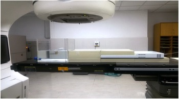
The Clinac 2100-C (Varian Medical Systems, Palo Alto, CA, USA) capable of producing 6 and 15 MV photons and 6, 9, 12 and 15 MeV electrons was used in this study. The two-dimensional linear array detector, IMatrixx which has 729 parallel plate ion chambers and separated with a distance of 7 mm, was used to measure the PD from the edge of the field and to a distance up to 45 cm from it. Solid water slabs of thickness 1 and 0·5 cm and water phantom were used to get the required geometry for the PD measurement and the arrangement is as shown in Figure 1.
Phantom setup for measurement of the peripheral dose.

The IMatrixx was first calibrated for the energies 6 and 15 MV as per the vendor protocol and the calibration yielded the user correction factor for both the energies. The IMatrixx was placed on a solid water phantom of thickness 5 cm, to provide sufficient backscatter. The slab of thickness 1·5 cm was placed over it, so that the detector lies at the depth of dmax, and was irradiated for 100 MU with the 6 MV photon beam. The percentage deviation of the dose measured by the IMatrixx and that of the dose measured with 1D water phantom as per the TRS-398 protocol were compared. The same procedure was repeated for 15 MV photon beams with a slab thickness of 3 cm.
The various factors that influence the PD are the field size, collimator angle, gantry angle, depth, source to surface distance (SSD), treatment technique and energy. The IMatrixx was irradiated such that the first row of detectors coincided with the edge of the optical field. The measurements were done for different field sizes of 5 × 5, 10 × 10, 15 × 15 and 20 × 20 cm2, collimator angle 0° and 90°, SSD 85, 90, and 95 cm, and for depths, 5 cm, dmax and surface. The influence of gantry angles, 30°, 60°, 300° and 330° and energies, 6 and 15 MV on the PD, was also studied.
The PD was measured for a distance of 23 cm from the edge of the field and then the IMatrixx was shifted by 23 cm and the same procedure was repeated. The IMatrixx was irradiated for 100 MU for each of the above-mentioned measurements, and the image was acquired in cine mode. The values were then exported in ASCII format, and the central axis values were identified. The values were converted into a percentage of the dose measured at the centre of the field. The % PD is calculated as, % PD = (PD (measured)/isocentre dose) × 100, where PD (measured) is the absolute dose in Gy at an appropriate point.
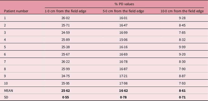
To validate the PD values generated using the IMatrixx, ten cervical cancer patient verification plans were chosen and TLD-100 chips were placed on the slab phantom at a distance of 1, 5 and 10 cm from the edge of the field. The % of the dose received at these points was recorded using the Thermo Scientific TLD 3500 reader. The PD values measured with the TLD were compared with the IMatrixx-measured values.
Results
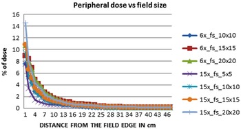
The data set of PD was acquired for field sizes from 5 × 5 cm2 to 20 × 20 cm2 in steps of 5 cm, collimator rotation 0° and 90°, SSD-90, 95 and 100 cm, gantry angles in steps of 30° and at depths surface, dmax and 5 cm as a function of distance up to 45 cm. The variation of PD with field size as a function of distance was studied for both 6 and 15 MV photon beams at the depth of dmax, and it is found to increase with field size due to the increase in the scatter volume and the same is shown in Figure 2.
Variation of peripheral dose with field size.

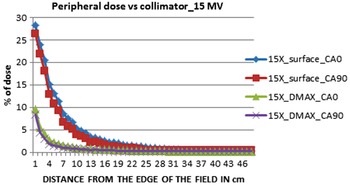
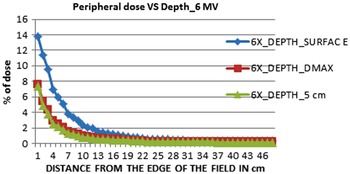
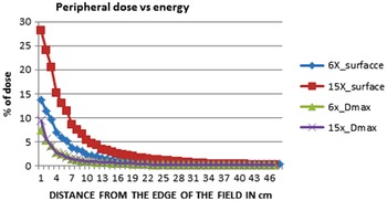
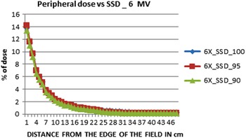
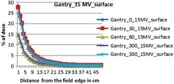
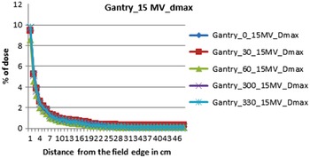
The influence of the collimator on the PD was studied at surface and dmax depth and is shown in Figure 3 for 15 MV beams and the same trend is noted for 6 MV beams. The depth has a major influence on the PD. The measurements were taken at three different depths: surface, dmax and 5 cm for both 6 and 15 MV beams, which are shown in Figure 4. The variation of PD with energy at surface and dmax depth is shown in Figure 5. The effect of SSD on the PD is shown in Figure 6 for 6 MV beams. The effect of gantry angle on the PD was studied for both energies and depths: surface, dmax and the results are shown in Figures 7 and 8, respectively.
Variation of peripheral dose with collimator.

Variation of peripheral dose with depth.

Variation of peripheral dose with energy.

Variation of peripheral dose with SSD.

Variation of peripheral dose with gantry angle at surface.

Variation of peripheral dose with gantry angle at dmax depth.

The TLD-100 chips-measured % PD along with the mean and standard deviation is shown in Table 1. The comparison between the TLD and IMatrixx % PD value is presented in Table 2. The results of the mean and standard deviation of the TLD measurements prove its efficacy and the percentage variation of PD values measured at 1, 5 and 10 cm depths using IMatrixx and TLD were matched well with a maximum deviation of 3·16%.
The % PD values measured with the TLD

Comparison of the % PD values measured with the TLD and IMatrixx

Discussion
The PD gains more importance as the low doses of radiation can induce secondary cancers in highly radiosensitive organs. Furthermore, the PD in children, pregnant women and patients with pacemakers can develop difficulties. The protocols for decreasing the probability of secondary cancer risk have been previously studied. Reference Cosset, Hetnal and Chargari8 The Biological Effects of Ionizing Radiation Report VII uses the linear no-threshold dose model for the estimation of secondary cancer risks. 9 Various techniques have been developed for the measurement of PD. Several authors have studied the influence of field size, depth, energy, treatment distance, gantry and collimator angle on the PD using detectors like the TLD, MOSFET, ionisation chamber and diodes. Reference Scarboro, Followill and Kerns5–Reference Knezevic, Stolarczyk and Bessieres7 The present study evaluated the PD measurements using the IMatrixx ion chamber array.
Klein et al. Reference Klein, Maserang and Wood10 concluded from his phantom study using micro and cylindrical ionisation chambers that the PD values for closer points were more for lower energies and larger field sizes and it was higher for high-energy beams for points more distant from the field edge. Similar results were obtained in this study for % PD values. Up to a distance of 10 cm from the field edge, the value of 6 MV was on the higher side compared to the 15 MV beam at dmax depth. For a 20 cm × 20 cm field, the value was 4·23% and 3·73% at a 5-cm distance from the field edge for the 6 and 15 MV, respectively. At any point, the % PD increased with an increase in field size due to an increase in scatter volume irrespective of the energy.
Mohsin et al. Reference Mohsin, Zakaria and Abdullah11 reported that the changes in PD with the depth are not as significant as the changes with the field size. It has been observed from our study that the % PD values are higher at the surface and decrease with depth for both the 6 and 15 MV. Covington et al. Reference Covington, Ritter and Moran12 have discussed various parameters that affect the PD measured using ionisation chamber in their technical report. They have observed moderately higher PD for collimator angle 0° for open fields and have also observed a decrease in the PD with increasing energy. We have also found a slight increase of 0·31% for the collimator angle 0° as compared to the collimator angle 90°. Gopiraj et al. Reference Gopiraj and Ramasubramanian13 have studied in detail the efficacy of the MOSFET as a detector for PD measurement. The effects of field size, collimator rotation, depth and beam modifiers were studied. This study has concluded that the ionisation chamber and MOSFET yield similar results.
The % PD decreased with an increase in energy and the % decrease was less at dmax as compared to surface measurements. The % PD increased with SSD from 90 cm to 100 cm for both energies due to the increase in the scattering medium as we move away from the source head. The % PD decreases as we move away from 0° to 60° gantry angle on either side. The influence of gantry angle is less at dmax than at the surface and also for lower energy. The % PD is high at the surface and decreases rapidly in the buildup region after which the decrease in % PD is less. The data obtained with the IMatrixx were comparable to well-established detectors in practice for the measurement of PD. In addition, the other detectors have been provided a single-point measurement, while the arrays of detectors in the IMatrixx are able to do measurements at multiple points simultaneously. The other advantages of the IMatrixx are its detector resolution and free from any positional error for multiple point measurements compared to other detectors.
A % PD dataset was developed similar to the percentage depth dose tables for easy reference in clinical practice. The % PD look-up tables for both 6 and 15 MV photon beams are as presented in Tables 3 and 4, respectively. These look-up tables can be used to get an idea about the PD to pacemaker and foetus in relevant patients.
Look-up table of % PD for 6 MV photon beams

Look-up table of % PD for 15 MV photon beams

Most of the published studies used one of the detectors at a time for the PD measurements. The comparison of PD measured using several detectors might improve the credibility and decrease the ambiguity in the PD measurements. On the other hand, the current developments in artificial intelligence and machine learning can be used for the accurate computation of PD. Nevertheless, these modern approaches need more resources and expertise. Further research with more detectors together as well as knowledge-based computation methods is desired to gain more information in the PD measurements.
Conclusion
The PD measurements give us a clear picture of the dose outside the treatment field. With the increase in workload on every Teletherapy unit in our country, it becomes difficult to get the machine time. The IMatrixx saves a lot of time and in a single exposure, it is possible to measure the PD for 23 cm. Further, the results are comparable to the well-established detectors used for the PD measurement. The look-up dataset generated can be used in the scoring of plans and in the reporting of the PD for clinical situations which involve highly radiosensitive organs, pacemakers and foetuses in pregnant women.
Financial Support
None.
Conflict of Interest
The authors declare no conflicts of interest.
Ethical Standards
This article does not contain any studies with human participants performed by any of the authors.