INTRODUCTION
Bone and antler are extremely challenging archaeological sample matrices for radiocarbon (14C) dating due to the relative complexity of the pretreatment chemistry required to isolate and purify a suitable carbon fraction. The majority of bone samples are neither cremated nor burnt and the protein (collagen) fraction is almost exclusively the material of choice for analysis. Many different methods for collagen extraction have been reported, with varying degrees of success, following the widely adopted method proposed by Longin (Reference Longin1971). The Longin method is designed to isolate the collagen from the mineral fraction (hydroxyapatite), as well as from any contaminants present from the post-depositional environment, while simultaneously trying to avoid adding contaminants from the laboratory environment. This is a delicate balance because, the more complex the chemistry that is applied to the sample, the more potential there is that additional contaminants may be inadvertently incorporated. Since Longin’s original method was published, a number of alternative pretreatment methods have been proposed, including:
-
1. ultrafiltration to remove non-collagenous molecules of <30 kDa from the extract (Brown et al. Reference Brown, Nelson, Vogel and Southon1988; Bronk Ramsey et al. Reference Bronk Ramsey, Higham, Bowles and Hedges2004; Higham et al. Reference Higham, Jacobi and Bronk Ramsey2006; Brock et al. Reference Brock, Bronk Ramsey and Higham2007; Hüls et al. Reference Hüls, Grootes and Nadeau2007, Reference Hüls, Grootes and Nadeau2009);
-
2. a ninhydrin reaction with the α-NH2 group on free amino acids from either degraded collagen or contaminants that ultimately leads to their decomposition and release of CO2 (Nelson Reference Nelson1991; Tisnérat-Laborde et al. Reference Tisnérat-Laborde, Valladas, Kaltnecker and Arnold2003);
-
3. protein hydrolysis followed by separation and dating of single amino acids, e.g. hydroxyproline, which is rare in most other animal proteins (Stafford et al. Reference Stafford, Hare, Currie, Jull and Donahue1991; McCullagh et al. Reference McCullagh, Marom and Hedges2010); and
-
4. a two-stage pretreament method, which employs an acid/alkali step to remove humics from the bone before using the Longin method for collagen extraction (Arslanov and Svezhentsev Reference Arslanov and Svezentsev1993).
The demand for 14C dating of bone material has increased within the last few years as bone offers some advantages over charred plant remains for assuring the security of archaeological associations. This is primarily because articulated bone groups are samples that are demonstrably not residual and can therefore provide 14C ages that may be included in Bayesian models to produce dating precision to within a few decades (e.g. Bayliss and Whittle Reference Bayliss and Whittle2007).
One area of study that has provided some concerns is that of background correction. The background associated with “infinite age” bone samples has been found to be somewhat greater than that typically associated with other sample types (e.g. Tisnérat-Laborde et al. Reference Tisnérat-Laborde, Valladas, Kaltnecker and Arnold2003; Wood et al. Reference Wood, Bronk Ramsey and Higham2010) and indeed, in the SUERC Radiocarbon Laboratory, it has been found that the bone background is approximately twice that of other background materials including inter-glacial wood and geological calcite. For example, in the case of a sample of 1 half-life in age, this makes a difference of around 30 years and for two half-lives, around 60 years. Therefore, it is particularly important when the age data are to be included in Bayesian models that the 14C determinations are accurate to within the quoted precision (Bayliss et al. Reference Bayliss, Bronk Ramsey, van der Plicht and Whittle2007: Figures 21–24) and ensuring this requires the use of appropriate and well characterized background samples.
Currently, the SUERC Radiocarbon Laboratory employs a mammoth bone (Mammuthus cf. trongontherii) from Latton Quarry, Wiltshire, England, as the bone background standard. This sample (LQH12) was supplied by Dr Katharine Scott (St Cross College, Oxford). It originates from a deposit that correlates with Marine Isotope Stage 7 (MIS7) (Scott and Buckingham Reference Scott and Buckingham2001; Lewis et al. Reference Lewis, Maddy, Buckingham, Coope, Field, Keen, Pike, Roe, Scaife and Scott2006) and should therefore be of infinite age with respect to radiocarbon. This was verified by Cook et al. (Reference Cook, Higham, Naysmith, Brock, Freeman and Bayliss2012).
The purpose of the study reported here was to gain a better understanding of the bone background in our system by (1) studying the background trend relative to the inter-glacial wood and instrument backgrounds, the latter through measurement of high purity natural graphite that requires no laboratory processing; (2) comparing the bone background following two different pretreatment schemes [modified Longin (Reference Longin1971) and modified-ultrafiltration (Brock et al. Reference Brock, Bronk Ramsey and Higham2007) as described in Dunbar et al. (Reference Dunbar, Cook, Naysmith, Tripney and Xu2016)]; and (3) on the basis of the above, establishing a suitable statistical procedure to estimate the background and associated error for each batch containing unknown age bone samples. The study used 69 graphite targets prepared from the Latton Quarry mammoth bone (LQH12), which were measured in 26 batches between January 2013 and August 2014 at the SUERC AMS Laboratory.
METHODS
Both SUERC spectrometers use 134-position MC-SNICS sources for running 14C samples, with two sources available on the NEC 5MV Tandem AMS and one on the 250kV SSAMS. Batches of samples are notionally divided into 13 groups of 10 samples, each containing (1) one oxalic acid II primary standard (SRM-4990C); (2) one humic acid secondary standard of less than 1 half-life in age (used in three international inter-calibration studies: C-14 Cross-check as the Peat Sample, VIRI Sample T, and SIRI Sample N); (3) either a modern secondary standard material (TIRI Sample A, barley mash) or a background standard; and (4) 7 unknowns. Each batch typically includes two samples comprising 1.5 mg of high-purity natural graphite powder (100 mesh) with a purity of 99.9995%, sourced from Alfa Aesar to monitor spectrometer background. These two samples represent the lowest F14C (fraction modern corrected for background) value that can be achieved by the AMS instrument at the time. The measured 14C/13C ratios of the natural graphite samples are generally less than 5×10–14 (equivalent to a 14C age of 65 kyr).
The SUERC laboratory employs a “background subtraction” method to calculate 14C ages and fraction modern values for all unknowns and standards in all batches measured in the AMS laboratory (Brown and Southon Reference Brown and Southon1997; Dunbar et al. Reference Dunbar, Cook, Naysmith, Tripney and Xu2016). This method uses the raw 14C/13C ratios and determines an appropriate, average background value for the sample types in the batch and subtracts this value from the unknowns and standards in the batch. Fractionation correction is then applied to all unknowns and standards (using a fractionation factor determined from the offline δ13C measurement) to calculate F14C.
For non-bone, organic samples, the Heidelberg wood used in the Fifth International Radiocarbon Intercomparison (VIRI Sample K) (Scott et al. Reference Scott, Cook, Naysmith, Bryant and O’Donnell2007, Reference Scott, Cook and Naysmith2010a, Reference Scott, Cook and Naysmith2010b) and termed “Heidelberg Wood” is used as the background standard. For carbonate samples, the Icelandic doublespar used in the Third International Radiocarbon Intercomparison (TIRI Sample F) (Scott Reference Scott2003) is employed. Typically, 7 samples of the Heidelberg wood are pretreated, combusted, graphitized, and analyzed to produce a mean background value (and standard deviation) for every batch of approximately 130 graphite targets (Xu et al. Reference Xu, Anderson, Bryant, Cook, Dougans, Freeman, Naysmith, Schnabel and Scott2004; Naysmith et al. Reference Naysmith, Cook, Freeman, Scott, Anderson, Xu, Dunbar, Muir, Dougans, Wilcken, Schnabel, Russell, Ascough and Maden2010; Dunbar et al. Reference Dunbar, Cook, Naysmith, Tripney and Xu2016). Icelandic doublespar and Latton Quarry mammoth bone backgrounds are added to the batch as required, depending on the nature of the unknown samples.
Outline details of pretreatment for wood and bone samples are given below, with full details in Dunbar et al. (Reference Dunbar, Cook, Naysmith, Tripney and Xu2016). Over the period 2013–2014, batches of samples sent to the AMS regularly included graphite targets synthesized from LQH12 mammoth bone collagen, regardless of whether or not the batch contained bone samples, to provide sufficient data for statistical analysis. The objective was to define a bone background value and its uncertainty that reflected measurement variability.
Wood Pretreatment
The most reliable fraction for determining the 14C incorporated into the wood at the time of growth is alpha cellulose (Hoper et al. Reference Hoper, McCormac, Hogg, Higham and Head1998) and it is this wood fraction that is used here as a basis for comparison with the bone background values. The alpha cellulose is prepared using a modification of the Hoper et al. (Reference Hoper, McCormac, Hogg, Higham and Head1998) technique (Dunbar et al. Reference Dunbar, Cook, Naysmith, Tripney and Xu2016). The procedure includes a three-step soxhlet extraction using chloroform/ethanol (CHCl3/C2H5OH) (2:1 by volume), then ethanol and finally water. This is followed by cold bleaching of the sample using sodium hypochlorite (NaClO) to remove the lignin fraction. Once the wood turns white in color, an acid/base/acid (ABA) pretreatment is carried out to break down hemicellulose and isolate the alpha cellulose fraction.
Bone Pretreatment
The laboratory uses several guideline criteria that must be verified in order to proceed with the dating of a bone sample. At the pretreatment stage, the total collagen yield is an important indicator of the preservation of the bone and its suitability for dating. If the amount of collagen extracted from the original sample is <1% of the starting weight of bone material, the sample is rejected. Between 1 and 5% there is a judgement call based on the initial visual inspection of the appearance of the bone and the extracted collagen. The collagen quality criterion is that the C/N atomic weight ratio, measured by CF-IRMS, should be in the range of 2.9–3.6 (De Niro Reference DeNiro1985). If a value falls outside this range (typically a higher value) the sample is rejected.
SUERC Modified Longin
A 1–2-g sub-sample of mammoth bone is taken and all the bone surfaces are cleaned to remove adhering soil and contaminant material using a Dremel multi-tool. After close visual inspection to ensure that the samples are relatively free from adhering material, they are demineralized using HCl, the solution decanted and the demineralized bone washed and placed in ultra-pure water. The collagen is then dissolved by heating, filtered and the solution reduced to <20 mL before being freeze dried to produce crystalline collagen powder. A full description of the laboratory’s bone method can be found in Dunbar et al. (Reference Dunbar, Cook, Naysmith, Tripney and Xu2016, Reference Dunbar, Naysmith, Cook, Scott, Xu and Tripney2017).
SUERC Modified Ultrafiltration
The collagen extraction procedure is the same as above up to the point of reducing the volume to <20 mL. At this point, the collagen solution is transferred to a pre-cleaned Vivaspin 20TM filter, centrifuged, transferred to a pre-weighed 20-mL vial and freeze dried to remove the solution, leaving a white crystalline collagen powder.
CO2 Production, Purification, and Graphitization
For both bone and wood samples, 10 mg of material are combusted in pre-cleaned quartz combustion tubes containing copper oxide and silver foil (Vandeputte et al. Reference Vandeputte, Moens and Dams1996). The CO2 is cryogenically collected, purified and converted to graphite using the Zn and Fe method of Slota et al. (Reference Slota, Jull, Linick and Toolin1987). The graphite is then pressed into aluminium cathodes for measurement at the SUERC AMS Laboratory.
Description of the Formal Statistical Analysis
The natural form of analysis used to explore the data generated in this study would be an analysis of variance with the inclusion of factors such as AMS instrument, sample type, or pretreatment scheme to explain the variation in F14C and to indicate whether these factors were statistically significant. Instead, a linear mixed effects model (sometimes called a random-effects model) was used for this analysis, introducing a random effect (rather than a fixed effect) for a batch. This is the appropriate model (rather than the standard linear model or 1-way ANOVA) as it could not be assumed that each measurement was independent, given that there are multiple samples within the same batch. This model introduces two components of variability, one that captures the within batch variability (residual), and another that considers the across (or between) batch variability. Fixed effects were then introduced for pretreatment, instrument, sample type and year.
Notionally, the statistical model would be represented by the following equation (where i denotes instrument and j denotes batch, each batch containing
 $k{\equals}1,\,\ldots\,,n_{j} $
cathodes):
 $$y_{{ijk}} {\equals}{\rm \rmu }{\plus}\alpha _{i} {\plus}\beta _{j} {\plus}{\rm {\varepsilon}}_{{ijk}} $$
Here y ijk is the F14C-value, μ is a fixed constant (the true population mean F14C value for the material), α i is the fixed instrument effect, βj is the random batch effect, and ε ijk is the random error on the measurement on cathode k in batch j on instrument i. It is assumed that ε ~ N(0, σ2), and β ~ N(0, η2). σ2 is described as the analytical uncertainty (variance) across the cathodes within the batch and η2 as the variability between batches. The total variability is then σ2 +η2.
One consequence of the random effects model is that observations are correlated. In the model stated above, observations in different batches are independent because they do not share any random components. For example, y 111 (the first observation from batch 1) depends on β 1 and ε 111 while Y 121 (the first observation from batch 2) depends on β 2 and ε 121. In contrast, measurements in the same batch are correlated; for example y 111 and y 112 each depend on β 1.
A series of such models were tested, examining the effects of AMS instrument, pretreatment scheme, sample type and year as fixed effects in the random effects model.
RESULTS AND DISCUSSION
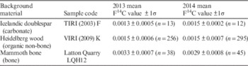
Table 1 shows the values for all 3 background types for 2013–2014 (Dunbar et al. Reference Dunbar, Cook, Naysmith, Tripney and Xu2016). It can be observed that there is no significant difference between the Icelandic doublespar and Heidelberg wood background values and the fact that very few unknown carbonate samples are received by the laboratory is reflected in the small number of carbonate background standards analyzed in 2013 and 2014.
Fraction modern values for different background types in 2013 and 2014.

However, the mammoth bone background is about twice that of the Heidelberg wood even though both materials require equally complex but different pretreatments (alpha cellulose fraction for wood and bone collagen extraction for the mammoth bone) (Dunbar et al. Reference Dunbar, Cook, Naysmith, Tripney and Xu2016). The F14C value for the Heidelberg wood background is more comparable to the Icelandic doublespar which requires no such complex pretreatment (a simple acid wash).
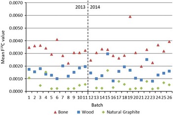
Figure 1 shows the average Heidelberg wood and mammoth bone background data plotted against the natural graphite data for each measured sample batch. The results show that the Heidelberg wood background values closely follow the same pattern as the natural graphite values but offset slightly higher. This indicates that there is some additional carbon in these samples, either in the form of residual contaminant that was not removed during pretreatment or added to the samples during the processing to graphite, but that this additional carbon is close to constant in terms of its influence on the 14C activity. In contrast, the mammoth bone background values do not follow the natural graphite values as closely, indicating that there is a non-uniform and further addition of contaminant carbon during sample processing, or the pretreatment process did not remove all contaminant carbon from the samples.
Plot of natural graphite, Heidelberg wood and mammoth bone backgrounds for all batches used in this trial. Batches 1–11 were run in 2013; 12–26 were run in 2014.

The plot also suggests that there is slightly more variability in the measurements of mammoth bone samples than Heidelberg wood, especially in 2014. The mammoth bone samples follow the same laboratory procedures as the wood with the only difference being the pretreatment methods.
As a side issue, we compared the natural graphite values in both spectrometers, the 5 MV AMS (separated into sources S1 and S2) and the SSAMS (single source). The data presented in Table 2 indicate that the instrument background for both spectrometers is very low and how little it contributes to the final background results.
Natural graphite F14C values on each AMS source over the period 2013–2014.

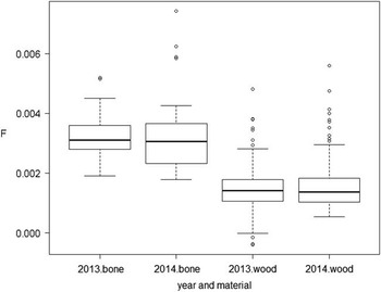
Figure 2 shows the distribution of F14C, emphasizing again the lower values for Heidelberg wood compared to mammoth bone, but demonstrates consistency of the median values for each material between 2013 and 2014. A small number of unusual observations are indicated (○in the figure) for both Heidelberg wood and mammoth bone.
All F14C values for mammoth bone and Heidelberg wood background standards for 2013–2014.

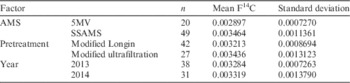
Within the statistical model, it is possible to examine the different factors and their effect on the observed variation in the results. The factors considered were (1) the AMS instrument (5MV versus SSAMS), (2) the bone pretreatment method (modified Longin versus modified ultrafiltration), and (3) the year of measurement (2013 versus 2014). Table 3 shows the summary statistics for F14C by each factor and the number of targets measured. The mixed effects model was then fitted and no factor was found to be statistically significant (p<0.05). However, the AMS factor was close to statistically significant, therefore two statistical models (with and without AMS) are presented along with their results.
Summary statistics for mammoth bone F14C by AMS, pretreatment, and year.

The first statistical model only has the random effect for the batch included, with the total variation then decomposed into “between batch variability” and “within batch variability.” Using this model, the residual error (standard deviation) is 0.000784, very broadly similar to the between batch error (standard deviation) of 0.0007032. The statistical model provides an overall estimate of the mammoth bone F14C as 0.0032725 with a standard error (standard deviation of the mean) of 0.0001739 (accounting for within and between batch variability, expressed as the precision at 1σ on the mean background).
The second statistical model, which includes the AMS component (even though not statistically significant), gives a mean F14C of 0.0029138 (the SSAMS offset is 0.0005871) and residual error of 0.0007828, which is now slightly greater than the between batch error at 0.0006665. Therefore, as may be expected, variability between batches is reduced when we keep the AMS component in the model, but the residual error essentially remains unchanged.
CONCLUSIONS
The analysis has confirmed that the mammoth bone background is systematically higher than the Heidelberg wood in the SUERC laboratory in 2013–2014. Further detailed investigation of the mammoth bone background targets over this two-year period, using a random effects model incorporating within and between batch variability, showed no systematic difference between pretreatment methods, but there is a suggestion of a difference in the mean background level between the SSAMS and the 5MV instrument. It was not possible to examine interactions between the AMS, year, and pretreatment, since at the time only small numbers of mammoth bone background targets were routinely and systematically run in every batch. However, following this study, the laboratory now routinely measures an appropriate number bone background targets in every batch such that the background material may be matched to the unknown sample material, thus a bone background subtraction is now applied to unknown bone samples.
Accumulating all the F14C values over the year, incorporating the variability between batches, the “best” estimate for the F14C of the mammoth bone is 0.0032725 and its standard error is 0.0001739. The analysis suggests that the variability within and between batches are very broadly of the same magnitude. (This is of relevance when considering what the contributing factors might be, and may reflect the variability in the individual targets but also the variability in instrument operation and stability from batch to batch.)
The significant benefits of preparing and running multiple background targets in every batch during routine operation is in the best estimation of the background value and its error for each sample type being run in each wheel.
From this small study, no statistical difference between either pretreatment methods used for analysing bone at the SUERC Radiocarbon Laboratory was observed.
Acknowledgments
Special thanks to Helen Kinch for bone collagen preparation and to Philippa Ascough for AMS measurements and Ross Brown for statistical analysis.