Introduction
Intrauterine exposure of the rodent to maternal protein restriction is well established as a protocol for inducing cardiovascular disease and risk factors for disease.Reference Langley-Evans 1 In rats, protein restriction is associated with early onset of high blood pressure,Reference Langley and Jackson 2 possibly mediated by changes in peripheral resistance.Reference Swali, McMullen and Langley-Evans 3 In mice, it has been shown that protein restriction programmes atherosclerosis.Reference Yates, Tarling, Langley-Evans and Salter 4 In addition to these disease-related outcomes, there is evidence that foetal undernutrition determines vascular reactivityReference Torrens, Brawley and Anthony 5 and the ability of the heart to recover contractile function following a period of ischaemia.Reference Elmes, Gardner and Langley-Evans 6
The effects of protein restriction upon the heart manifest early in postnatal life. Cheema et al.Reference Cheema, Dent, Saini, Aroutiounova and Tappia 7 showed that neonates exposed to low protein (LP) diet in utero had impaired cardiac function. Depressed ejection fraction was associated with left ventricular hypertrophy and evidence of cardiomyocyte apoptosis. Corstius et al.Reference Corstius, Zimanyi and Maka 8 reported that hearts from maternal LP diet-exposed rats culled at birth had fewer cardiomyocytes and suggested that deficits of cell number present at birth may be irreversible and predispose to abnormal function later in life.
Changes in the structure of the heart, effectively remodelling basic anatomy, may be a consequence of diet-induced changes to the epigenome. Slater-Jefferies et al.Reference Slater-Jefferies, Lillycrop and Townsend 9 reported that in both adult and neonatal hearts from rats exposed to protein restriction in utero, methylation of the peroxisome proliferator-activated receptor α was reduced, suggesting that epigenetic regulation of cardiac energy metabolism may operate in undernourished pregnancies. Although there is great interest in the contribution maternal undernutrition makes to the setting of epigenetic marks which hence determine long-term gene expression,Reference Gluckman, Hanson, Buklijas, Low and Beedle 10 there is also an extensive literature that suggests that undernutrition during pregnancy may facilitate increased transfer of glucocorticoids from the mother to the foetus.Reference Seckl and Holmes 11 Glucocorticoids are powerful regulators of gene expression acting directly via the glucocorticoid receptor (GR). They have also been shown to determine patterns of DNA methylation during development.Reference Bogdarina, Haase, Langley-Evans and Clark 12
The aim of this study was to investigate whether the impact of maternal protein restriction could be detected in cells cultured in vitro. Our hypothesis was that the diet encountered in utero establishes a cellular memory of early events that would manifest in altered cardiomyocyte differentiation and function. As glucocorticoids have been implicated in the early life programming of cardiovascular disease, cell cultures were treated with dexamethasone to examine the relationship between maternal protein restriction and glucocorticoid sensitivity. Establishing the existence of long-term programming effects in cell culture offers a number of advantages, including the reduced need for large numbers of animals and the elimination of confounding effects of developing pathologies.
Method
Animals
All experiments were conducted under licence and in accordance with the Home Office Animals (Scientific Procedures) Act, 1986. Wistar rats were supplied by Harlan Laboratories (UK) and pair-housed in wire mesh and Perspex cages in a controlled environment (21°C, 55% humidity, 12:12 light:dark cycle). A total of 20 females were used to generate the neonatal material used in the experiments. In brief, virgin females weighing 210–260 g were mated with a stock stud male and fed either a control diet (18% casein based, n = 10) or a LP diet (9% casein based, n = 10) from conception until littering, as previously described.Reference McMullen, Gardner and Langley-Evans 13 At delivery of litters, all dams were culled and eight neonates per litter (four male and four female) were culled within 18 h of birth for collection of hearts and preparation of cardiomyocyte cultures.
Culture protocol
Neonatal hearts (all eight per litter combined) were dissected in ice-cold calcium and magnesium-free Hanks Balanced Salt Solution under sterile conditions. Cells were then prepared using the Worthington neonatal cardiomyocyte isolation kit (Lorne Laboratories, UK) following the manufacturer's instructions. Cells were seeded onto 24-well plates at 500 cells/mm2 (100,000 cells per well) and grown in Dulbecco's Modified Eagle Medium (Invitrogen Gibco 17.5 mM glucose product code VX12634028, with 10% heat inactivated foetal bovine serum) for 10 days, incubated at 37°C (5% CO2). Medium was changed every 48 h. Cultures at 10 days were used as the baseline time point for all assays and experimental treatments. By this stage, the cultures were confluent and there was evidence that cells were beating in the culture plates. All cultures were switched to serum-free medium for the last 48 h of each experiment. On day 10 in culture, cardiomyocytes were either frozen (−80°) for baseline measures, or were treated with dexamethasone for a period of 48 h and then frozen at −80°C. Dexamethasone treatments were performed in two separate sub-studies. In the first sub-study, where the target end point was glucose uptake (five litters per maternal dietary group), dexamethasone was added to cultures to give a final concentration of either 0.1 or 10 μM. In the second, where gene expression was the main focus (five litters per maternal dietary group), dexamethasone was added at either 0.01 or 0.1 μM. Corticosterone concentrations in late gestation rat foetuses are approximately 1 μM.Reference Baram and Schultz 14 Prior to all measurements, cell counts were performed using a haemocytometer, with trypan blue staining used to determine live cell number. Cell viability varied between 40% and 70% and, although significantly lower (P < 0.05) at 12 days compared to 10 days, was unaffected by maternal diet or dexamethasone treatment.
Cardiomyocyte staining and cell size
Cardiomyocytes that are not terminally differentiated are mononucleated, whereas fully differentiated cells become binucleate.Reference Corstius, Zimanyi and Maka 8 To assess differentiation, cultures were stained with the nuclear stain, bisbenzamide, and then viewed under a microscope. Numbers of bi- and mono-nucleate and total numbers of cardiomycocytes were counted in five separate microscope fields per well. As previous reportsReference Cheema, Dent, Saini, Aroutiounova and Tappia 7 , Reference Corstius, Zimanyi and Maka 8 suggested that LP diet was associated with cardiomyocyte hypertrophy, we examined two indices of cell size in culture. To obtain an indicator of cell size (DNA:protein ratio), unstained cells were lysed. Protein was determined by the method of Bradford,Reference Bradford 15 and DNA by using the Hoechst fluorimetric assay.Reference Labarca and Paigen 16 Cellular protein concentration was determined by calculating the protein content per million cells.
Glucose uptake assay
Basal and insulin-stimulated glucose uptake were determined by measuring the uptake of 2-deoxy-D-[2,6-3H] glucose, according to the method of Kozma et al.Reference Kozma, Baltensperger and Klarlund 17 Insulin-stimulated uptake was assessed through the addition of 200 nM bovine insulin to cultures. All uptake measures were expressed per million cells.
Gene expression
To determine the expression of key target genes in culture (GR; glucose transporter 1, GLUT1; glucose transporter 4, GLUT4), RNA was extracted from cultured cardiomyocytes using a Qiagen RNeasy minikit according to the manufacturer's instructions. RNA quality was confirmed using a Nanodrop ND1000 spectrophotometer and cDNA was prepared as previously described.Reference Cornock, Langley-Evans, Mobasheri and McMullen 18 Real-time PCR was performed in 384-well plates using a Roche 480 Lightcycler and primers designed for the genes of interest using Primer Express (GR; NM_012576.2, GLUT1; NM_138827.1, GLUT4; NM_01275.1). All expression data were normalized to rat cyclophilin A (NM_017101) expression. The expression of the housekeeping gene was not altered by maternal diet or dexamethasone treatment.
Statistical analysis
All data are presented as mean ± s.e.m. and were analysed using either two-way ANOVA (effects of diet × dexamethasone) or Student's t-test as appropriate. In all experiments, eight hearts from a litter were pooled for cardiomyocyte isolation and as such n represents a whole litter in the analyses. All end points were measured using material from triplicate cultures from each litter. P < 0.05 was accepted as statistically significant.
Results
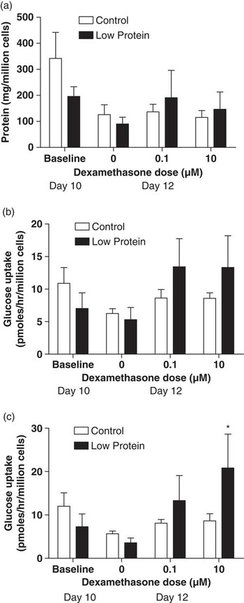
Initial seeding of cultures was set at 100,000 cells per well, and by 10 days in culture there were no significant differences in live cell number between control (82,400 ± 16,786 cells/well) and LP diet (79,800 ± 15,970 cells/well) derived cultures. There was no significant diet-related difference in the relative proportions of mono- and binucleated cells (control 91.1 ± 2.0% mononucleated, LP 92.0 ± 0.8% mononucleated). Apoptotic cell death was assessed using the Roche Cell Death ELISAPLUS kit, but no significant effects of any treatments were noted (data not shown). As DNA concentration gives a proxy for cell number and protein for metabolically active tissue size, changes in the DNA:protein ratio, in the absence of differences in DNA concentration, provide an indicator of cell size. Analysis of the DNA:protein ratio of the lysed cell cultures indicated no significant difference in cell size (control: 0.079 ± 0.042 LP: 0.059 ± 0.028). As shown in Figure 1a, there was no effect of maternal diet on cellular protein concentration. The protein concentration tended to be lower at day 12 than day 10, reflecting reduced cell viability. At the baseline time point (day 10 in culture), there was no significant difference in either basal (Fig. 1b) or insulin-stimulated glucose uptake (Fig. 1c) between cultures prepared from control or LP-exposed animals. Over the subsequent 2 days, in the absence of dexamethasone, glucose uptake (basal and insulin-stimulated) remained relatively stable. Basal uptake was not responsive to dexamethasone at either the 0.1 or 10 μM concentration, but the higher concentration elicited an increase in insulin-stimulated glucose uptake. This effect was seen only in cultures from the LP-exposed animals, where insulin-stimulated glucose uptake was 4-fold higher with 10 μM dexamethasone than in the absence of dexamethasone (Fig. 1c). LP-derived cultures at both concentrations of dexamethasone had greater insulin-stimulated glucose uptake than controls. Dexamethasone treatment had no significant effect upon cell number in culture, binucleation, the DNA:protein ratio (data not shown) or the cellular protein concentration (Fig. 1a).
Glucose uptake by neonatal cardiomyocytes following 10–12 days in culture. (a) Protein concentration of cells in culture. (b) Basal glucose uptake of cultured cardiomyocytes. (c) Insulin-stimulated glucose uptake of cultured cardiomyocytes. Data are shown as mean ± s.e.m. for five observations per group. Day 10 comparisons between control and low protein were analysed by t-test. Day 12 comparisons were analysed using two-way ANOVA. * indicates significantly different to 0 μM dexamethasone treatment of the same maternal diet group. ANOVA indicated an effect of dexamethasone treatment upon insulin-stimulated glucose uptake (P = 0.042).

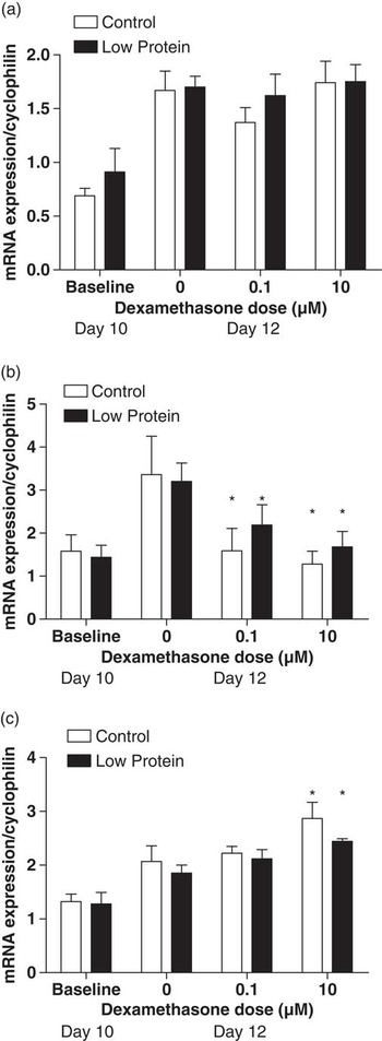
Given that there were maternal diet-related differences in the response of glucose uptake to dexamethasone treatment, the expressions of glucose transporters and GRs were determined in further cultures. As shown in Figure 2, at 10 days in culture, there were no significant effects of maternal diet upon expression of mRNA for GLUT1, GLUT4 or GR. With a further 2 days in culture, expression of the glucose transporters tended to increase, but there were still no differences in expression between the control and LP groups, in the absence of dexamethasone. For analysis of gene expression, lower doses of dexamethasone were added to cultures than had been used in the glucose uptake study (0.01 and 0.1 μM). GLUT1 expression was not influenced by either dose, but GLUT4 expression was suppressed by glucocorticoid treatment at both doses. Expression of GR was not influenced by maternal diet and was increased by dexamethasone treatment at 0.1 μM.
Gene expression in cultured cardiomyocytes. (a) GLUT 1 expression. (b) GLUT 4 expression. (c) glucocorticoid receptor expression. Data are shown as mean ± s.e.m. for five observations per group. Day 10 comparisons between control and low protein were analysed by t-test. Day 12 comparisons were analysed using two-way ANOVA. * indicates significantly different to 0 dexamethasone treatment of the same maternal diet group. ANOVA indicated an effect of dexamethasone treatment upon GLUT 4 expression (P = 0.008) and GR expression (P = 0.009).

Discussion
The aim of this study was to determine whether maternal protein restriction in rat pregnancy had lasting effects upon the neonatal heart, which would manifest in cardiomyocyte cultures. We have clearly shown that this is the case and that cells cultured from LP-exposed animals had an enhanced sensitivity to glucocorticoids in vitro. With high-dose dexamethasone, cells derived from the LP litters exhibited greatly enhanced insulin-stimulated glucose uptake when measured in a relatively hyperglycaemic environment. This supports our hypothesis that maternal undernutrition establishes a cellular memory of early life events. The physiological consequences of upregulated glucose uptake would be expected to be a greater utilization of glucose as a substrate and possibly lower utilization of fatty acids. This pattern of substrate utilization has been shown in mice which over-express cardiac GLUT1.Reference Yan, Young and Cui 19 Such animals are more prone to contractile dysfunction, and it is noteworthy that rats exposed to LP diet in utero are more prone to cardiac dysfunction after ischaemia-reperfusion.Reference Elmes, Gardner and Langley-Evans 6
In contrast to previous reports,Reference Cheema, Dent, Saini, Aroutiounova and Tappia 7 , Reference Corstius, Zimanyi and Maka 8 , Reference Aroutiounova, Fandrich, Kardami and Tappia 20 our study found no evidence that cardiomyocytes from protein-restricted litters were more prone to apoptosis or differed in rates of proliferation and differentiation, and the data did not therefore support our original hypothesis. Like Corstius et al.,Reference Corstius, Zimanyi and Maka 8 we found no difference in the proportions of binucleate cells present in culture at any stage of our experiments, and nor did the live cell number vary with maternal diet or dexamethasone treatment. Although we did not assess apoptosis directly, there was no evidence of difference in the rates of necrotic or apoptotic cell death as the cultures grew from initial seeding to the measurement stages of 10 (baseline) and 12 days. Although Cheema et al.Reference Cheema, Dent, Saini, Aroutiounova and Tappia 7 suggested greater hypertrophy in cardiomyocytes from LP-exposed rats compared to control neonatal rats, we could find no evidence of this in the 10- or 12-day cultures, based upon DNA:protein ratios or cellular protein concentrations. Porrello et al.Reference Porrello, Meeker, Thomas, Widdop and Delbridge 21 showed that glucocorticoids can suppress growth of neonatal cardiomyocytes through interaction with angiotensin II receptors. Lister et al.Reference Lister, Autelitano, Jenkins, Hannan and Sheppard 22 suggested that hypertrophy itself increases expression of GR and enhances sensitivity to glucocorticoids. In the absence of hypertrophy, this would not explain the findings of this study.
On incubation with high-dose dexamethasone (10 μM), cultures from the LP-exposed litters exhibited a 4-fold increase in insulin-stimulated glucose uptake, whereas control cultures showed no response to glucocorticoid treatment. A similar trend was seen with 0.1 μM dexamethasone, but this failed to achieve statistical significance. We infer from these results that cardiomyocytes from protein-restricted rats are more sensitive to glucocorticoids. However, the mechanism behind this differential sensitivity remains unclear as we could detect no difference in expression of GR between control and LP-exposed cardiomyocytes. Similarly, the means through which glucose uptake was enhanced was not identified in our study, as we could detect no difference in expression of mRNA for the glucose transporters. GLUT 1 is the major glucose transporter in foetal tissue, and expression declines in early postnatal life,Reference Studelska, Campbell, Pang, Rodnick and James 23 giving way to GLUT4. We showed in vitro that GLUT4 was downregulated by dexamethasone, but there was no evidence that maternal diet impacted upon this process. The explanation of how insulin-stimulated glucose uptake is influenced by maternal diet may lie in the insulin-signalling pathway, which has been shown to be a target for programming by protein restriction in a number of tissues, including skeletal muscle.Reference Fernandez-Twinn, Wayman and Ekizoglou 24 , Reference Ozanne, Olsen and Hansen 25 The PI3 kinase-Akt-mTOR pathway is a key regulator of the translocation of glucose transporters to the cell membrane and thus glucose uptake, and therefore may be an interesting target for further investigation. The apparently paradoxical observation that insulin-stimulated glucose uptake was upregulated by dexamethasone in the LP-exposed cells, whereas dexamethasone downregulated GLUT4 mRNA expression, may be explained by programming of the longevity of GLUT4 proteins, or programmed differences in translation from the message. Further investigation will consider expression of glucose receptors at the level of protein.
Prenatal protein restriction is associated with abnormalities of cardiac function and anatomy in the rat, which may give rise to hypertension and enhanced susceptibility to ischaemia-reperfusion injury in adult life.Reference Langley and Jackson 2 , Reference Elmes, Gardner and Langley-Evans 6 This study has demonstrated that a remodelling of the heart structure may not be the sole driver of such processes, as cultured cardiomyocytes manifest maternal diet-related differences after 10–12 days in vitro. One explanation of the findings may be that a variation in maternal diet sets patterns of gene expression that determine the growth, development and metabolic capacity of the cells for at least the short-term duration of this experiment. Alternatively, it may be inferred that sensitivity to endocrine signals may be permanently programmed by undernutrition. Such an interpretation would add weight to the argument that a variation in maternal diet sets in train processes, either through changes in the sequence of developmentally mediated gene expression or at the level of the epigenome, which impact upon long-term gene and protein expression and metabolic functions.
Acknowledgements
This work was funded by the European Union FP7, FOOD-CT-2005-007036 (EARNEST project).