When the Senning procedure was introduced almost 50 years ago for treatment of patients with transposition,Reference Senning1 the prognosis and quality of life for these patients improved dramatically. A technically less demanding, but conceptually similar, procedure was introduced a few years later by Mustard, and became more widely used.Reference Mustard2 There are, unfortunately, several long term complications related to these interventions, such as arrhythmia, failure of the systemic ventricle, baffle leaks, and obstructions in the newly created venous pathways.Reference Trusler, Williams and Duncan3–Reference Wilson, Clarkson and Barratt-Boyes5 The contemporary surgical approach is the arterial switch procedure, which after primarily successful operations carries an excellent prognosis.Reference Losay, Touchot and Serraf6 There are, however, still many adult patients undergoing long-term follow-up after atrial redirection procedures who are exposed to the risk for late complications.Reference Marelli, Mackie and Ionescu-Ittu7 Thus, there is an ongoing need for high quality imaging in the periodic follow-up of these patients.
Imaging in such patients is demanding, and requires experience, expertise and, as a basis for this, an adequate case load, which may only be available in regional centres caring for adults with congenitally malformed hearts. Current imaging modalities may allow for decentralised investigation, with interpretation of the study in, or in collaboration with, the regional centre. In the periodic out-patient follow-up of patients with congenitally malformed hearts, echocardiography is often the first choice of imaging, as it is easy accessible. In evaluation of the atrial compartments after atrial redirection procedures, echocardiographic examination may be less suitable. Indirect signs of obstruction, such as increased velocities of flow, may be detected, but anatomical evaluation is difficult.Reference Therrien, Gatzoulis, Webb and Daubeney8 Standard cardiovascular magnetic resonance imaging is technically demanding and time-consuming in patients with complexly malformed hearts, such as transposition, defined on the basis of concordant atrioventricular and discordant ventriculo-arterial connections.Reference Sampson, Kilner and Hirsch9 Three-dimensional time resolved contrast-enhanced magnetic resonance angiography, or better described as dynamic angiography, is safe and relatively easy to perform. It is usually available in a modern general centre for imaging, and may thus be suitable for this strategy. It has not yet been determined whether dynamic angiography can identify haemodynamic significant obstructions in the venous pathways in the population of patients with atrial redirection procedures. We have, therefore, compared dynamic angiography with a standard non-contrast cardiovascular magnetic resonance protocol known to be useful after the atrial switch,Reference Dorfman and Geva10 using the latter as the golden standard.
Material and methods
Patients
All subjects gave their written informed consent for the study. The protocol was approved by the local ethics committee. We recruited 36 consecutive adults, older than 16 years, of whom 13 were women, who had been referred for a clinical cardiovascular magnetic resonance examination, all having undergone the Mustard procedure. They were studied at the Cardiovascular Magnetic Resonance Unit, Royal Brompton Hospital, London. Beside the referral for cardiovascular magnetic resonance, there was no additional selection for the study.
Study design
Dynamic angiography was compared against standard cardiovascular magnetic resonance as a golden standard in the detection of obstruction of the venous pathways and other cardiovascular lesions associated with the underlying cardiac malformation and its surgical treatment. The dynamic angiographies were interpreted by a single investigator, RHM, who had access only to the dynamic angiographic acquisitions.
Standard cardiovascular magnetic resonance
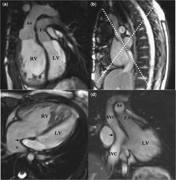
All investigations were performed on a Siemens Sonata 1.5 T magnetic resonance scanner, manufactured by Siemens A/G, Erlangen, Germany. The protocol included transaxial stacks of half-Fourier acquisition single-shot turbo spin-echo images, and coronal and sagittal steady state free precession multislice stacks covering relevant parts of the chest, steady state free precession cines including long-axis views of both ventricles, “four-chamber view”, ventricular outlets, a short-axis stack covering both ventricles and oblique planes aligned with the superior caval, inferior caval and pulmonary venous atrial pathways (Fig. 1), with oblique cross cuts as necessary to achieve alignment with all parts of the paths. Expiratory breath-hold phase-velocity maps with through-plane velocity encoding were located to transect any suspected narrow region. If jet formation was visible in a narrowed path, the velocity map was located to transect the jet. Velocity maps were then used to measure the peak velocity and record the time course of flow, with the initial velocity encoding set on 1.0 m/s and adjusted as appropriate. Azygos veins with diameters greater than 5 mm as seen on transaxial images was regarded as dilated.
Unobstructed atrial pathways after atrial redirection surgery as seen by standard cardiovascular magnetic resonance, here represented by steady-state free precession cine images (a–d). Figure 1a demonstrates the ventriculo-arterial connections in transposition. The morphologically right ventricle (RV) serves as the systemic ventricle, and is connected to the aorta (Ao). The morphologically left ventricle (LV) is the subpulmonary ventricle connected to the pulmonary trunk (PA). Figure 1b shows the planning for images (dotted lines) of the atrial compartments as seen in 1c and 1d. Oxygenated blood from the pulmonary veins drains to the region of the original left atrium (asterisk in 1c). The atrial septum has been removed and pulmonary venous blood is now directed anteriorly by the baffle (arrowed in 1c and 1d) to the systemic ventricle and then to the aorta. Deoxygenated blood from the superior caval vein (SVC) and inferior caval vein (IVC) passes on the other side of the baffle to the left ventricle, then to the pulmonary trunk. The atrial pathways are unobstructed in this case (1b–d).

Dynamic angiography
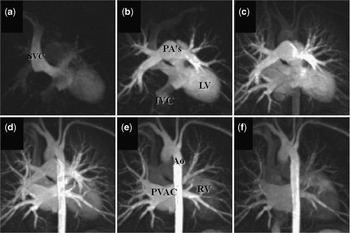
Dynamic angiography relies on fast acquisition of images of a pre-selected volume during bolus administration of the contrast agent Gadolinium-DTPA, Magnevist®, produced by Schering, Berlin, Germany. A pre-contrast acquisition is performed to ensure proper positioning, and artefact-free acquisition using 3-dimensional fast gradient-echo sequence acquired in the coronal slab. This was also used for subsequent subtraction from the post-contrast images to eliminate signal from body tissue. A 3-dimensional fast gradient echo sequence was used and the post-contrast acquisition was started 3 seconds after the start of contrast injection and the sequence was repeated 6 times during a breath hold. Each repetition requires 5 to 6 seconds (Fig. 2). The contrast was injected via a pump at a rate of 2–3 ml/s into the right (or left when access on the right side was difficult) antecubital vein followed by a flush with 15 ml of normal saline. The contrast dose was typically 0.2 mmol/kg. Imaging parameters were as follows: Field of view 40 × 32 × 13 cm, voxel size 1.3× 1.6 × 3.0 mm, TR/TE 2.54 ms/0.97 ms, flip angle 30°, bandwidth 780 Hz/pixel. Sequential k-space filling was used. To speed up acquisition, parallel imaging using generalised autocalibrating partially parallel imaging applied with acceleration factor 2 in the primary phase encoding direction with 32 reference lines. The standard Siemens phase-array receiver coil was used with 6 anterior and 6 posterior elements selected. Field of view over sampling was 13% in the phase encoding direction and 9% in the slice direction. Partial Fourier of 3/4 was applied in phase encode direction. The contrast-enhanced angiography study were processed and analysed by an experienced investigator using the raw data and 3-dimensional reformatting including maximum signal intensity projection and multiplanar reconstruction.
Unobstructed atrial pathways after atrial redirection surgery as seen by dynamic angiography and maximum signal intensity projection (a–f). SVC = superior caval vein, IVC = inferior caval vein, PA’s = pulmonary arteries, Ao = aorta, PVAC = pulmonary venous atrial compartment, RV = right ventricle.

Definitions of obstructions to venous flow
In standard cardiovascular magnetic resonance, the reference method in this investigation, obstruction of an atrial pathway, specifically the superior caval, the inferior caval, or the pulmonary venous pathways, was defined on the basis of mild stenosis representing evidence of a mild narrowing on cine images, but with pulsatile flow and a peak velocity in early diastole) not exceeding 1 m/s. Moderate stenosis was defined on the basis of narrowing on cine images, with pulsatile flow and a peak velocity above 1 m/s, except in one case of narrowing of one of the two the caval venous channels, when the elevated velocity was explained by increased flow due to narrowing of the other caval venous channel, and shunting of blood via a dilated azygos vein. Severe stenosis was defined as narrowing on cine images with a peak velocity of over 1.5 m/s, and a damped and continuous flow curve. In dynamic angiography, obstruction was defined as anatomical narrowing with thin contrast signal or absence of contrast distal to the obstruction. Furthermore, it was possibly to evaluate the flow direction in the azygos vein. Reversed flow indicates obstruction of flow in the superior caval venous pathway.
Results
Identification of patients with obstruction in the systemic venous channels
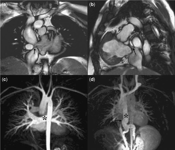
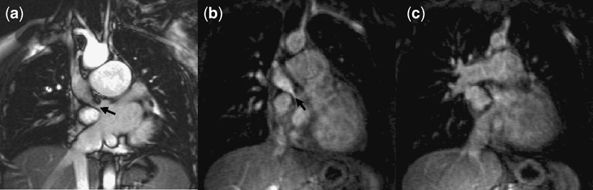
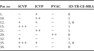
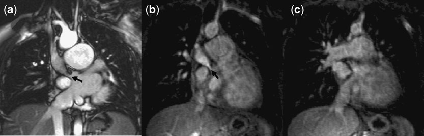
Moderate or severe obstructions were detected in 4 (11%) patients, and correctly identified by dynamic angiography with a sensitivity of 100%, specificity of 100%, positive predictive value of 100%, and negative predictive value of 100%. In 4 additional patients, mild obstructions were identified, with 2 of these detected with dynamic angiography. Thus, 6 of 8 patients with any obstruction were correctly identified, and only mild obstructions were missed. Overall sensitivity was 75%, with 100% specificity, a positive predictive value of 100%, and negative predictive value of 93%. In 2 patients, obstructions were found in the systemic venous channels. The standard CMR and dynamic angiography findings are illustrated in Figures 3 and 4, and an overview is shown in Table 1.
Steady-state free precession cine (a–b) and dynamic angiography (c–d) images illustrating a case with evidence of moderate obstruction of the inferior caval venous pathway (arrowed) (3a, b, d) and an unobstructed pulmonary venous atrial compartment on cine imaging. Dynamic angiography shows the pulmonary venous returns (asterisk in 3c), and late filling of the dilated azygos vein (asterisk in 3d) from below.

Severe narrowing of the superior caval venous pathway seen (arrow) on steady-state free precession cine (4a), unprocessed angiogram (4b), and normal dimesions of the inferior caval venous pathway in the same patient seen on cine image (4a) and unprocessed angiogram (4c).
Eleven obstructions in the atrial channels in nine patients detected by standard cardiovascular magnetic resonance as a reference compared with detection by dynamic angiography.
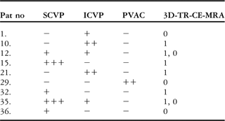
− = no obstruction, +mild obstruction, ++ = moderate obstruction and +++ severe obstruction/occlusion detected by standard CMR. 0 = obstruction not detected by 3D-TR-CE-MRA and 1 = obstruction (any degree) detected by dynamic angiography SCVP = superior caval venous pathway, ICVP = inferior caval venous pathway, PVAC = pulmonary venous atrial compartment.
Superior caval venous pathway
We identified 5 obstructions in the superior pathways, of which 2 were severe and 3 mild. Both severe obstructions, and 2 of the mild obstructions, were correctly identified by dynamic angiography. The third mild obstruction, in a patient with a persistent left as well as a right superior caval vein, was not clearly seen due to inadequate opacification of the right superior caval venous pathway, the contrast having been injected into a left antecubital vein.
Inferior caval venous pathway
There were 5 obstructions of the inferior pathways, 2 moderate and 3 mild. Of the patients with mild obstructions, 2 also had severe obstruction of the superior channel. The mild obstructions were not detected by dynamic angiography, albeit that the severe obstructions of the superior pathway were detected by dynamic angiography. In 1 patient, a stent had been inserted, without residual obstruction. This was identified by both methods, however without velocity data in dynamic angiography.
Pulmonary venous pathway
In 1 patient, there was moderate anatomical obstruction of the pulmonary venous pathway, with peak velocity 1.6 m/s. This lesion was not readily seen by dynamic angiography.
Follow-up
We have follow-up data in 4 of the patients with significant obstruction of the caval venous pathway. Stents were inserted in 2 patients with significant stenosis. Exercise capacity improved in both after the procedure. In one of these patients, symptoms of flushing resolved after the intervention. An additional patient, where follow up imaging showed further progression of stenosis, is under consideration for stenting, but currently remains well and asymptomatic. A patient with complete obstruction of the inferior caval venous pathway remains well, and is also under continued follow-up.
Azygos vein
In 3 subjects, 2 with severe superior caval venous obstruction, and 1 with mild right superior caval venous obstruction and persistence of the left superior caval vein, the direction of flow in the azygos vein was towards the abdomen. The vein itself was dilated in 9 patients, these findings being readily visible on dynamic angiography (Fig. 3), and also identifiable on standard cardiovascular magnetic resonance.
Subpulmonary stenosis
Mild subpulmonary stenosis, with peak velocity of flow of approximately 3 m/s, was seen in 4 subjects. All 4 were identified as narrowing in the subpulmonary outflow tract by dynamic angiography.
Ventricular septal defect
In one patient, there was a small ventricular septal defect, producing a ratio of pulmonary to systemic flows of from 1.4 to 1.6 to 1, which was identified by dynamic angiography.
Baffle leak
No baffle leaks were observed with either method.
Safety of cardiovascular magnetic resonance
All investigations were technically successful. No scan had to be terminated due to claustrophobia or other discomfort suffered by the patient. There were no complications related to injection of contrast or contrast media.
Discussion
In this investigation, we have studied the ability of dynamic angiography to detect obstructions in the atrial channels in patients having had previous atrial redirection procedures for transposition. All moderate to severe lesions in the systemic venous channels were detected, and hence all clinically significant obstructions, whereas some of the mild lesions remained undiagnosed. All investigations were technically successful, without adverse events, were relatively fast to perform, and there were no false positive results. The technique of dynamic angiography may be used alone, or in conjugation with standard cardiovascular magnetic resonance and/or other non-invasive imaging methods, such as echocardiography, to obtain a robust non-invasive diagnosis of clinically significant obstruction of the venous channels after atrial redirection procedures. Alignment of prescribed imaging planes in complex congenitally malformed hearts can be challenging, particularly after atrial redirection procedures, and the approach using dynamic angiography allows acquisition without specialist knowledge, although interpretation still requires appropriate understanding of underlying anatomy and physiology. An advantage is that it may be performed locally according to a standard protocol, and interpreted in collaboration with experts working in centres dealing with adults having congenitally malformed hearts.
The use of dynamic angiography has recently been reported in other patients with congenital cardiac disease.Reference Fenchel, Saleh and Dinh11–Reference Mohrs, Petersen and Voigtlaender13 In a series of 81 children and adults with a broad spectrum of lesions, only 5 had transposition corrected by means of atrial redirection.Reference Fenchel, Saleh and Dinh11 The authors excluded any major baffle leak in this group. In another report on 20 adults with congenital cardiac disease, 1 patient had undergone a previous Mustard operation without any additional pathology reported at dynamic angiography.Reference Goo, Yang and Park12 The technique has also been used to evaluate the cavo-pulmonary connections in 15 patients with a Fontan circulation.Reference Mohrs, Petersen and Voigtlaender13 To the best of our knowledge, however, there is no previous systematic study using dynamic angiography in the evaluation of obstruction of the venous pathways.
Non-invasive diagnosis of systemic venous obstructions is often difficult to obtain in adult patients by echocardiography, although indirect signs, such as increased velocities of flow, may be seen. The standard cardiovascular magnetic resonance investigation with cines and velocity maps is usually diagnostic, but is rather time consuming and technically demanding. The approach with dynamic angiography is easier to perform, but is not as sensitive as standard cardiovascular magnetic resonance in detecting clinically insignificant mild obstructions. On the other hand, it produced no false positive results. Our data, therefore, supports the utility of this method in evaluating the systemic venous channels to detect clinically significant obstructions, alone or in conjugation with other imaging methods and modalities.
Only 1 of our patients had a significant obstruction in the pulmonary venous compartment. In this individual, the dynamic angiography did not detect the lesion. This was probably due to the antero-posterior course of this channel, not ideal for visualisation in our acquisitions, which are more suitable for the more prevalent obstructions in the systemic venous channels. On the basis of this result, we cannot recommend dynamic angiography for evaluation of the pulmonary venous compartment.
Although sought for, we did not detect any baffle leaks using either method. These leaks are usually small, and cardiovascular magnetic resonance is probably not ideal their detection. In one patient, nonetheless, a small ventricular septal defect was visualised using dynamic angiography. Sub-pulmonary left ventricular outflow obstruction is not uncommon in this group of patients due to leftward displacement of muscular ventricular septum, and was detected in 4 patients in our series by both dynamic angiography and standard cardiovascular magnetic resonance methods.
In conclusion, dynamic angiography is valid in the detection of clinically significant, obstructions in the systemic venous pathways in patients having a previous atrial redirection operation, but is less sensitive in detecting mild stenoses compared with standard two dimensional cardiovascular magnetic resonance techniques. Dynamic angiography can be particularly useful in centres with a low case load, where expertise in detailed image acquisitions is not available. The technique may also be of value in the evaluation of associated lesions, such as ventricular septal defects and subpulmonary stenosis.
Acknowledgement
Dr Sonya Babu-Narayan was supported by The British Heart Foundation and Dr Bengt Johansson was supported by the Swedish Heart and Lung Foundation and the Heart Foundation of Northern Sweden.
Dr Bengt Johansson and Dr Sonya Babu-Narayan contributed equally to this work. There are no conflicts of interest related to this work.