Introduction
Harsh gastric environment and first-pass metabolism are the two major reasons for reduced bioavailability of the drugs through oral route. Peptide/protein drugs are especially vulnerable to the oral route of delivery due to obvious physiological reasons. Second, oral route requires multiple dosage administration, which is cumbersome for the patients. Parenteral route, on the other hand, is invasive and is not patient compliant. It is associated with acute pain and local site morbidity. Second, parenteral route is especially troubling for the patients who need regular injections such as those suffering from insulin-dependent diabetes or those requiring certain other regular injectable drugs such as glucagon-like peptide-1 (GLP-1) analog and amylin [Reference Shojaei1, Reference Bagan, Paderni, Termine, Campisi, Lo Russo and Di Fede2]. Swallowing tablet or capsule is usually problematic for dysphagic patients, geriatric people, and pediatric population. Children and geriatric patients very often are particular about the taste of the formulation they are consuming. Dysphagic patients, irrespective of age, are very prone to aspiration of the drug formulation, which might lead to another medical emergency [Reference Stegemann, Gosch and Breitkreutz3, Reference Mennella, Spector, Reed and Coldwell4]. Therefore, to avoid such difficulties and to reach patient compliance, alternative routes of drug administration have been explored in last few years. Buccal and sublingual routes are most patient complaint and guarantee a completely noninvasive route of delivery.
Transbuccal drug delivery has gained a lot of attention and importance in last few years for its easy accessibility and high vascularity. High vascularity ensures systemic absorption of the drug without encountering any harsh physiological environment and first-pass metabolism. These features ensure a high bioavailability as compared to the most common oral route. The oral mucosa is composed of stratified squamous epithelium (outermost layer) followed by basal membrane below which is the innermost layer called lamina propria. The buccal mucosa epithelium is almost 40–50 cell layers thick while the sublingual mucosa has lesser layers. The thickness of the buccal mucosa is reported to be 500–800 μm. Since the buccal and sublingual mucosae do not experience mechanical stress, they have nonkeratinized epithelia as opposed to that of gingiva and hard palate which are keratinized. The high vascularity of buccal mucosa is a great attraction for transmucosal drug delivery since the blood vessels directly drain into the internal jugular vein, enabling fast systemic delivery [Reference Barua, Kim, Jo, Seo, Park, Lee, Yun, Oh and Lee5, Reference Sudhakar, Kuotsu and Bandyopadhyay6].
Presence of mucus is another key anatomical component of buccal mucosa that is an important requirement for any transbuccal drug formulation. The mucus is primarily composed of water (∼95–99%), but the macromolecular ingredient that provides the signature viscosity is a class of glycoprotein known as mucin that accounts for 1–5% of mucus. Mucins are large protein with a molecular weight ranging from 0.5 MDa to almost 20 MDa. These mucins are the key components that are responsible for making adhesion with the various mucoadhesive polymers used in mucosal drug delivery. The mucus in the oral cavity is secreted as a part of saliva by the salivary glands. It has been reported that almost 70% of mucins in saliva are secreted by the minor salivary glands. Saliva is mostly water with around 1% inorganic and organic components including a few enzymes. The salivary pH range is from 5.5 to 7 and that depends upon the flow rate. At high flow rate, as reported, the bicarbonate and sodium level increase resulting in an increased pH toward 7. The mucin carries the overall negative charge at the physiological pH due to the presence of sialic acid moieties and sulfate residues that play an important role in mucoadhesion [Reference Madhav, Shakya, Shakya and Singh7]. Mucins, in terms of origin and presence, are classified into monomeric (attached to the epithelial surface) and oligomeric (secreted). The latter are responsible for imparting the rheological behavior to the saliva and mucus. The major mucin oligosaccharides present are N-acetylgalactosamine, N-acetylglucosamine, sialic acid, and fucose while sulfate groups are also present prominently. There are a total 19 mucin genes identified in humans (MUC) among which MUC2, MUC5AC, MUC5B, and MUC6 have cysteine-rich motifs in their N- and C-terminal domain that account for the oligomerization [Reference Thornton8, Reference Bansil and Turner9]. Linden et al. [Reference Linden, Sutton, Karlsson, Korolik and Mcguckin10] have reported the physiological presence of various mucins and their classification as secreted or cell surface attached. MUC2, MUC5AC, MUC5B, MUC6, and MUC19 are secretory and form gel while MUC7 is a secretory nongel-forming mucin. MUC1, MUC3A/B, MUC4, MUC12, MUC13, MUC15, MUC16, MUC17, and MUC20 are the cell surface–attached mucins. Among these mucins, MUC1 has been reported to be predominantly present on the buccal epithelium while MUC5B and MUC7 are the primarily secreted mucins in the saliva [Reference Zalewska, Zwierz and Gindzieñski11]. The daily saliva production in healthy humans is 1–1.5 L. The total stimulated saliva flow rate ranges from 1 to 3 mL/min while the saliva flow rate of unstimulated saliva is 0.25–0.35 mL/min. The normal salivary pH ranges from 6 to 7, but it changes as per the salivary flow (almost 5.8 during low flow and 7.8 during high flow) [Reference De Almeida, Grégio, Machado, De Lima and Azevedo12].
Porcine (pigs) are considered one of the most suitable animal models for in vitro and in vivo testing of mucoadhesive buccal formulation because of very close anatomical and physiological similarity to that of human buccal mucosa. Kulkarni et al. [Reference Kulkarni, Mahalingam, Pather, Li and Jasti13] have reported that the porcine buccal mucosa resembles human counterpart in ultrastructure (nonkeratinized), enzyme localization and activity, and permeability. Since human buccal samples are not easily available for in vitro and in vivo testing, porcine mucosa remains the best feasible model due to easy availability. Apart from pig, other large animals that have been used for buccal drug formulation testing are monkey, apes, and dogs due to highly similar structure and function of the respective tissue. For initial in vitro studies, the buccal tissue needs to be harvested and the animals need to be sacrificed for the same. The processing of the buccal tissue after harvesting is even tedious [Reference Janet Hoogstraate and Bodd14]. Cui et al. [Reference Cui and Mumper15], however, reported in one of their articles that the buccal mucosa of rabbit is very similar to that of humans among all the laboratory rodents commonly available. In an important article by Thirion-Delalande et al. [Reference Thirion-Delalande, Gervais, Fisch, Cuiné, Baron-Bodo, Moingeon and Mascarrel16], the team mentioned that the rabbit buccal mucosa is mostly nonkeratinized similar to that of humans. The buccal epithelium of rabbit is 30–40 layers thick which is almost exact as that of monkeys and very similar to that of humans (40–50 layers as mentioned previously). Even the connective tissue density and vascularization were similar to that of monkeys. Rabbits are more accessible than dogs and monkeys when comparing the availability of large and small animals. But the primary problem and question that remain here is sacrificing the animal even for simple in vitro studies such as mucoadhesion.
To address the issue of difficult in access to the suitable animal for mucoadhesion study and further preparation of excised buccal tissue, synthetic polymeric structures were fabricated to mimic the buccal mucosa to test the mucoadhesion of drug formulations. In this regard, as per our literature search, Khutoryanskiy et al. [Reference Hall, Khutoryanskaya and Khutoryanskiy17, Reference Cook, Smith and Khutoryanskiy18, Reference da Silva, Khutoryanskiy, Bruschi and Cook19] have carried out a considerable amount of work. They fabricated mucosa mimetic-surface by two initial approaches viz. layer-by-layer deposition of interpolymeric complex of methylcellulose and polyacrylic acid (PAA) on a chemically modified glass surface at a temperature around 120 °C and copolymerization of 2-hydroxyethyl methacrylate (HEMA) with other polymers such as N-vinylpyrrolidone, sorbitol methacrylate, N-acryloyl-glucosamine (AGA), and 2-hydroxyethyl acrylate. When compared with a natural mucosal tissue, the complex hydrogel of HEMA-AGA rendered almost similar mucoadhesion to the drug formulation. Apart from developing a complete polymer-based mucosa-mimetic material, Jacobsen et al. [Reference Jacobsen, van Deurs, Pedersen and Rassing20, Reference Jacobsen, Nielsen, Brøndum-Nielsen, Christensen, Olin, Tommerup and Rassing21] have developed filter-grown human buccal carcinoma cell line TR146.
Mucoadhesive polymers have been widely used to develop various mucosal drug delivery systems. Forms such as tablets, gels, nanoparticles, patches, and films have been synthesized and tested till date. Various natural, seminatural, and synthetic polymers have been widely validated to have strong mucoadhesive properties. Some commonly used polymers among the lot such as chitosan, alginate, PAA, sodium carboxymethyl cellulose (Na-CMC), hyaluronic acid (HA), and thiomers have shown promising characteristics to develop drug formulations [Reference Khutoryanskiy22, Reference Russo, Selmin, Baldassari, Gennari, Caviglioli, Cilurzo, Minghetti and Parodi23]. Chitosan oligosaccharide (COS) is the hydrolyzed product of chitosan with various properties different from that of chitosan. The most important aspect is its water solubility that was lacking with chitosan. COS has largely been an unexplored polymer with usage reports primarily as nanoparticles or as coating on other nanoparticles [Reference Muanprasat and Chatsudthipong24, Reference Zhang, Huang, Sun, Xing, Yamamoto and Gao25].
In this work reported, we have fabricated two novel things that, to the best of our knowledge, have not been reported till date. The first one is COS-based blended films as a proposed vehicle for buccal drug delivery while the second one is a very low-cost eggshell membrane (ESM)–based mucosa-mimetic surface using bovine submaxillary mucin (BSM). COS is a water-soluble version of chitosan which makes it easy to handle and is more appropriate than chitosan for biomedical applications. Second, we have utilized its reported mucoadhesive nature to formulate a blended patch that can be used to deliver drugs through buccal mucosa. Another attraction of the work is the fabrication of artificial buccal mucosa–mimetic surface using low-cost ESM which is normally a food waste. The mucosa-mimetic surface was fabricated using the commercially available BSM that mimicked the mucin present along the buccal mucosal surface. The COS-poly vinyl alcohol (PVA)–blended patch displayed much better mucoadhesive nature as than the COS and alginate blended patch.
Results and discussion
Fabrication of COS-blended films
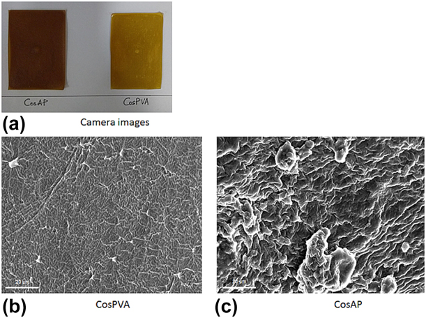
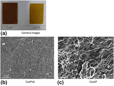
The fabricated COS-blended films are shown in Fig. 1(a) in a camera-captured image. The dimension of the parent films was 8 × 5 cm.
(a) Camera image of the COS-based films; (b) HR SEM images of CosPVA (sample 1); (c) HR SEM images of CosAP (sample 2).

Film thickness
The average film thickness for 2 × 2 cm2 was measured to be 1.24 mm (sample 1) and 1.29 mm (sample 2).
Folding endurance
The folding endurance test was performed as described in the methods section. Sample 1 (CosPVA) film did not crack even after folding at a single place for 200 times while sample 2 (CosAP) developed crack after average 20 foldings. Therefore, we can say that CosPVA film has a better elasticity than CosAP film. The possible reason for this observation could be the polyelectrolyte interaction between cationic COS and anionic sodium alginate, while the interaction was much stable between COS and uncharged PVA.
Surface morphology analysis
Figures 1(b) and 1(c) show a combined scanning electron microscope (SEM) images at 400×, 1600×, and 3000× for sample 1 and sample 2, respectively. SEM images revealed a much smoother surface for the CosPVA films than the CosAP films. The possible reason for this difference in surface morphology could be due to the chemical interaction among the polymers. In sample 1, COS is a cationic polymer blended well with PVA which is an uncharged polymer. But in sample 2, the cationic COS formed a polyelectrolyte structure with anionic sodium alginate leading to formation of a rough surface due to improper chemical blending.
Polymer interaction analysis by XRD and FTIR
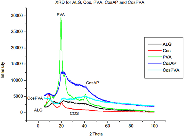
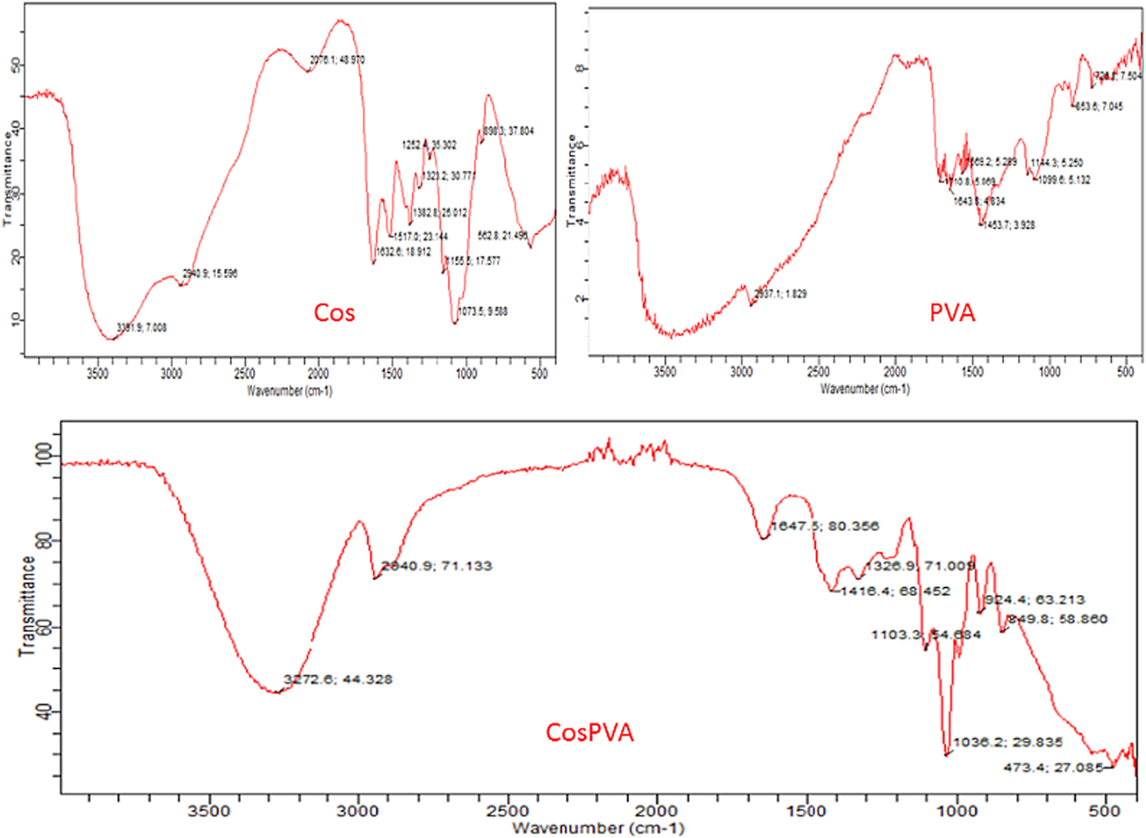
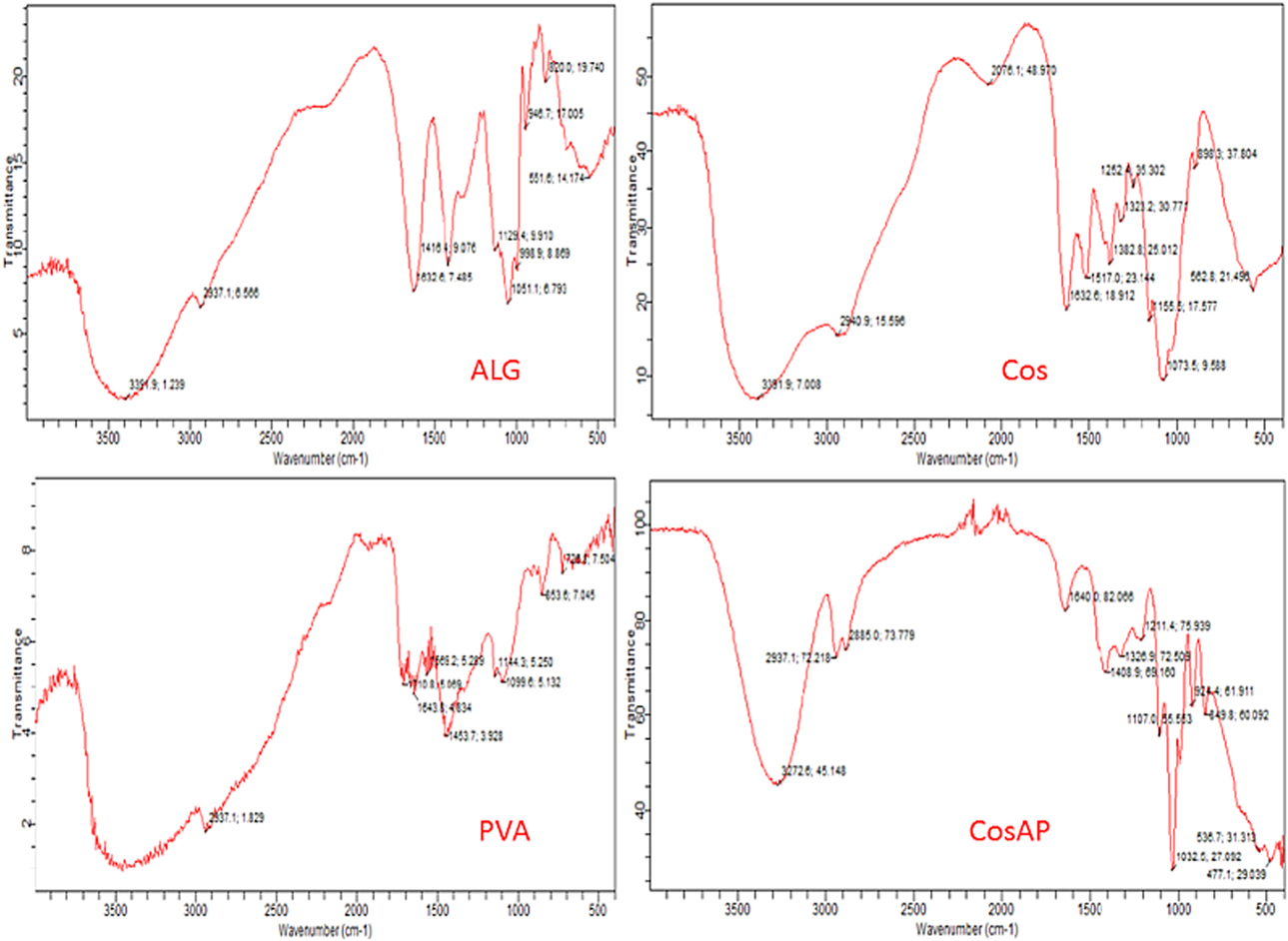
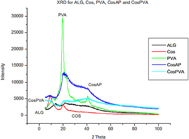
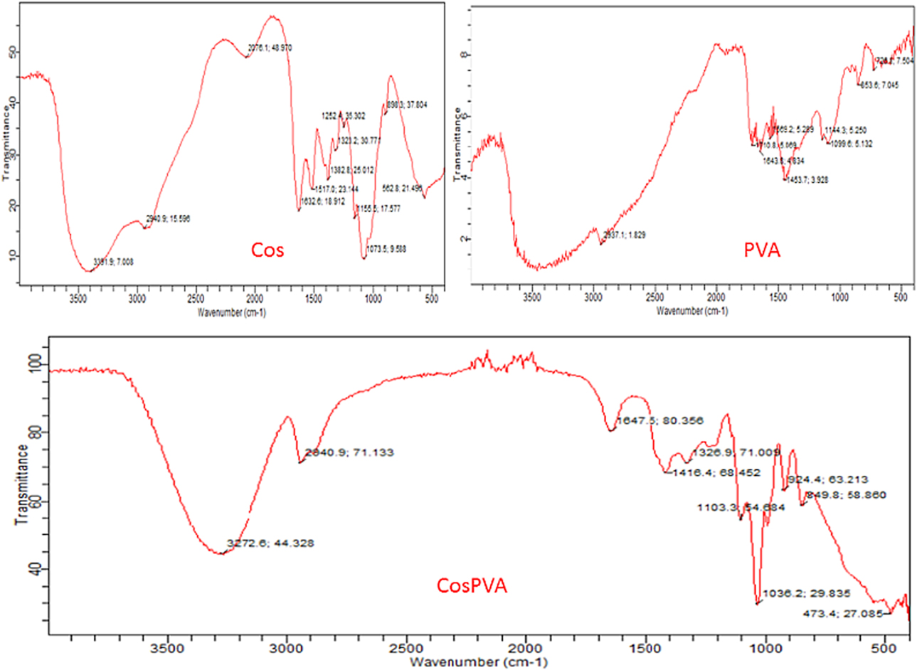
Both X-ray diffraction (XRD) and Fourier transform infrared (FTIR) analysis revealed a change in peaks among the polymers and ingredients. Figure 2 shows the XRD image of both the films and the individual ingredients, while Figs. 3 and 4 are the FTIR images for sample 1 and sample 2, respectively. The peak and bond characteristics of both sample 1 and sample 2 are completely different from those of the individual ingredients. The XRD image clearly reveals the presence of crystallinity in the individual polymers, while it is no longer present in the films. Second, the change in peak intensity indicates a clear blending of the polymers. COS shows peaks at 2θ value 10 and 20 while sodium alginate shows two small peaks at around 15 and 20. PVA shows a sharp peak at 2θ value 20 and another small peak at 2θ value 40. The blended films display different sets of peaks that are comparatively blunt, indicating that the films have lost crystallinity. Sample 1 shows two blunt points at 2θ value 20 and 40, while sample 2 displays a blunt point at 2θ value around 10 and another around 20.
XRD analysis of interactions in the samples.
FTIR analysis of interactions in CosPVA (sample1).
FTIR analysis of interactions in CosAP (sample 2).
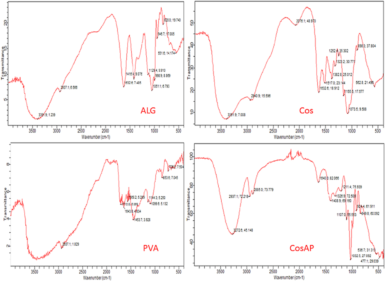
The FTIR spectrum of COS displayed peaks at 1632, 1517, and 1382 cm−1 that corresponds to the signature bands of amide I, amide II, and amide III, respectively. The peak at 1073 corresponds to the C–O stretching at the C6 position. For sodium alginate, a band at 3391 cm−1 corresponds to O–H stretching while C–H stretching is revealed by a band at 2937 cm−1. The band at 1632 cm−1 (asymmetric carboxylate stretching) and 1416 cm−1 (symmetric carboxylate stretching) are characteristic bands for sodium alginate. The band at 1129 cm−1 (C–C stretching) and 1051 cm−1 (C–O stretching) shows that alginate is a polysaccharide. These characteristic peaks are clearly lost in the blended films further revealing a chemical interaction between COS, alginate, and PVA in the respective films.
Hardness testing of the films
The hardness, as tested using the durometer Shore A hardness tester according to the ASTM D2240 standard, was found to be 30 for sample 1 and 20 for sample 2. Since this was an indentation-based test, the value obtained shows that the durometer instrument can make deeper indentation in sample 1 (hence more flexible) than in sample 2 (rigid).
Mucosa-mimetic surface fabrication
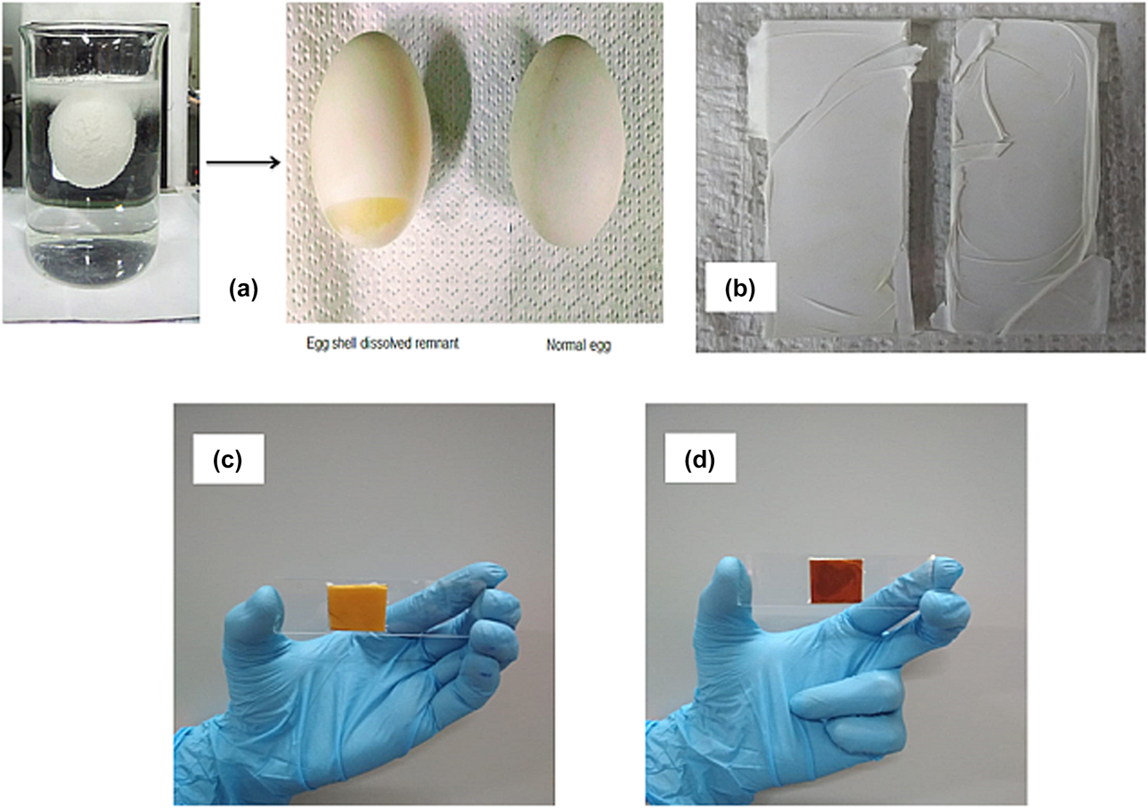
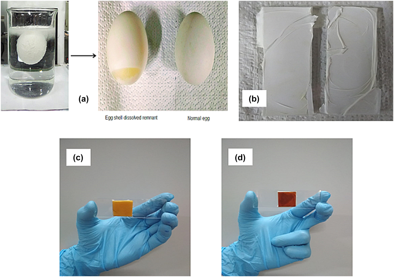
As described in the section “Mucosa-mimetic surface fabrication” in the materials and methods section, a very low cost and simple ESM-BSM surface was fabricated using ESM, glass slide, double-sided adhesive tape, and BSM. Figure 5(a) shows the dissolution of the outer calcium carbonate shell of the egg leaving behind the bouncy ESM enclosing the yolk. Figure 5(b) shows the ESM-BSM mucosa–mimetic surface, while Figs. 5(c) and 5(d) show the films attached on the fabricated mucosa-mimetic surface.
(a) ESM extraction by dissolution of outer shell of the egg; (b) ESM-BSM mucosa-mimetic surface; (c) Sample 1 adhered to the ESM-BSM surface; and (d) Sample 2 adhered to the ESM-BSM surface.
Formulation of simulated saliva
A modified SAGF medium, as described in the section “Formulation of simulated saliva”, was formulated. The pH was adjusted to 6.8 as reported since this value mimics the physiological saliva. This saliva was further used in the mucoadhesive timing testing of the fabricated COS-based patches.
Mucoadhesion residence time testing
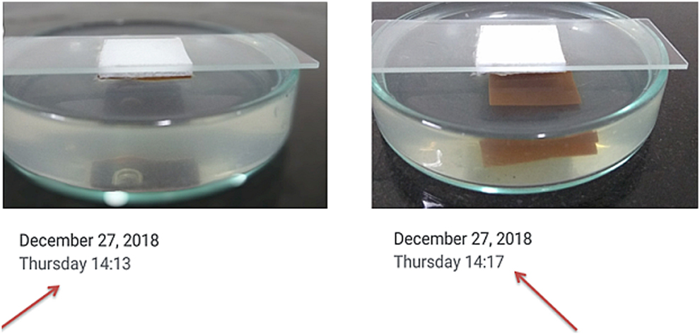
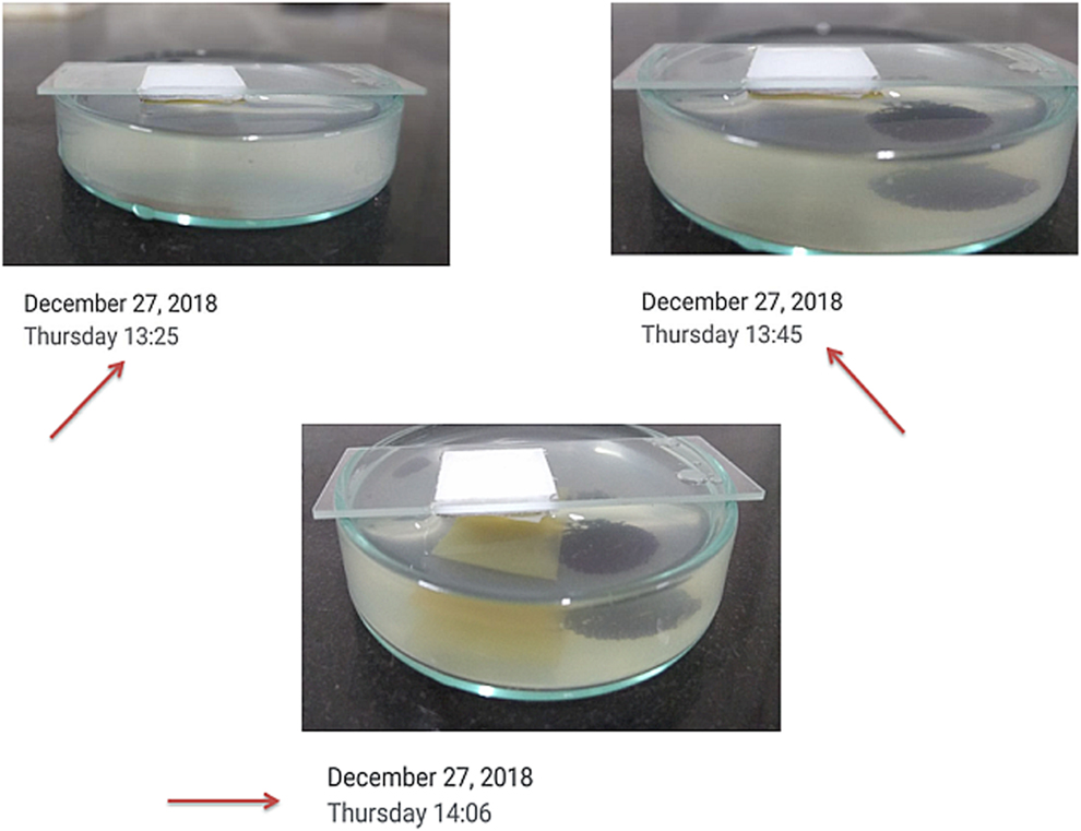
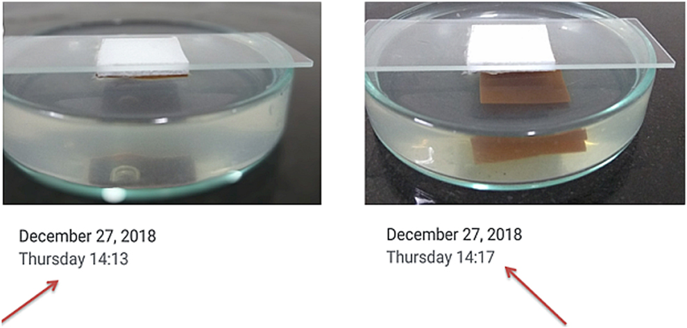
Figures 6 and 7 show the whole setup of testing the mucoadhesion time. To our surprise, sample 1 (CosPVA film) showed a mucoadhesion time of around 40 min while sample 2 (CosAP) film detached after almost 4 min. The image clearly shows the attached films (with time) and detached films (with time). Although sample 2 contained two reported mucoadhesive polymers, the polyelectrolyte system possibly reduced the mucoadhesion. This is because the functional groups responsible to impart the mucoadhesive characteristic to the polymers have largely interacted among themselves and are hardly available to interact with the mucin.
Mucoadhesion residence time for sample 1.
Mucoadhesion residence time for sample 2.
Conclusion and future aspects
Porcine (pig) are being used as one of the animal models closest to humans in physiological terms to go ahead with testing the buccal drug formulations [Reference Kulkarni, Mahalingam, Pather, Li and Jasti13]. Excised animal mucosa, especially porcine, is commonly reported to be used for in vitro testing of buccal drug delivery formulations. There are two possible limitations of using this animal model: First, the buccal mucosa extraction from the excised tissue is itself a tedious job and requires a lot of expertise and second, it is very difficult to access pig slaughter houses in cities (many cities in India) where it is considered a very uncommon practice. Apart from pig, rabbit buccal mucosa is considered and reported to be very similar to that of humans with slight keratinized regions known as para-keratinized regions [Reference Cui and Mumper15]. Rabbit is the animal of choice where it is difficult to access porcine, dogs, or primates. These animals, in any case, need to be sacrificed to carry out the study. Fabrication of synthetic mucosa-mimetic surface has been considered within the last decade to replace the use of animal mucosa for such experimental purposes. Kutoryanskiy et al., as per our extensive literature search, has perhaps carried out most significant work in fabricating mucosa-mimetic surfaces. The only problem, as per our observation, in such a fabrication is the application of synthetic polymers, reagents, and equipment that can add up to the cost of the experiment. The use of buccal epithelium cell lines grown on filter is a closely mimicking concept but the maintenance of cells in culture facility takes time and requires investments. Therefore, fabrication of a low-cost buccal mucosa–mimetic surface is the need of the hour to reduce the animal sacrifice for conducting the in vitro formulation studies.
Herein, we reported two novel things that, as per our extensive search, have not been reported till date. We fabricated and analyzed COS-blended films, proposed to be used as buccal drug delivery film. Second, we fabricated a novel low-cost ESM- and BSM-based buccal mucosa–mimetic surface and tested it preliminary to be used as an alternative to the animal buccal mucosa tissue. In our work, we found that COS blended well with PVA, alginate, and glycerol. CosPVA films displayed much better mucoadhesion residence time as than the CosAP films. The possible reason for such a result could be the polyelectrolyte interaction between COS and sodium alginate. A polyelectrolyte formation generally reduces the free amine group in COS and free carboxylic group in alginate that are essential to interact with mucin.
The low-cost, easily fabricated mucosa-mimetic surface reported in this work can also be used to mimic the gastrointestinal mucosal surface by incorporating essential manipulations such as changing the submaxillary mucin to gastric mucin. The synthetic surface, further, can be used even for formulations such as mucoadhesive tablets, capsules, and microparticles. We can, therefore, say that this mucosa-mimetic artificial ESM-BSM surface could help in reducing the cost of in vitro mucoadhesion testing. Second, such as fabricated surface could help reducing animal sacrifice for harvesting the mucosal tissue for in vitro analyses.
Materials and methods
Materials
Chitosan oligosaccharide (COS; CAS No. 148411578) was procured from SRL Chemicals, Mumbai, India. Sodium alginate (SA; CAS No. 9005383) and polyvinyl alcohol (PVA; CAS No. 9002895) was procured from Loba Chemie, Mumbai, India. Glycerol (CAS No. 56815) was purchased from HiMedia, Mumbai, India. Sodium chloride (NaCl), magnesium chloridehexahydrate (MgCl2·6H2O), calcium chloride (CaCl2), potassium chloride (KCl), potassium dihydrogen phosphate (KH2PO4), urea, sodium sulfate (Na2SO4), and xanthan gum were procured from HiMedia, Mumbai, India, Merck Millipore, Mumbai, India, and Loba Chemie, Mumbai, India. Bovine submaxillary mucin (BSM, Type 1-S; CAS No. 84195528) was procured from Merck, Mumbai, India. All the chemicals and reagents were of analytical grade. ESM was isolated from the poultry eggs procured from local market.
Fabrication of COS blended films
The films were fabricated using the solvent casting method at parameters reported earlier [Reference Kumar and Kumar26]. Briefly, 2% (w/v) COS solution and 2% (w/v) sodium alginate solutions were prepared in distilled water. PVA solution (5% w/v) was prepared in water at 80 °C. Two blended films were casted in a combination of (COS + PVA; Sample 1) and (COS + SA + PVA; Sample 2) in a plastic container (dimension 8 × 5 cm). Glycerol was used as the plasticizer in both the films. The casted films were peeled of gently after their fabrication. Films of area 2 × 2 cm2 were cut from both the casted parent films for further analyses.
Thickness measurement
The thickness of the films was measured by a Vernier caliper with a least count of 0.1 mm. Thickness was measured at 4 different points and average was calculated.
Folding endurance
Folding endurance is the most common test performed to check the breaking point and flexibility of the polymeric films. The films were continuously folded at the same point for 200 times.
Surface morphology study
The surface of the films were gold coated by sputter coating followed by imaging using high-resolution scanning electron microscopy (HR-SEM) [Nova Nano SEM 450, FEI (Thermo Fisher), Hillsboro, Oregon] performed at central instrumentation facility, IIT-BHU, India. The images were captured at 400×, 1600×, and 3000×. The analysis was carried out at a voltage of 15 kV.
Polymer interaction analysis by FTIR and XRD
The interaction between polymeric ingredients in the film was characterized using FTIR (FTIR Cary 630, Agilent, Santa Clara, California) and XRD (X’pert powder, PANalytical, Netherlands) at Department of Chemistry and Department of Metallurgical Engineering, NIT Raipur, respectively. The films were analyzed using the attenuated total reflectance (ATR) mode of the instrument while the individual polymeric powders were analyzed by forming KBr pellets. FTIR analysis was performed in a range of 4000–400 cm−1. In XRD, the analysis was performed in the 2θ range from 10° to 90°. The X-ray source was incident at 40 kV and 25 mA.
Indentation hardness study
The indentation hardness of the films was measured using a durometer (Shore A hardness test) as per ASTM D2240 standards. The test was performed at CIPET Raipur, Chhattisgarh. The method is based on assessing the resistance to indentation for materials such as polymers and elastomers.
Mucosa-mimetic surface fabrication
ESM was extracted using an in-house developed method. The poultry egg was dipped in a 500 mL solution of glacial acetic acid and water (40% glacial acetic acid v/v) for 5 h. The outer calcium carbonate shell dissolved completely after 5 h leaving behind a bouncy ball-like remnant. It was gently pricked using a needle and the yolk oozed out in the solution leaving behind the ESM. The membrane was carefully removed, washed with distilled water, and kept in a container with fresh water. To prepare the mucosa-mimetic surface, a glass slide was wiped with alcohol and a 2 × 2 cm2 double-sided adhesive tape was fixed at its center. The ESM was carefully attached to the other exposed surface of the adhesive tape and matched with its periphery. It was air dried for 2 h. 4% (w/v) BSM solution was prepared, and it was carefully evenly spread on the dried ESM surface (ESM-BSM). This is a novel and very low-cost method for developing mucosa-mimetic surface for mucoadhesion testing.
Formulation of simulated saliva
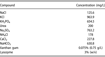
Simulated saliva is an integral part of in vitro testing of buccal and sublingual drug formulations. As per our extensive literature search for simulated saliva, the most similar salivary solution, in terms of inorganic compositions, was found to be described by Gal et al. [Reference Gal, Fovet and Adib-Yadzi27] as SAGF medium. We modified this SAGF medium to impart it with a viscosity similar to our natural saliva. The viscosity of the saliva was induced by adding xanthan gum. The enzymatic environment of the artificial saliva was slightly mimicked by adding commercially available lysozyme. The composition detail is presented in Table I.
Composition of fabricated simulated saliva.

Mucoadhesion residence time testing
To test the mucoadhesion time, COS-based films (sample 1 and sample 2) were applied on to the ESM-BSM surface and pressed for two minutes to let the films attach to the surface. The whole setup was then kept upside down in a petri-plate containing 25 mL simulated saliva up to the brim. The initial time was noted as soon as the film was just immersed in the simulated saliva. The final time was noted when the film got detached. The experiment was performed in triplicate and the average of the three times was calculated.
Acknowledgments
The authors are grateful to the Ministry of Human and Resource (MHRD, Government of India) and National Institute of Technology (NIT), Raipur (India) for providing research fellowship and necessary facilities for conducting the research. We also thank to Central Instrumentation Facility, IIT-BHU, India and CIPET Raipur, India for their co-operation to perform certain part of the experiments in their laboratory.
Abbreviations
- AGA
N-acryloylglucosamine
- ASTM
American Society of Testing and Materials
- ATR
Attenuated total reflectance
- BSM
Bovine submaxillary mucin
- COS
Chitosan oligosaccharide
- ESM
Egg-shell membrane
- FTIR
Fourier transfer infra red
- GLP-1
Glucagon like peptide-1
- HA
Hyaluronic acid
- HEMA
2-Hydroxyethyl methacrylate
- HR-SEM
Hig resolution scanning electron microscopy
- MUC
Mucin
- Na-CMC
Sodium carboxymethyl cellulose
- PAA
Poly acrylic acid
- PVA
Poly vinyl alcohol
- SEM
Scanning electron microscope
- XRD
X-ray diffraction