Introduction
An ideal method of laryngeal examination would afford an accurate assessment of disease, making it possible to differentiate normal variations, and to document findings in a clear, concise and reproducible manner. Ideally, it would be relatively inexpensive, time efficient and well tolerated by the patient.Reference Kelly1
The first successful attempt to visualise the vocal folds was achieved by Manuel Garcia, a Spanish music teacher, who was able to observe his own vocal folds using a dental mirror in 1854.Reference Garcia2 Since then, there have been significant technological advances and several techniques have been used in the evaluation, diagnosis and documentation of laryngeal pathology in clinical practice. Broadly, these methods are classified into: optical (mirror laryngoscopy), laryngeal endoscopic (rigid and flexible), radiographic, acoustic and electrophysiological.
Since the first successful photographic documentation of the larynx was achieved by Thomas French in 1884 (with a laryngeal mirror and a primitive camera), many different methods of laryngeal photography have been developed. Modalities include fibre-optic, telescopic and microscopic laryngeal techniques, as well as the use of dynamic video recordings.Reference Yanagisawa and Yanagisawa3 Yutaka Tachiki performed the first successful videographic documentation of the larynx in 1954. This has since been advocated as a method for documenting all endoscopic procedures in otolaryngology.Reference Tachiki4
Photo-documentation of laryngeal disease has progressed steadily from primitive black and white film still photography to high quality colour digital images that can be easily produced in an office-based laryngoscopic examination. The Hopkins rigid, rod-lens, wide-angle telescopes increase light transmission, magnify the image and provide an almost infinite depth of focus. Coupled with a camera, the clear optics of these telescopes offer an excellent method of both still and dynamic documentation. Fibre-optic laryngoscopes were first introduced in 1976, providing excellent visualisation of the vocal folds, with the advantage of being small in diameter and flexible.Reference Silberman, Wilf and Tucker5 This was followed by the development of the charge-coupled device for distal-chip endoscopes. The charge-coupled device has basically replaced the optical fibres at the distal end of the fibre-optic laryngoscopes. With advances in fibre-optic and distal-chip technology, flexible laryngoscopic examination techniques also lend themselves well to photo-documentation. Many papers, surgical atlases and textbooks have featured the results of photographic techniques using both flexible and rigid fibre-optic instruments.Reference Benjamin6–Reference Yanagisawa8
With advancing technology and the decreasing cost of the equipment required for digital photo-documentation, the advantages of digital photography outweigh their disadvantages. Advantages include, but are not limited to, compatibility with current lighting equipment, ease of focus and colour balance adjustment, instant playback and review, rapid delivery of a printed image, mass storage capacity, and ease of electronic integration into medical records. In addition, unwanted information can be immediately deleted. Furthermore, images and video can easily be copied and transmitted electronically for communication with other clinicians, as well as being integrated into both teaching and research presentations. The ability to edit and adjust the size, colour and contrast of the original image is also very useful.
An extensive literature review provided a thorough description of various methods of laryngeal photography and its development over time; however, none of the studies that were identified compared the quality of still images (not video or stroboscopic images) obtained by rigid versus distal-chip flexible laryngoscopes.
Although the rigid Hopkins rod telescopes have been perceived by many to produce a superior image to flexible laryngoscopes, with the advent of newer ‘distal-chip’ flexible endoscopes, the difference in perceived image quality has markedly narrowed. Hence, the current study aimed to compare the quality of still images taken by rigid laryngoscopes versus flexible distal-chip laryngoscopes in photographs of the same pathology.
Materials and methods
Study cases
The study utilised a prospective single-blind design. A total of 10 early stage glottic cancer cases (T1 and T2 stage tumours), diagnosed by a head and neck surgeon, were selected. Final diagnosis and extent of disease were confirmed intra-operatively by microlaryngoscopy (all cases were candidates for transoral laser microsurgery). We excluded: post-operative and irradiated larynges, subtle pathologies not easily diagnosed by a still image, and poorly exposed or focused images.
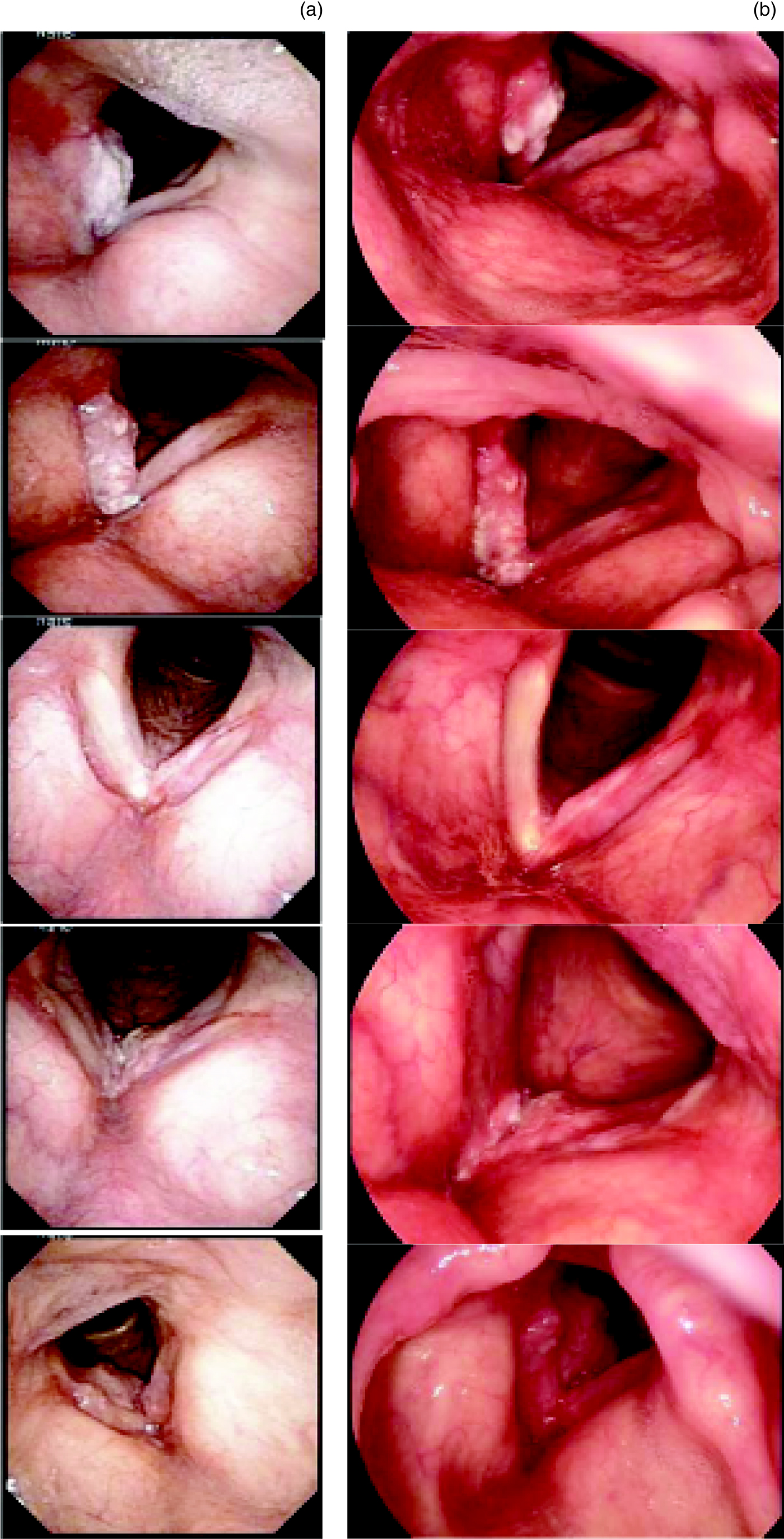
For each case, a staff laryngologist took two still photographs (during the course of the sequential rigid and distal-chip flexible laryngoscope examination) that showed the best view of the pathology (Figure 1). Efforts were made to produce images (from each of the two modalities) that were of similar size and orientation. Hence, there were 2 photographs for each pathologic larynx (a total of 20 photographs).
Selection of photographs taken by: (a) rigid (70 degree) laryngoscope and (b) flexible distal-chip laryngoscope.

A code number was assigned to each photograph, and a complete pathological description was documented for each case and presented in a reference table. This description was based on intra-operative microlaryngoscopy findings regarding: the tumour side (right or left), involvement of the anterior commissure, anatomical sub-site (glottic alone, glottic and subglottic, glottic and supraglottic, subglottic alone, supraglottic alone, transglottic) and tumour stage (T1a, T1b, T2), as well the modality used to take the photograph. Obviously, vocal fold mobility could not be assessed on the basis of a still image. An answer key reference table was created that included the above-mentioned pathological description for each case.
Instruments
Two instrument modalities were used for this study: rigid and distal-chip flexible laryngoscopic techniques. Rigid examination involved the Kay Pentax laryngoscopy tower (Kay RLS 9100B; Lincoln Park, New Jersey, USA), which incorporated: Kay Pentax digital strobe 9200 software, a 70 degree Karl Storz laryngoscope (Tuttlingen, Germany), a Hopkins rod laryngeal telescope and a Toshiba three chip colour charge-coupled device camera head. The flexible examination included the use of an Olympus® distal-chip flexible nasopharyngoscope (model ENF V2), and an Olympus (Exera II CLV-180) Xenon light source and processor. During the video laryngoscopic examination, frozen frames taken by each laryngoscope technique (rigid and flexible) were selected and printed out using a Hewlett Packard colour laser printer (model 2600n), with a resolution of 600 dpi on standard 8.5 × 11 inch printer paper.
Participants and procedure
The study participants comprised 9 practising otolaryngologists (excluding laryngologists), and 10 trainee (resident) otolaryngologists from the Division of Otolaryngology-Head and Neck Surgery, Dalhousie University. Each participant was shown the laryngoscopic photographs by the main investigator. All photographs were placed in a binder in a random sequence and accompanied by a standard questionnaire worksheet which was to be completed by the participants.
The reviewers were blind to the patient history, diagnosis and type of laryngoscope (rigid or flexible) used to acquire each photograph. Participants were asked to document their findings (based on the photographs) on the questionnaire worksheet provided (Appendix 1). They were also given the tumour–node–metastasis (TNM) staging system for glottic cancer.
Questions were divided into three categories: clinical, photograph quality and overall satisfaction with the image quality (based on self-reported ability to make a confident diagnosis with the images obtained). Clinical questions included: tumour side (right or left, indeterminable), anterior commissure involvement (yes, no, indeterminable), anatomical sub-site (glottic alone, glottic and subglottic, glottic and supraglottic, subglottic alone, supraglottic alone, transglottic, indeterminable) and tumour stage (T1a, T1b, T2, indeterminable). The questions regarding photograph quality were based on a 5-point rating scale (1 = poorest, 5 = best). Quality assessment was based on three main attributes: colour fidelity, focus and definition, and exposure and lighting.
We calculated the degree of accuracy between the answer key reference table and participants' worksheet answers for each corresponding case. McNemar's test was used to compare accuracy rates for the rigid laryngoscope images versus the flexible distal-chip laryngoscope images.
Results
The study comprised a total of 19 participants: 9 staff members, 4 senior residents and 6 junior residents. The participating staff members included two general otolaryngologists, two otologists, two paediatric otolaryngologists, one rhinologist, and two head and neck surgeons. A total of 10 cases were included in the study; each was represented by a photograph taken via the distal-chip flexible laryngoscope and another photograph taken using the rigid laryngoscope. All photographs were assigned a code. We used codes from 1 to 10 for the distal-chip flexible endoscope photographs and codes from A to J for the rigid endoscope photographs. The clinical descriptions for the 10 cases included: sidedness (5 right, 5 left), anterior commissure involvement (7 yes, 3 no), anatomical sub-site involvement (4 glottic only, 2 glottic plus supraglottic, 3 glottic plus subglottic, 1 transglottic) and tumour stage (3 for T1a, 1 for T1b, 6 for T2) (Table I). All participants answered the worksheet questionnaire for all 20 photographs. The results obtained are described in the following subsections.
Answer key

Codes 1 to 10 represent the distal-chip flexible endoscope photographs and codes A to J indicate the rigid endoscope photographs.
Clinical questions
We calculated the overall accuracy for the answers given to each clinical question, and determined whether there were any statistical differences in the accuracy rates between the flexible and rigid laryngoscope photographs. The p value was considered significant if it was less than or equal to 0.05. The results are shown in Tables II and III.
Accuracy based on participant type

Data represent percentages.
Accuracy based on laryngoscope type

All data (apart from the probability row) represent percentages. NS = non-significant
With regard to sidedness, the overall accuracy was 94.7 per cent, and there were no statistically significant differences between the rigid and flexible laryngoscope images (p = 0.238). For anterior commissure involvement, the overall accuracy was 53.7 per cent. Although the difference between the rigid and flexible techniques was not statistically significant, there was a trend favouring the flexible over the rigid laryngoscope images (p = 0.052, just over the significance cut-off point). With regard to the anatomical sub-site involved, overall accuracy was 46.6 per cent, and there were no statistically significant differences between the rigid and flexible laryngoscope images (p = 0.92). As for tumour stage, overall accuracy was 47.1 per cent, and there were no statistically significant differences detected between the rigid and flexible laryngoscope images (p = 1.00).
Photograph quality and overall satisfaction
There were no statistically significant differences between the rigid and distal-chip flexible laryngoscope techniques with regard to photograph quality (colour, focus and exposure) as rated by both staff and residents. Likewise, there was no statistically significant difference in the reviewers' overall satisfaction with the photographs. (See Tables IV and V.)
Photograph quality

All data (apart from the probability column) represent rating scores for photograph quality (out of a maximum possible score of 190 (number of participants multiplied by the number of photographs) for each parameter). Flex = flexible; NS = non-significant
Overall satisfaction

All data (apart from the probability column) represent overall satisfaction rating scores (out of a maximum possible score of 190 (number of participants multiplied by the number of photographs)). Flex = flexible; NS = non-significant
Discussion
Photo-documentation has replaced hand sketched drawings of pathology and has enhanced classical textual descriptions in many aspects of medicine. Photo-documentation provides valuable material in lectures, teaching, research and publications. Moreover, it can represent an ideal and unbiased view of lesion progression. In this study, we assessed the use of still images in the evaluation and diagnosis of early glottic cancer (tumour stages T1a, T1b and T2), which can be diagnosed without the need to evaluate vocal fold mobility. The still images were not accompanied by any clinical description except for the recent TNM staging system for early glottic cancer.
The overall accuracy rate for lesion sidedness was 94.7 per cent; however, the accuracy rates for anatomical sub-site involvement, anterior commissure involvement and tumour staging were 46.6 per cent, 53.7 per cent and 47.1 per cent, respectively. Surprisingly, despite the best available visual imaging techniques, our data showed that – at least with regards to single, still images – otolaryngology clinicians may fail to accurately detect critical oncologic variables of early laryngeal cancer. Although not evaluated in this study, the possibility that dynamic video may enhance accuracy remains to be tested. The use of still images as the sole documentation method in medical charting and in correspondence with other physicians appears to be insufficient.
Several papers have been published that compare the use of rigid versus flexible laryngoscopes (both fibre-optic and distal-chip) in the evaluation of laryngeal pathology.9–11 In 1993, Yanagisawa et al. showed that rigid video stroboscopy was superior to fibre-optic imaging in the evaluation of vocal fold lesions.Reference Yanagisawa and Yanagisawa9 In 2008, Eller et al. compared the use of fibre-optic versus distal-chip devices, and also compared the use of flexible distal-chip versus rigid laryngoscopes in the evaluation of vocal fold masses.Reference Eller, Ginsburg, Lurie, Heman-Ackah, Lyons and Sataloff10 The investigators concluded that, despite advances in flexible laryngoscope distal-chip technology, rigid laryngoscopes were still superior with regards to image quality for diagnostic purposes. However, both of these studies compared recorded segments of videolaryngoscopic examinations, not still images. In our study, we compared the overall accuracy in diagnosis and clinical description between distal-chip flexible laryngoscope photographs and rigid laryngoscope photographs of the exact same pathology. We also compared the participants' evaluation of the photograph quality and their overall satisfaction with each photograph (based on their ability to reach a diagnosis using the images obtained).
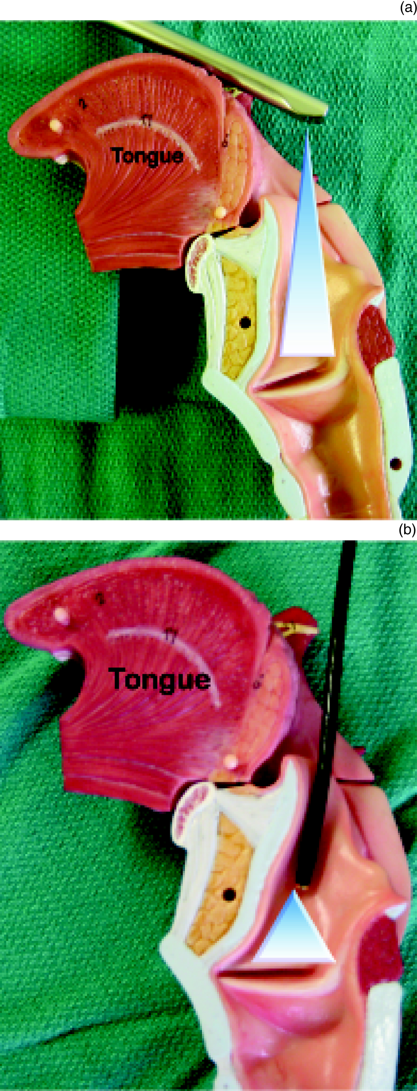
Overall, the images taken by both the rigid and distal-chip flexible laryngoscopes yielded comparable scores. No statistically significant differences were noted in terms of disease sidedness, anatomical sub-site involvement or tumour stage. Although not quite statistically significant, there was a trend towards favouring the flexible over the rigid laryngoscope with regards to anterior commissure involvement. This may be due to the fact that the tip of the flexible laryngoscope can be positioned to enable a slightly different angle of visualisation with respect to the anterior commissure (Figure 2).
Positioning and visualisation of: (a) rigid (70 degree) laryngoscope (arrowhead) and (b) flexible distal-chip laryngoscope (arrowhead).

Photographic quality (colour, focus and exposure) was rated as slightly above average (ratings of 3 and 4 were achieved) for both laryngoscope modalities. However, overall satisfaction rates were scored slightly higher for the flexible laryngoscope. Hence, images obtained from distal-chip flexible laryngoscopes appear to be at least as useful in diagnosing laryngeal malignant pathology as those taken from traditional rigid methods.
• Flexible fibre-optic nasopharyngoscopes and rigid Hopkins rod endoscopes are commonly used to document laryngeal pathology
• Hopkins rod telescope images have been perceived as superior to those produced via flexible fibre-optic endoscopy
• With the advent of newer ‘distal-chip’ flexible endoscopes, the image quality difference has narrowed
• This novel study compared the quality of still images obtained by rigid versus distal-chip flexible laryngoscopes
• There were no differences between the techniques for clinical accuracy and subjective photograph quality
• There was a trend favouring the flexible laryngoscope in evaluating anterior commissure involvement (p = 0.052)
The limitations of our study include the usage of an analogue camera system, two-dimensional photography, and a non-specialised photo-printer and photo-paper. In addition, image orientation and the relative size of the images were not always perfectly comparable. Future studies could include: a comparison of images obtained in the clinic with those taken under microscopy in the operating theatre, a comparison of dynamic video techniques with still images, and the potential use of narrow-band imaging techniques.
Conclusion
Our data showed no significant differences in terms of overall clinical accuracy and the photograph quality achieved using rigid versus distal-chip flexible laryngoscopes. There was a trend approaching significance favouring the distal-chip flexible laryngoscope in the evaluation of anterior commissure involvement (p = 0.052), which could be attributed to the fact that this laryngoscope can be positioned at different angles, potentially enabling better visualisation of the anterior commissure. Future studies should incorporate comparisons with dynamic digital video recordings, which are increasingly available in many centres.
Appendix 1. Laryngology research participant working sheet
Dear Doctor,
Please look at this laryngoscopic image of a pathological larynx. This is an early untreated laryngeal cancer. T3 lesions have been excluded. Take a moment to review carefully the abnormal findings and tick with a check mark (☑) in the appropriate boxes below for the most likely answer.
Thank you for your participation.
Case code …
1. Primary location:
□ Right hemilarynx
□ Left hemilarynx
□ Cannot determine
Anterior commissure involved (does the lesion cross the midline)?:
□ Yes
□ No
□ Cannot determine
Cancer is:
□ Glottic alone
□ Supraglottic alone
□ Subglottic alone
□ Glottic plus supraglottic
□ Glottic plus subglottic
□ Transglottic
□ Cannot determine
2. Staging:
□ T1a
□ T1b
□ T2
□ Cannot determine
3. Photograph quality (on a scale from 1 (poorest) to 5 (best)). Circle one only:
Colour fidelity 1–2–3–4–5
Focus/ definition 1–2–3–4–5
Exposure/ lighting 1–2–3–4–5
4. Overall satisfaction with this photograph to make your diagnosis:
□ Fully satisfied
□ Partially satisfied
□ Not satisfied
5. Comments: