Introduction
Lipases are serine hydrolases that are ubiquitous enzymes in all organisms. Lipases can be divided into six families, neutral (PF00151), acid (PF04083), lipase2 (PF01674), lipase3 (PF01764), GDSL (PF00657) and hormone-sensitive lipases (PF06350), defined by sequence relationships within the α/β hydrolase fold superfamily of proteins (Holmquist, Reference Holmquist2000; Aoki et al., Reference Aoki, Inoue, Makide, Saiki and Arai2007). All six families appear to use the same, two-step reaction mechanism based on a catalytic triad of residues (usually Ser–His–Asp/Glu) that generates a charge relay system and a highly nucleophilic serine (Zimmermann et al., Reference Zimmermann, Strauss, Haemmerle, Schoiswohl, Birner-Gruenberger, Riederer, Lass, Neuberger, Eisenhaber, Hermetter and Zechner2004). These three amino acid residues always appear in the relative positions in the tertiary structure, but are distant from each other in the lipase primary sequence. The catalytic triads in the lipase tertiary structure are positioned within a close distance to each other to catalyze the hydrolysis reaction of carboxylic acid esters in aqueous medium or the reverse reaction in organic solvents (Reetz, Reference Reetz2002). A characteristic α/β hydrolase fold was found in all solved lipase crystallographic structures, including bacterial (Pseudomonas and Bacillus) lipases, fungal Rhizomucor lipases, and horse and human pancreatic lipases (van Pouderoyen et al., Reference van Pouderoyen, Eggert, Jaeger and Dijkstra2001).
Lipases play a crucial role in fat metabolism by catalyzing the hydrolysis of triacylglycerol to free fatty acids and glycerol at the interface of lipid and water (Jaeger & Reetz, Reference Jaeger and Reetz1998; Gupta et al., Reference Gupta, Gupta and Rathi2004). Although lipases are highly chemo-, regio- and chiral-selective enzymes, they also process esterolytic activities for the carboxyl ester bond cleavage of water-insoluble esters (Jaeger & Reetz, Reference Jaeger and Reetz1998; Reis et al., Reference Reis, Holmberg, Watzke, Leser and Miller2009; Treichel et al., Reference Treichel, de Oliveira, Mazutti, Di Luccio and Oliveira2010). Lipases can also catalyze the reverse reaction in the presence of a low water concentration (Park et al., Reference Park, Lee, Chi and Jeong2005; Franken et al., Reference Franken, Marcon, Treichel, Oliveira, Freire, Dariva, Destain and Oliveira2010; Severac et al., Reference Severac, Galy, Turon, Monsan and Marty2011). Insect lipases have key roles in lipid acquisition, storage and mobilization, and are also fundamental to many physiological processes such as insect reproduction, development, defense against pathogens, oxidative stress and pheromone signaling. The six major lipase families were found in the sequenced genomes of five species (Drosophila melanogaster, Anopheles gambiae, Apis mellifera, Bombyx mori and Tribolium castaneum), and are also found in other organisms (Horne et al., Reference Horne, Haritos and Oakeshott2009; Ranjbar et al., Reference Ranjbar, Zibaee and Sendi2015). The different roles required of lipases at the four discrete life stages of holometabolous insects might also contribute to the diversity of lipases required by insects (Horne et al., Reference Horne, Haritos and Oakeshott2009).
Compared with vertebrates, insects rely on an innate immune system to defend themselves against infections by pathogenic microorganisms (Ishii et al., Reference Ishii, Hamamoto, Kamimura, Nakamura, Noda, Imamura, Mita and Sekimizu2010). During the fungal penetration through the host cuticle, hydrolytic enzymes, such as proteases, chitinases and lipases, are secreted (Yang & Yeh, Reference Yang and Yeh2005; Ali et al., Reference Ali, Huang and Ren2009). The importance of any one of these enzymes depends upon the characteristics of the cuticle of the target insect and the invasion route and mechanism used by the fungus. The participation of proteases and chitinases in the host infection by entomopathogenic fungi has been demonstrated (Ortiz-Urquiza & Keyhani, Reference Ortiz-Urquiza and Keyhani2013). Lipases could be involved in the immune breach process by hydrolyzing the ester bonds of lipoproteins, fats and wax esters (Ali et al., Reference Ali, Ren and Huang2014). In this study, we selected a lipase-related protein in Antheraea pernyi (ApLRP) and studied its tissue distribution and expression profiles under immune challenge by a Gram-negative bacterium, a Gram-positive bacterium, a fungus and a virus. We also expressed and purified the recombinant ApLRP, and tested the effects on its activity of temperature, pH, metal ion, inhibitors and detergents. Our results indicated that ApLRP might be involved in the innate immune response.
Materials and methods
Insect rearing
A. pernyi was provided by the Sericultural Research Institute of Henan, China. A. pernyi larvae were fed on fresh oak leaves and reared at room temperature with a 14:10 h light:dark photoperiod until they pupated.
Cloning and sequence analysis of ApLRP
Through transcriptome sequencing of A. pernyi, one putative lipase gene was predicted by the BlastX program (NCBI). Open reading frames (ORFs) were searched using DNAstar software (version 5.02). Based on the ORF sequences, a pair of primers (ApLRP-SP and ApLRP-AP) (table 1) was designed to amplify the ORF of the ApLRP gene. The target PCR band was purified, ligated into vector pGEM T-Easy (Promega, WI, USA), and then sequenced using a 3730 XL DNA analyzer (Applied Biosystems, CA, USA). SignalP 4.1 server (http://www.cbs.dtu.dk/services/SignalP-4.1/) was used to search for a signal peptide. The molecular weight of the ApLRP protein was calculated using ExPASy (http://web.expasy.org/compute_pi/). A multiple sequence alignment was performed using ClustalX2 (http://www.ebi.ac.uk/tools/clustalw2/index.html). A phylogenetic tree was constructed by MEGA 5.1 using the neighbor-joining algorithm (Tamura et al., Reference Tamura, Peterson, Peterson, Stecher, Nei and Kumar2011), with statistical analysis by the bootstrap method using 1000 repetitions.
Sequences of primers used in this study.

Note: Restriction sites are underlined.
Expression and purification of recombinant of ApLRP protein
Total RNA was extracted from fat body of A. pernyi larvae using the Trizol reagent (Takara, Dalian, China), and reverse-transcribed to first-strand cDNA using PrimeScript™ One step RT–PCR Kit Ver.2 (Takara), following the manufacturer's instructions. To express the recombinant ApLRP protein, specific primers (pET-ApLRP-SP and pET-ApLRP-AP) (table 1) were designed to amplify the nucleotide sequence encoding the ApLRP mature protein without its signal peptide. PCR was performed using an amplification program comprising 4 min at 94°C; 35 cycles of 94°C for 30 s, 58°C for 30 s, 72°C for 1 min, and a final elongation step of 72°C for 10 min. The PCR product was separated on a 1% agarose gel and purified using a DNA Gel Extraction Kit (Axygen, Hangzhou, China). The purified PCR product and vector pET-28a (+) were both digested with BamH I and Hind III (Takara) at 37°C for 4 h, and then ligated using T4 ligase (Takara) at 4°C for 12 h. The recovered recombinant plasmid was transformed into competent Escherichia coli (transsetta DE3) cells (TransGen Biotech, Beijing, China). After adding isopropyl-β-D-thiogalactopyranoside (IPTG) for induction for 4 h at 30°C, the bacterial cells were harvested by centrifugation at 8000 × g for 10 min at 4°C and washed with phosphate-buffered saline (PBS) (pH7.4) twice. The recombinant protein was analyzed by 12% SDS–PAGE. The pellets were resuspended in binding buffer (50 mM NaH2PO4, 300 mM NaCl, 10 mM imidazole, pH 8.0) and disrupted by sonication on ice. After centrifugation at 12,000 × g for 20 min at 4°C, the recombinant protein was purified using an Ni-NTA Agarose column (QIAGEN, China) under native conditions, according to the manufacturer's protocol. The purity and identity of the eluted fractions were analyzed by SDS–PAGE. The concentrations of purified ApLRP were determined using an EasyII Protein Quantitative Kit (TransGen Biotech), using bovine serum albumin as a standard. Purified ApLRP samples were aliquoted into microcentrifuge tubes and stored at −80°C for future use.
Preparation of ApLRP polyclonal antibodies
Anti-ApLRP polyclonal antibodies were prepared by HuaAn Biotechnology Co., Ltd (Hangzhou, China). Purified recombinant ApLRP protein (total 2.5 mg), as an antigen with adjuvant, was injected into a New Zealand white rabbit by three times. Blood was collected from the pre-immunized rabbit from the marginal vein of the ear as a negative control. The antibodies against ApLRP were divided into microtubes and stored at −80°C
Quantitative RT–PCR analysis
Total RNA of each tissue was extracted using the TRIzol reagent (Takara), according to the manufacturer's instructions. The RNA samples were then treated with RQ1 RNase-free DNase (Promega, WI, USA) to remove genomic DNA contamination. First strand cDNA was synthesized from one microgram of total RNA using PrimeScript™ RT Master Mix (Takara), following the manufacturer's instructions. All the primers for qRT–PCR were designed using the online tool Primer3 (http://bioinfo.ut.ee/primer3-0.4.0/) (table 1). The A. pernyi 18s gene (GenBank accession No: DQ347469) was used as an internal control. Real-time PCR was performed in a total volume of 20 µl, containing 10 µl 2 × SYBR Premix Ex TaqII (Tli RNase Plus) (Takara), 1 µl of each primer, 1 µl of 1:8 diluted cDNA templates and 7 µl RNase-free H2O. Quantitative RT–PCR was performed using a CFX96™ real-time detection system (Bio-Rad, CA, USA), using the following procedure: initial denaturation at 95°C for 30 s; followed by 40 cycles of denaturation at 95°C for 5 s, annealing at 60°C for 30 s; and a final extension at 72°C for 30 s. A melting curve analysis (65–95°C) was determined to confirm the unique and specific PCR product for each reaction. The relative expression level was determined according to the 2−ΔΔCt method (Livak & Schmittgen, Reference Livak and Schmittgen2001). Each biological treatment was repeated three times. The data were presented as the means ± standard error (SE).
Immunoblot analysis
Total protein samples from various tissues were ground in liquid nitrogen and dissolved in radio immunoprecipitation assay (RIPA) lysis buffer (Aidlab Biotech, Beijing, China) for protein extraction. After the protein concentration was determined using the bicinchonininc acid (BCA) method, total protein samples (20 µg each) were subjected to 12% SDS–PAGE and then transferred onto a polyvinylidene difluoride (PVDF) membrane (Millipore, MA, USA) using a Mini Trans-Blot electrophoretic transfer system (Bio-Rad). After blocking with 5% non-fat milk (diluted with PBS containing 0.1% Tween-20) (PBST), the membranes were washed in PBST three times and then incubated with the prepared anti-ApLRP polyclonal antibody (diluted 1:200 with 5% non-fat milk in PBST) for 2 h at room temperature, and glyceraldehyde-3-phosphate dehydrogenase (GAPDH) antibody (Beyotime) was used as a loading control primary antibody. After washing with PBST, the membranes were incubated with goat anti-rabbit IgG (Beyotime, Shanghai, China) (diluted 1:2000 with 5% non-fat milk in PBST) for 1 h at room temperature. The immunoblot signal was detected using an HRP-DAB Detection Kit (Tiangen, Beijing, China).
Tissue expression distribution
To determine the tissue-specific distribution of ApLRP transcripts and proteins, six tissues (midgut, silk gland, fat body, malpighian tubules, integument and hemocytes) were dissected from larvae at the third day of the fifth instar stage. RNA extraction and quantitative RT–PCR for ApLRP transcripts analysis, SDS–PAGE and immunoblot analysis for ApLRP protein were performed as described above.
Pathogen-induced expression analysis of ApLRP
Before induction, larvae from the third day of the fifth instar stage were placed in a Petri dish without food to ensure hunger and then chilled on ice for 20 min. Heat-inactivated Gram-negative bacterium (E. coli Trans1-T1, 8 µl, 109 cfu ml−1), Gram-positive bacterium (Micrococcus luteus 8 µl, 0.5 mg ml−1), fungus (Beauveria bassiana 8 µl, 109 cfu ml−1) and virus (nuclear polyhedrosis virus 8 µl, 109 PIBs ml−1) were injected into the abdomen of the larvae. All the microorganism pellets were diluted in the sterilized PBS. Larvae injected with sterilized PBS were used as the negative control. The wounds of the larvae were sealed with Vaseline immediately after injection. The midguts from individual samples were collected after 1.5, 3, 6, 12, 24 and 48 h. The tissues of three larvae were collected as one sample; and the biological sampling was repeated three times. The midgut samples were tested by qRT–PCR to detect the ApLRP mRNA expression profiles.
Activity assays of ApLRP
Colorimetric assay: ApLRP activity was determined using a modified protocol using p-nitrophenyl palmitate (pNPP) as a substrate (Ali et al., Reference Ali, Ren and Huang2014). The assay mixture comprised 200 µl of sample and 1800 µl of substrate solution (10 mg pNPP dissolved in 1 ml of 2-propanol diluted in 9 ml of 50 mM Tris–HCl pH 8.0 containing 40 mg of Triton X-100 and 10 mg of gum Arabic). The assay mixture was incubated at 37°C for 30 min and then the absorbance was measured at 410 nm. One unit of enzyme activity was defined as the amount of enzyme that liberated 1 nmol p-nitrophenol min−1 under the assay conditions.
Effect of temperature on the activity of purified ApLRP
The effect of temperature on ApLRP activity were studied at 15, 25, 35, 45, 55 and 65°C using the above colorimetric assay method. Assay mixture and sample were incubated at various temperatures for 20 min before the start of the reaction.
Effect of pH on the activity of purified ApLRP
The effect of pH on ApLRP activity was tested using 50 mM buffers at various pH values from 3 to 12. Glycine–HCl buffer was used for the pH range from 3 to 6; pH values from 7 to 9 were achieved using Tris-buffer; and glycine–NaOH buffer was used for pH values from 10 to 12. The sample was incubated with the various pH buffers at room temperature for 20 min before initiation of the reaction with the pNPP substrate.
Effect of various substances on ApLRP stability
The purified ApLRP was incubated in the presence of various chemical compounds including metal ions (ZnCl2, MgSO4, Fe3(SO4)2, MgCl2, CuSO4, NaCl or CaCl2, 1 mM each), detergents (glycerol, Tween-20 or SDS, 0.1% v/v each), and inhibitors (EDTA, b-mercaptoethanol or PMSF, 1 mM each) at room temperature for 20 min. ApLRP activity was determined by the pNPP method, as described above.
All experiments were carried out in triplicate. Data were analyzed using one-way ANOVA analysis by DPS software (version 9.50) (Tang & Zhang, Reference Tang and Zhang2013). The values reported are means of the three replicates ± standard deviation (SD). A P value <0.05 was considered statistically significant.
Results
Cloning and characterization of ApLRP gene
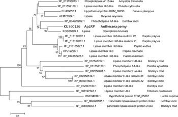
The ApLRP cDNA had a 1095-bp ORF encoding a 364-residue polypeptide (GenBank No: KU360126). The protein contains a signal peptide of 21 residues (MATLSAIALTSIVIISAVNAA). The molecular weight of the protein was calculated from the deduced amino acid sequence as 40.22 kDa, with a predicted isoelectric point (pI) of 8.53. The ApLRP protein was most similar to a lipase from Operophtera brumata (KOB66899.1, 81% identity), followed by phospholipase A1-like form B. mori (XP_004926202.1, 68% identity). Phylogenetic analysis indicated that the Ap protein was clustered with the O. brumata lipase (fig. 1).
Phylogenetic analysis of ApLRP with other lipases. The phylogenetic tree was constructed using the neighbor-joining algorithm, and bootstrap values (1000 repetitions) of the branches are indicated. The name and GenBank No. of the lipases are indicated.

Expression and purification of the recombinant ApLRP protein
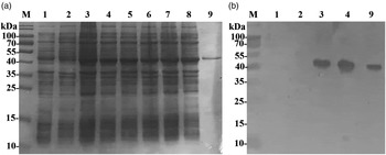
To produce sufficient protein for functional studies, we expressed the mature ApLRP protein in E. coli (transetta DE3) cells. Recombinant ApLRP protein was successfully expressed as a soluble protein and was not influenced by the IPTG concentration from 0.2 to 1 mM (fig. 2a). The apparent molecular mass was 45 kDa, which was higher than the predicted mass of mature ApLRP (38.2 kDa) and His-tag (fig. 2a). SDS–PAGE analysis showed that purified proteins appeared as a single band on the gel (fig. 2a). Western blotting analysis showed that recombinant ApLRP proteins were recognized by the anti-His antibody (fig. 2b).
Expression and purification of recombinant ApLRP. (a) Analysis of recombinant ApLRP proteins on 12% SDS–PAGE gels. (b) Western blotting analysis of recombinant proteins using anti-His-tag antibodies. Bacterial proteins were collected after 4 h of induction with different IPTG concentrations. M, molecular weight marker; Lane 1, untransfected E. coli (transetta DE3) cells; Lane 2, pET-28a (+) control vector in E. coli; Lane 3, non-induced E. coli (pET-ApLRP); Lane 4, induction of E. coli (pET-ApLRP) by 0.2 mM IPTG; Lane 5, induction of E. coli (pET-ApLRP) by 0.4 mM IPTG; Lane 6, induction of E. coli (pET-ApLRP) by 0.6 mM IPTG; Lane 7, induction of E. coli (pET-ApLRP) by 0.8 mM IPTG; Lane 8, induction of E. coli (pET-ApLRP) induction by 1.0 mM IPTG; and Lane 9, purified recombinant ApLRP.

Tissue distribution of ApLRP
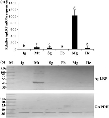
The qRT–PCR results showed that ApLRP was particularly highly expressed in the midgut, with low levels in malpighian tubules, silk gland, hemocytes, integument and fat body (fig.3a). Western blotting showed that the ApLRP protein could be detected only in the malpighian tubules (fig. 3b). Comparing the qRT–PCR results with the western blotting results, we found that ApLRP mRNA expression profiles were inconsistent with the protein content profiles, suggesting that ApLRP could be expressed by midgut and other tissues, and then transferred to and be accumulated in the malpighian tubules of A. pernyi.
Tissue distribution of ApLRP in the fifth instar larvae of A. pernyi. (a) Analysis of ApLRP mRNA expression in tissues of larvae using qRT–PCR. The ApLRP mRNA level in the fat body was used as the calibrator. Bars represent mean ± SE (n = 3). Bars labeled with different letters are significantly different (P < 0.05). (b) Analysis of ApLRP protein expression in tissues of larvae by Western blotting. ApLRP was detected using anti-ApLRP rabbit polyclonal antibodies. And GAPDH antibody was used as a loading control primary antibody. Ig, Integument; Mt, Malpighian tubule; Sg, Silk gland; Fb, Fat body; Mg, Midgut; Hc, Hemocyte.

Pathogen-induced expression pattern of ApLRP in midgut and malpighian tubules
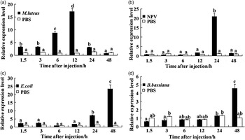
To further understand the induced expression profiles of ApLRP in response to microbial infection, qRT–PCR was carried out. Compared with the control, the mRNA expression level of ApLRP in the midgut was significantly upregulated and peaked at 12 h after infection with M. luteus, it then decreased from 24 h, reaching normal levels at 48 h (fig. 4a). The transcription level of ApLRP was significantly upregulated only at 24 h after infection with NPV, and then quickly dropped to normal levels at 48 h (fig. 4b). The ApLRP transcription level was significantly upregulated from 24 to 48 h when treated by E. coli (fig. 4c). The ApLRP mRNA expression was just strongly upregulated at 48 h compared with the control (fig. 4d). These results suggested that ApLRP might play an important role in defending against microorganism infection.
Expression profiles of ApLRP mRNA induced in the midgut of the fifth instar larvae of A. pernyi. Larvae from the third day of the fifth instar were injected with heat-inactivated Gram-negative E. coli (Trans1-T1 8 µl, 109 cfu ml−1), Gram-positive M. luteus (8 µl, 0.5 mg ml−1), fungi B. bassiana (8 µl 109 cfu ml−1) or virus NPV (8 µl, 109 PIBs ml−1). PBS (8 µl) was injected as a control. Midgut and malpighian tubules were collected at 1.5, 3, 6, 12, 24 and 48 h post-injection. The expression of ApLRP mRNA in the midgut was determined by qRT–PCR. The ApLRP mRNA level in the PBS-injected midgut was designated as the calibrator. Panels (a), (b), (c) and (d) represent the mRNA transcript levels of ApLRP in midgut after challenge with E. coli, B. bassiana, NPV and M. luteus, respectively.

Effect of temperature and pH on ApLRP activity
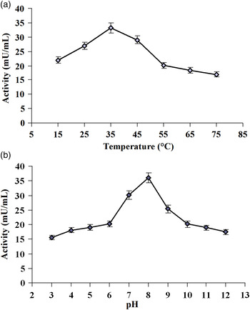
The lipase activity of the purified ApLRP protein was measured at various temperatures from 0 to 80°C. ApLRP was active from 15 to 75°C (fig. 5a). The optimal activity was observed at 35°C, at which the activity reached 33 mU ml−1 at pH 8.0. ApLRP activity gradually increased with increasing temperature until it reached 35°C, and then decreased with increasing temperature. The activity decreased significantly at higher temperatures with 60 and 49% relative activity remaining at 55 and 75°C, respectively.
Effect of temperature (a) and pH (b) on ApLRP activity. Average values from both experiments from three replications were plotted in solid circles with SD values represented as error bars.

Buffers of pH 3–12 were used to determine the pH optimum for ApLRP activity. Significant variation was seen for the purified ApLRP at different pHs. ApLRP showed its maximum activity at pH 8.0 in the Tris buffer system, when the activity reached 36 mU ml−1 at 35°C (fig. 5b). ApLRP was active in buffers ranging from pH 7 to 9, with a slight preference toward the alkaline pH buffers.
Effect of various substances on ApLRP stability
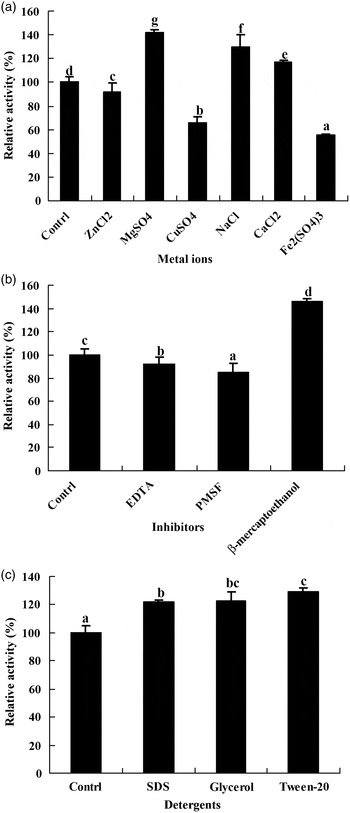
To determine whether metal ions could stimulate or inhibit the activity of ApLRP, Zn2+, Mg2+, Na+, Ca2+,Cu2+ and Fe3+ were tested. The results showed that metal ions affected ApLRP activity significantly (fig. 6a). ApLRP activity was enhanced in the presence of MgSO4, NaCl and CaCl2. Conversely, ZnCl2, CuSO4 and Fe2(SO4)3 inhibited the enzyme at similar concentrations (fig. 6a).
Effect of various substances on the activity of purified ApLRP. (a) Effect of divalent salts. (b) Effect of protein inhibitors. (c) Effect of surfactants. The control value was measured without the addition of metal ions, inhibitors or detergent. Metal ion, inhibitors or detergent solutions at a final concentration of 1 mM were added to the enzyme assay mixture. Average values from three experiments were plotted with error bars representing the SD.

Incubation of the purified ApLRP with different protein inhibitors had different effects on the enzyme activity (fig. 6b). The activity of ApLRP was activated by b-mercaptoethanol, but inhibited by EDTA or PMSF. The three selected detergents (Tween-20, glycerol or SDS) enhanced ApLRP activity and the highest increase in ApLRP activity (30% more) was observed with the addition of Tween-20 when compared with the control (fig. 6c).
Discussion
Lipases are widespread in insects, but little is known about the characteristics of insect lipases. In this study, we characterized an ApLRP. The ApLRP protein contains an Esterase_lipase superfamily domain and clusters with the O. brumata lipase. This indicated that ApLRP may act on carboxylic esters, and catalytic reaction involves three residues (a serine, a glutamate or aspartate and a histidine).
The ApLRP mRNA expression was the highest in midgut; however, the ApLRP protein was solely located in the malpighian tubules. Lipases in other organisms have been detected in different tissues, including the midgut, fat body and some Hymenopteran venom. The Bmlipase-1 gene from B. mori was expressed in the anterior and middle portions, but not in the posterior portion of the midgut (Ponnuvel et al., Reference Ponnuvel, Nakazawa, Furukawa, Asaoka, Ishibashi, Tanaka and Yamakawa2003). The Lip-1 gene, a D. melanogaster lipase gene, was demonstrated to be expressed at the foregut–midgut boundary in embryos by whole-mount in situ hybridization (Pistillo et al., Reference Pistillo, Manzi, Tino, Boyl, Graziani and Malva1998). In addition to the midgut, insect lipase genes and proteins have reported in the embryo, ovaries, male accessory gland, gonad, pheromone gland, fat body, antennae, salivary gland and venom gland. The presence of lipases in different tissues indicated their different functions in lipid digestion, lipid storage and mobilization, yolk protein and immune responses (Horne et al., Reference Horne, Haritos and Oakeshott2009). Malpighian tubules are osmoregulatory organs that maintain the ionic balance and remove toxic substances from the body, and act as autonomous immune-sensing organs in response to invading microbial pathogens (Tapadia & Verma, Reference Tapadia and Verma2012). The expressions of the ApLRP mRNA in the midgut were induced by pathogenic microorganisms, and the expression patterns differed depending on the pathogen (E. coli, B. bassiana, M. luteus and NPV). Our results indicated that ApLRP might mediate the immune response of A. pernyi during pathogen challenge. Besides the acquisition of dietary lipids, lipase BmorN12 from B. mori midgut had strong antiviral activity in larvae infected with BmNPV (Ponnuvel et al., Reference Ponnuvel, Nakazawa, Furukawa, Asaoka, Ishibashi, Tanaka and Yamakawa2003). Two lipases in D. melanogaster (CG6675 and CG4267) were upregulated in the Toll pathway or immune deficiency (IMD) pathway (De Gregorio et al., Reference De Gregorio, Spellman, Tzou, Rubin and Lemaitre2002). Two lipase genes in Lymantria dispar larval midgut were significant downregulated in response to infection by Bacillus thuringiensis kurstaki (Sparks et al., Reference Sparks, Blackburn, Kuhar and Gundersen-Rindal2013). Lipases in entomopathogenic fungi are involved in the infection process of insect surfaces by hydrolyzing the ester bonds of lipoproteins, fats and waxes present on the surface and in the body (Ali et al., Reference Ali, Ren and Huang2014). DONGLE (DGL) in Arabidopsis thaliana, which encode a lipase involved in jasmonic acid biosynthesis, was induced upon wounding (Rudus et al., Reference Rudus, Terai, Shimizu, Kojima, Hattori, Nishimori, Tsukagoshi, Kamiya, Seo, Nakamura, Kepczynski and Ishiguro2014). Thus, further research is required to clarify the immune-related functions and mechanism of ApLRP.
Recombinant ApLRP protein was expressed in E. coli for activity measurement. The bands corresponding to ApLRP was found in non-induced and IPTG induction (fig. 2). Non-induced background expression was occurred. The expression vector pET-28a (+) containing lac promoter was induced by lactose or IPTG. Some non-induced expression cases has been reported when addition of yeast extraction (Donovan et al., Reference Donovan, Robinson and Glick1996; Marbach & Bettenbrock, Reference Marbach and Bettenbrock2012). Maybe a trace of lactose existed in yeast extraction for LB medium. And recombinant ApLRP protein expression was little variation in productivity from 0.2 to 1 mM IPTG concentration. Optimizing the IPTG concentration for gene expression was conducted in many studies, and some results also indicated protein yields were not affected by IPTG concentration (Collins et al., Reference Collins, Azevedo-Silva, da Costa, Branca, Machado and Casal2013, Liu et al., Reference Liu, Wu, Li, Liu and Wang2013). High concentration of IPTG increases toxicity or stress response in E. coli cells, and inhibits cell growth to affect recombinant protein expression (Donovan et al., Reference Donovan, Robinson and Glick1996; Liu et al., Reference Liu, Wu, Li, Liu and Wang2013).
Lipase activity can be affected not only by the carbon source used but also by physico-chemical factors, such as temperature, pH, metal ion and detergent. The optimal temperature for the ApLRP was 35°C, which was similar to that of Isaria fumosorosea lipase (Ali et al., Reference Ali, Ren and Huang2014). Most of the previously studied lipases have temperature optima in the range of 30–60°C, although some extreme lipases exhibit high activity at low or high temperatures (Nawani & Kaur, Reference Nawani and Kaur2007; Joseph et al., Reference Joseph, Ramteke and Thomas2008; Jeon et al., Reference Jeon, Kim, Kim, Kim, Lee, Kang, Kim and Lee2009). Higher activity of purified ApLRP was detected between 25 and 45°C and a reduction in enzyme activity was observed with increasing temperature, similar to what has been noted by others (Lima et al., Reference Lima, Krieger, Mitchell and Fontana2004). Higher ApLRP activity was seen in the range (pH 6–10), with an optimum of 8.0 and a slight preference toward alkaline pH buffers. The optimal temperature and pH of lipase in the Ectomyelois ceratoniae midgut were 30°C and pH 7, respectively (Ranjbar et al., Reference Ranjbar, Zibaee and Sendi2015). An optimal pH of 11, and optimal temperature at 30°C were revealed in a digestive lipase from the larval midgut of Pieris brassicae (Zibaee, Reference Zibaee2012). The pH optimum of B. terrestris lipase BT-1 was 8.3, and the temperature optimum was 50°C (Brabcova et al., Reference Brabcova, Prchalova, Demianova, Bucankova, Vogel, Valterova, Pichova and Zarevucka2013). Optimal pH of a soluble lipase from Bactericera oleae was found to be 4 and 6 but membrane-bound lipase showed pH 4 as optimal value (Delkash-Roudsari et al., Reference Delkash-Roudsari, Zibaee and AbbaciMozhdehi2014). In some entomopathogenic fungi, lipases display high stability between pH 7.0 and 9.0, and the alkaline stability of lipases from entomopathogenic fungi may be an important characteristic for lipase application as an insect control agent (Saxena et al., Reference Saxena, Davidson, Sheoran and Giri2003). Lipases from Bacillus stearothermophilus SB-1, B. atrophaeus SB-2 and B. licheniformis SB-3 are active over a broad pH range (pH 3–12) (Bradoo et al., Reference Bradoo, Saxena and Gupta1999). In contrast, several other fungal lipases appear to be more stable between pH 4.0 and 7.0, but unstable at alkaline pH regimes, including the lipases from Mucor sp. and Rhizopus oryzae (Yu et al., Reference Yu, Qin and Tan2007).
In our study, ApLRP activity was enhanced in the presence of MgSO4, NaCl and CaCl2, inhibited by ZnCl2, CuSO4 and Fe2(SO4)3. The activity of digestive lipase from P. brassicae was inhibited by Na+, Mn+, Fe2+ and Cu2+, and increased by Ca2+ and Mg2+ (Zibaee, Reference Zibaee2012). The lipolytic activity of extracellular lipase from I. fumosorosea was enhanced in the presence of Ca+2, Mg+2, Na+ and NH4 + salts, while Zn+2, Fe+2 and Cu+2 inhibited enzyme activity, which is similar to ApLRP (Ali et al., Reference Ali, Ren and Huang2014). By binding to fatty acids, magnesium and copper ions stimulated lipase activity, driving the reaction to completion (Ali et al., Reference Ali, Ren and Huang2014). The function of Ca2+ in lipase is a matter of debate: it has been hypothesized that the released fatty acids associate with Ca2+ to form calcium salts, thus relieving product inhibition by free fatty acids. Two potential mechanisms of the ion's effect on lipase activity have been proposed; one is direct action at the catalytic site, and the other involves the formation of complexes between the metal ions and ionized fatty acids in a manner that changes the solubility of the substrates and their behavior at interfaces (Lee & Rhee, Reference Lee and Rhee1993).
Inhibition of the purified ApLRP by PMSF in this study indicated that ApLRP belongs to the class of serine hydrolases. The lipase from E. ceratoniae midgut, I. fumosorosea lipase and Nomuraea rileyi lipase were similarly inhibited by PMSF (Supakdamrongkul et al., Reference Supakdamrongkul, Bhumiratana and Wiwat2010; Ali et al., Reference Ali, Ren and Huang2014; Ranjbar et al., Reference Ranjbar, Zibaee and Sendi2015). The ApLRP activity was activated by β-mercaptoethanol, as were Yarrowia lipolytica and Metarhizium anisopliae lipases (Yu et al., Reference Yu, Qin and Tan2007; Silva et al., Reference Silva, Santi, Berger, Pinto, Guimaraes, Schrank and Vainstein2009). By contrast, the activity of N. rileyi lipase was inhibited by β-mercaptoethanol (Ali et al., Reference Ali, Ren and Huang2014).
Detergents were reported to increase the activity of lipases by increasing and/or facilitating the contact between the lipase and its substrate (BoekeMa et al., Reference BoekeMa, Beselin, Breuer, Hauer, Koster, Rosenau, Jaeger and Tommassen2007; Reis et al., Reference Reis, Holmberg, Watzke, Leser and Miller2009). In our study, addition of SDS, Tween-20 and glycerol (0.1% v/v) enhanced ApLRP activity. The effects of detergents on lipase activity have been studied, with different results. The purified lipase from P. brassicae midgut was inhibited by SDS (Zibaee, Reference Zibaee2012). Tween-20 inhibited the activity of LipA by competing with the LipA active site in the assay system (Quyen et al., Reference Quyen, Le, Nguyen, Oh and Lee2005).
Taken together, our results suggest that ApLRP in malpighian tubules might participate in the immune responses against pathogen infection. The results increase our understanding of the mechanism of the A. pernyi disease resistance, and provide a novel target to improve the disease resistance of A. pernyi. However, the details of ApLRP's activity in malpighian tubules during the immune response are unclear. The involvement of insect lipases in insect immunity will be addressed in future studies.
Acknowledgements
This work was supported by the earmarked fund for Modern Agro-industry Technology Research System (CARS-22-SYZ10), National 863 plans projects of China (2011AA100306), National Nature Science Foundation of China (Grant no. 31301715, 31472147), the Anhui Provincial Natural Science Foundation of China (Grant no. 1308085QC60), Sericulture Biotechnology Innovation Team (2013xkdt-05), PhD programs in Biochemistry and Molecular Biology (xk2013042).