Major abnormalities in the coronary arteries may present in the first few months of life or remain dormant until the exertional demands of adolescence unmask symptoms of myocardial ischaemia. The advent of state-of-the-art modalities of imaging seems, at times, to have supplanted the electrocardiogram in making the diagnosis of potentially serious coronary artery abnormalities, especially in asymptomatic patients. However, as is also the case for a detailed history and physical examination, the electrocardiogram provides a potentially insightful look at the coronary arteries. Furthermore, the past decade has witnessed an increase in the use of the electrocardiogram as a screening tool in the assessment of the risk of sudden cardiac death in athletes in high school. This phenomenon is especially true in Italy, where long-standing cardiac screening has reduced the incidence of sudden cardiac death by nearly 90% over the past 20 years.Reference Corrado, Basso, Pavei, Michieli, Schiavon and Thiene1 This paper will review the benefits and limitations of both the resting electrocardiogram and the stress electrocardiogram in identifying a variety of congenital coronary arterial anomalies when these lesions are not associated with other congenital cardiac defects. The following congenital coronary arterial anomalies will be discussed:
• anomalous origin of the left coronary artery from the pulmonary artery,
• anomalous aortic origin of a coronary artery from the wrong aortic sinus of Valsalva,
• atresia of the left main coronary artery,
• myocardial bridges, and
• Coronary arterial fistulas.
Anomalous origin of the left coronary artery from the pulmonary artery
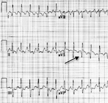
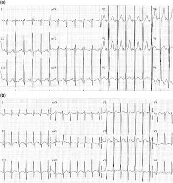
The anomalous origin of the left coronary artery from the pulmonary artery is a rare congenital lesion accounting for 0.5% of all congenital cardiac defects.Reference Keith2 The presentation of patients with the anomalous origin of the left coronary artery from the pulmonary artery is quite variable and ranges from incidental detection to the child presenting with congestive cardiac failure, left ventricular dysfunction, severe mitral regurgitation, and/or cardiac ischaemia. Clinical and electrocardiographic manifestations are silent in the foetus as high pulmonary arterial pressures preserve myocardial flow. As pulmonary arterial pressure declines, antegrade flow into the left coronary artery is reduced with eventual reversal into the pulmonary arteries, resulting in a coronary arterial steal. Late presentations are rare and usually are the result of a dominant right coronary arterial system with a well-preserved collateral flow. In a series of 15 patients with anomalous origin of the left coronary artery from the pulmonary artery diagnosed at a mean age of 3.5 months, the resting electrocardiogram revealed ischaemia in nine patients and hypertrophy in 10 patients. In fact, only one patient had a normal electrocardiogram.Reference De Wolf, Vercruysse, Suy, Blom, Matthys and Ottenkamp3 The classic electrocardiogram of anomalous origin of the left coronary artery from the pulmonary artery is depicted by deep broad q waves in the lateral leads (I, aVL, V6) with absent q waves in the inferior leads (Fig 1). However, variations in the pulmonary arterial pressures may account for the subtle, but noteworthy, electrocardiographic differences. Changes on the surface electrocardiogram may relate to a drop in the pulmonary arterial pressures (Fig 2a and b) antecedent to the classic lateral ischaemic manifestations. T-wave flattening in the lateral leads (I, aVL, V6) may also be observed before true T-wave inversion concordant with the electrocardiographic loss of mid-precordial R waves from a decline in the pulmonary arterial pressure.Reference Brekke, DeGroff and Schaffer4
A six-lead electrocardiogram that shows sinus tachycardia with deep Q waves in the lateral leads I and aVL (arrow). ST segament changes are also observed in the inferior leads.

(a) A 12-lead electrocardiogram of a 6-month-old child with anomalous origin of the left coronary artery from the pulmonary artery that exhibits the typical Q wave in V6, without manifestation in leads I and aVL. Note the prominent mid-precordial voltages (V1–V3) consistent with pulmonary hypertension (25 millimetres per second; 10 millimetres per milliVolt). Reprinted with permission from the Wolters Kluwer Health (Brekke et alReference Brekke, DeGroff and Schaffer4). (b) A 12-lead electrocardiogram taken in same patient 3 days later exhibits a small Q wave in V6; however, the Q waves are manifest in leads I and aVL. A marked reduction in the mid-precordial R waves is also seen, consistent with a drop in the pulmonary arterial pressure (25 millimetres per second; 5 millimetres per milliVolt). Reprinted with permission from the Wolters Kluwer Health; Brekke et alReference Brekke, DeGroff and Schaffer4.

Noteworthy considerations of alternative diagnosis, such as myocarditis or dilated cardiomyopathy, are critical, as misdiagnoses have been reported. Although colour Doppler has increased the yield in “ruling-in” anomalous origin of the left coronary artery from the pulmonary artery, “ruling-out” the diagnosis is more onerous. In distinguishing the anomalous origin of the left coronary artery from the pulmonary artery from a dilated cardiomyopathy, a QT pattern in aVL in which the Q wave is greater than or equal to 3 millimetres deep with an inverted T wave, can be seen in 83% of patients with the anomalous origin of the left coronary artery from the pulmonary artery as opposed to only 5% of patients with dilated cardiomyopathy.Reference Chang and Allada5 Careful attention to the clinical history and electrocardiographic abnormalities should invoke suspicion for the diagnosis with confirmation by non-invasive measures.
Anomalous aortic origin of a coronary artery from the wrong aortic sinus of Valsalva
Sudden cardiac death in highly conditioned athletes, deemed by society to be infallible, has sparked a veritable interest in identifying at-risk patients before their terminal event. Cardiovascular screening before participation in order to identify “at-risk” athletes, in conjunction with continued emphasis on education about cardiopulmonary resuscitation and increased availability of automated external defibrillators has been effective in reducing sudden cardiac death.Reference Berger6, Reference Corrado, Basso, Schiavon, Pelliccia and Thiene7
Incidence
Estimates of sudden cardiac death vary depending on the age, gender, and the population being sampled. The prevalence of sports-related deaths has been estimated at 0.5–1.6 per 100,000 individuals.Reference Corrado, Basso, Schiavon and Thiene8, Reference Maron, Gohman and Aeppli9 In the United States of America, hypertrophic cardiomyopathy is the most common cause of sudden cardiac death, whereas in Italy, arrhythmogenic right ventricular cardiomyopathy ranks the highest.Reference Maron, Epstein and Roberts10–Reference Maron, Shirani and Poliac13 Coronary arterial anomalies, (coronary artery arising from the wrong sinus of Valsalva with an inter-arterial, intramural and/or intra-conal course) are the second most common diagnosis behind hypertrophic cardiomyopathy, accounting for 17% of all reported non-traumatic sudden cardiac deaths.Reference Maron, Doerer, Haas, Tierney and Mueller14
A variety of coronary arterial abnormalities have been identified. The most common deviation exists in situations in which the circumflex coronary artery arises from the right sinus of Valsalva or the right coronary itself (incidence 0.37–0.6%).Reference Click, Vleitrstra and Kosinski15, Reference Davis, Cecchin and Jones16 The second most observed anomaly is the origin of the right coronary artery from the left facing sinus of Valsalva. Less common, but of even greater physiological consideration, is the emergence of the left main coronary artery from the right sinus of Valsalva. In a series of more than 100,000 adults undergoing cardiac catheterisation with coronary angiography, 0.17% had an anomalous right coronary artery and 0.047% had anomalous left coronary artery.Reference Yamanaka and Hobbs17 The prevalence of an anomalous coronary artery in a prospective study of 2388 asymptomatic children using two-dimensional echocardiography was 0.17%, with two patients with anomalous left coronary artery from the wrong sinus of Valsalva and two patients with an anomalous right coronary artery from the wrong sinus of Valsalva.Reference Davis, Cecchin and Jones16 Anomalous left coronary artery from the right sinus of Valsalva is rarer than its counterpart, anomalous right coronary artery, and anomalous left coronary artery from the right sinus of Valsalva is more often associated with sudden cardiac death than anomalous right coronary artery. Both, however, have been implicated in varying degrees as causing sudden collapse at studies performed during necropsy.Reference Eckart, Scoville and Campbell18–Reference Taylor, Rogan and Virmani22 In a large review of the autopsies performed at the Armed Forces Institute of Pathology, coronary arterial abnormalities were identified in 142 patients. Anomalous origin of the left coronary artery from the right facing sinus was deemed to be causative of sudden death in 57% of the cases compared with the anomalous right coronary artery, which caused sudden death in 25% of the patients.Reference Taylor, Rogan and Virmani22 Interestingly, sudden cardiac death was observed in 10% of cases in which the left circumflex coronary artery arose from the right sinus of Valsalva.Reference Taylor, Rogan and Virmani22 Younger patients, those less than 30 years of age, were more likely to die suddenly and had higher rates of exercise-related deaths. Unfortunately, many of the patients with this abnormality are asymptomatic before the terminal event.Reference Taylor, Rogan and Virmani22, Reference Virmani, Rogan and Cheitlin23
Pathophysiology
In both the anomalous right coronary artery from the wrong sinus of Valsalva and the anomalous left coronary artery from the wrong sinus of Valsalva, the origin of the coronary artery may share a common orifice or have a distinct and separate opening. The inter-arterial course between the great vessels, with or without an intramural course, leads to a potentially more than a single pathophysiological explanation of ischaemia and sudden death. Non-invasive imaging should focus not just on the course of the vessel, but also the angle of opening and potential presence of a slit-like orifice. Kinking at the acute angle, the spasm of the coronary artery, and compression between the great vessels may all lead to an insufficient coronary arterial flow, decreased myocardial reserve during exercise, ventricular tachycardia, and ultimately sudden death.
It is these haemodynamic perturbations that lead to the electrocardiographic manifestations. Yet, the symptoms are not present each time a patient exercises. An electrocardiogram that reveals marked infero-lateral ST segment depression during peak exercise with resolution in recovery may prove to be completely normal on a different day.Reference Brothers, Carter, McBride, Spray and Paridon24 Timely identification of anomalous aortic origin of the coronary artery from the wrong sinus of Valsalva is predicated on attention to the history, as the ischaemia leading to sudden cardiac death may be episodic.Reference Brothers, Carter, McBride, Spray and Paridon24–Reference Cheitlin, De Castro and McAllister26 Unfortunately, many of the patients with anomalous aortic origin of the coronary artery from the wrong sinus of Valsalva are diagnosed at autopsy.Reference Kragel and Roberts27 A detailed history is critical to the evaluation of the patient before any non-invasive imaging.
Screening history
Cardiovascular symptoms of pain in the chest, exertional dyspnoea, pre-syncope, or syncope during or initially after exercise have been reported in 18–30% of the cases.Reference Maron, Shirani and Poliac13, Reference Liberthson28 Cardiovascular screening before participation in sports, with a detailed history and physical examination, certainly “does not have sufficient sensitivity to guarantee” detection of all cardiovascular abnormalities associated with sudden cardiac death in adolescent athletes.Reference Maron, Thompson and Ackerman29 However, ignoring even a single event of exertional pain in the chest or syncope can be risky. It is Important in the process of screening to pay absolute attention to warning signs and symptoms. Of the 21 military recruits reviewed by Eckart et alReference Eckart, Scoville and Campbell18 who died suddenly, related to anomalous aortic origin of the left coronary artery from the wrong sinus of Valsalva, more than half of the individuals had a prodrome of cardiac-related symptoms, with syncope in six patients, pain in the chest in five patients, and dyspnoea in four patients.
The process of screening, however, should not be limited to the athlete in high school or college. Such a myopic view will miss the many children who, while not elite athletes in high school or college, are playing competitive sports or participating in intense recreational activities long before high school commences. It is vital that the appropriate questions be asked.
Questions:
• Has your child ever fainted during practice or a sporting competition?
• Has your child ever fainted immediately upon termination of exercise?
• Has your child ever developed significant dizziness during exercise?
• Has your child ever developed palpitations or an irregular pulse, such as skipped beats, during exercise?
• Has your child ever developed pain in the chest during or immediately upon termination of exercise?
• Has your child ever seen a provider of healthcare for complaints of pain in the chest, dizziness, or fainting? If yes, are those records available?
• Is there any familial history of sudden cardiac death, unexplained accidents in the car, or drownings at less than 40 years of age?
In an attempt to address the clinical history associated with anomalous aortic origin of the coronary artery from the wrong sinus of Valsalva, Basso et alReference Basso, Maron, Corrado and Thiene30 reviewed two large-scale studies of young competitive athletes who died suddenly in the United States of America (study inception 1990) and Italy (study inception 1979). Twenty-seven athletes, 22 male and five female, ranging in age from 9 to 22 years (16 plus or minus5), were identified. The majority, 25 out of 27, died during exercise, with nearly half being in the midst of a physically strenuous but prolonged period of intense activity. Of the 27 athletes, clinical information was available for 12 patients, with exertional syncope present in 25% and exertional pain in the chest in 25% of the patients. Six of the athletes who died suddenly had undergone a maximal stress test in the 6–18 months before the fatal event, and, in all instances, there were no concerning ST segment or T-wave changes. Interestingly, premonitory symptoms were only appreciated in those individuals with anomalous aortic origin of the left coronary artery from the wrong sinus of Valsalva. Anomalous aortic origin of the right coronary artery from the wrong sinus of Valsalva proved to be clinically silent. A comprehensive review of the medical literature identified 18 patients less than 35 years in age with an anomalous aortic origin of the coronary artery from the wrong sinus of Valsalva, who had undergone a maximal exercise stress test as part of their evaluation. Although cardiovascular symptoms were antecedent in greater than 90% of the patients, only 4% out of 18% had an abnormal exercise stress test. Ventricular ectopy, which has been touted as a marker of concern, especially if progressing during an exercise stress test and multiform in nature, is a rare occurrence in the anomalous aortic origin of a coronary artery from the wrong sinus of Valsalva. In the meta-analysis review of 18 published studies, premature ventricular contractions were only mentioned in three of the papers.Reference Corrado, Thiene, Coco and Frescura31–Reference Zeppilli, Della Russo and Santini33 In a separate single paediatric institution, of 16 children undergoing surgical repair of anomalous aortic origin of a coronary artery from the wrong sinus of Valsalva, nine had exertional pain in the chest and four had exertional pre-syncope/syncope, yet no patient had ischaemic changes on a maximal treadmill.Reference Brothers, McBride and Seliem34
Over the past 20 years, the Italian data has substantiated the role of the electrocardiogram in screening before participation in sports. The role of the electrocardiogram in screening before participation in sports has been especially well documented for many of the channelopathies and cardiomyopathies. However, in patients with an anomalous aortic origin of a coronary artery from the wrong sinus of Valsalva, a detailed history is the first tier of screening, and arguably more pivotal than either a resting or stress electrocardiogram. A resting electrocardiogram, in fact, is unlikely to be beneficial in identifying children with anomalous aortic origin of a coronary artery from the wrong sinus of Valsalva. Any history of exertional pain in the chest or exertional syncope warrants further imaging to rule out anomalous aortic origin of a coronary artery from the wrong sinus of Valsalva, even when the stress test is unremarkable.
Case presentation
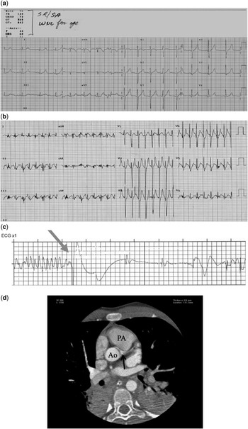
In February, 2007, a 14-year-old boy presented for medical care following a syncopal episode on the basketball court. Towards the end of a highly competitive game, feeling weak and tired, he sat down in the half-court and slumped forward without any tonic–clonic movements. He was taken to a local emergency room and received intravenous hydration and was subsequently discharged home. Ten months later, while running sprints at practice for basketball, he fell to ground without any antecedent palpitations or pain in the chest. He was referred to a paediatric cardiologist where he had a normal physical examination and normal resting electrocardiogram, which is shown below in Figure 3a.
(a) This 12-lead electrocardiogram was recorded at baseline and exhibits normal sinus rhythm without hypertrophy or ischaemia. (b) This 12-lead electrocardiogram was recorded at maximal sinus rate on a standard treadmill stress test. (c) This rhythm strip was recorded during defibrillation of ventricular fibrillation. (d) A cardiac computerized axial tomographic (CT) scan subsequently identified the left coronary artery arising from the right-sided sinus of Valsalva. The computerized axial tomographic (CT) image shows the left coronary artery (arrow) coursing between the aorta (Ao) and the pulmonary artery (PA).

A detailed familial history was unrevealing in suggesting other obvious familial causes of sudden cardiac arrest, including either a primary channelopathy or hypertrophic cardiomyopathy. A two-dimensional echocardiogram was reportedly normal. An exercise stress test to a maximal heart rate of 208 beats per minute revealed no ectopy or evidence of ischaemia, as shown below in Figure 3b.
The patient was cleared to resume unrestricted participation in sports. Nearly 8 months later, he collapsed while running sprints at practice for football. The recorded strip below (Fig 3c) shows his rhythm at this time.
Paramedics were present at the practice and began cardiopulmonary resuscitation. The paramedics used a defibrillator, with resultant return of a perfusable rhythm, followed by a documented sinus rhythm thereafter.
A cardiac computerised axial tomogram subsequently identified an anomalous left coronary artery arising from the right aortic sinus of Valsalva, as shown in Figure 3d. Surgical unroofing was performed with resultant full cardiac recovery, as well as neurologic recovery. The computerised axial tomogram (Fig 3d) shows the left coronary artery (arrow) coursing between the aorta and the pulmonary artery.
Left main coronary artery atresia or stenosis
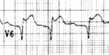
Atresia of the left main coronary artery is a rare condition in which the left coronary arterial system is fed via collateral flow from the right coronary arterial system. Often the collateral circulation is inadequate to meet the metabolic needs and myocardial ischaemia becomes evident. Nearly all patients present with syncope, failure-to-thrive, or myocardial ischaemia.Reference Musiani, Cerniagliaro, Sansa, Maseli and DeGasparis35 The electrocardiogram displays features consistent with lateral ischaemia (Fig 4) when the inversely disproportionate collateral vascularisation cannot provide adequate flow of blood to the ventricle, especially during times of high metabolic demands. Patients with myocardial ischaemia may have significant degrees of complex ventricular ectopy, up to and including ventricular fibrillation.
An electrocardiogram that shows profound elevation of the ST segments in lateral lead V6 consistent with myocardial ischaemia. A q wave is present.

Myocardial bridges
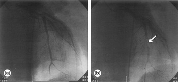
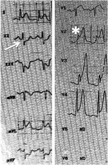
Myocardial bridges exist where the epicardial coronary arteries descend towards the ventricle with a muscular band over the coronary arteries. The left anterior descending coronary artery is the most common coronary artery affected by myocardial bridges. The bridges are most prevalent between the proximal and middle third of the left anterior descending coronary arteryReference Bezerra, Pratesd and Didio36 (Fig 5). Although these congenital anomalies are often benign, systolic compression may occur when intra-myocardial tension exceeds intra-coronary luminal pressure. In this situation, a segment of the myocardium may become ischaemic with the patient manifesting ventricular arrhythmias, ischaemic pain in the chest, or sudden cardiac death. Compression of the left anterior descending coronary artery by overlying muscle may result in the elevation of the ST segments in the anterior (V1 and V2) leads, often mimicking an anterior myocardial infarctionReference Bashour, Espinosa and Blumenthal37 (Fig 6). Other electrocardiographic findings may be seen in conjunction with the signs and symptoms of myocardial ischaemia, including the inversion of the T waves in all anterior leads, beyond the typical juvenile pattern.Reference Bestetti, Finzi, Amarl, Secches and Oliveira38 Reports of the formation of a thrombus and myocardial infarction have been reported in patients with myocardial bridges.Reference Agirbasli, Martin and Stout39 Depending on the extent of the formation of the thrombus, the electrocardiogram will reflect the coronary arteries involved. Treatment with beta-blockers and consideration for thrombolytics may be warranted.Reference Ortega-Carnicer and Fernández-Medina40
The left coronary arterial angiogram from a young adult who presented with pain in the chest during a match of soccer. The mid-left anterior descending coronary artery is normal in diastole (a) and compressed secondary to a myocardial bridge in systole (arrow; b). Reprinted with permission form the Elsevier; Ortega-Carnicer and Fernández-MedinaReference Ortega-Carnicer and Fernández-Medina40.

The electrocardiogram, taken at the time of presentation in the patient described in Figure 5, shows sinus tachycardia with elevation of the ST segments in the anterior leads (*) and reciprocal depression of the ST segments in the inferior leads (II, III and aVF). Leads V5 and V6 were not recorded. Reprinted with permission form the Elsevier; Ortega-Carnicer and Fernández-MedinaReference Ortega-Carnicer and Fernández-Medina40.

Coronary arterial fistulas
Coronary arterial fistulas exist as a link between the coronary artery, typically the distal coronary artery, and either a large vessel or a cardiac chamber. Approximately 25% of these fistulas drain into the pulmonary circulation and are associated with structural cardiac disease, most notably, tetralogy of Fallot and its variants.Reference Bauer, Bauer and Alexi-Meskishvilli41, Reference Holzer, Johnson, Ciotti, Poszzi and Kitchiner42 Most children are asymptomatic with normal tolerance of exercise despite a potentially moderate overload of volume. In reviews of children with coronary arterial fistula, the electrocardiogram is most commonly normal in more than 50% of the children with coronary arterial fistulas, whereas approximately 25% will show signs of either left atrial enlargement or ventricular hypertrophy.Reference Holzer, Johnson, Ciotti, Poszzi and Kitchiner42 Ischaemic changes have not been observed in patients with coronary arterial fistula. The electrocardiographic findings as such are essentially non-specific and are secondary to the loading of volume. Unlike the previously discussed isolated congenital coronary abnormalities, coronary arterial fistulas in children are associated with a very low morbidity and mortality. A host of transcatheter devices are available for use to close these fistulas in symptomatic patients or those in whom the fistulas do not spontaneously regress.
Summary
Congenital coronary arterial abnormalities as isolated lesions are exceedingly rare. The electrocardiogram, while a reasonable adjunct in the diagnosis of coronary arterial abnormalities, should not supplant a good history and physical examination. Careful attention must be devoted to any signs or symptoms of ischaemic pain in the chest or syncope, which must not be overlooked. Exertional pain in the chest and exertional syncope should prompt an extensive evaluation by both the echocardiographer and the electrophysiologist. Clearance for participation in sports should be curtailed until a complete evaluation has ruled out the presence of any of the following disorders: a channelopathic mutation, a cardiomyopathy, or a congenital coronary arterial anomaly.