Introduction
The frontal assessment battery (FAB) was developed to evaluate frontal lobe function easily and quickly at the bedside (Dubois et al., Reference Dubois, Slachevsky, Litvan and Pillon2000). The FAB consists of six subtests that explore the different aspects of frontal lobe function. The sensitivity and validity of this assessment tool have been demonstrated in various bradykinetic rigid syndromes associated with frontal lobe dysfunction (Dubois et al., Reference Dubois, Slachevsky, Litvan and Pillon2000; Paviour et al., Reference Paviour2005; Matsui et al., Reference Matsui2006). Comparisons of the FAB scores of patients with various types of dementia in many countries have also been reported (Iavarone et al., Reference Iavarone2004; Mok et al., Reference Mok2004; Slachevsky et al., Reference Slachevsky, Villalpando, Sarazin, Hahn-Barma, Pillon and Dubois2004; Lipton et al., Reference Lipton, Ohman, Womack, Hynan, Ninman and Lacritz2005; Castiglioni et al., Reference Castiglioni2006; Kugo et al., Reference Kugo2007). Although the clinical usefulness of the FAB in dementia is well documented, the neural substrates involved in Alzheimer's disease (AD) remain unclear. There are a few functional neuroimaging studies of the brain regions related to the FAB performance of the patients with AD (Guedj et al., Reference Guedj2008; Yoshida et al., Reference Yoshida2009; Kume et al., Reference Kume2011). In the present study, we investigated the relationship between the FAB score and regional cerebral blood flow (rCBF) to assess the neural substrates of FAB performance among patients with AD.
In most neuroimaging studies in which rCBF was evaluated, stereotaxic analysis of regions of interest (ROIs) was used. However, the reproducibility and objectivity of the results are controversial because small ROIs were manually placed on selected slices of single photon emission computed tomography (SPECT) images of the patient. Image analysis using ROIs has been criticized for the serious shortcoming that information outside the ROIs is not obtained. To overcome the above disadvantages, in the present study we conducted a comparison using statistical parametric mapping (SPM), which has the advantage of making no a priori anatomical hypotheses.
Methods
The instrument
The FAB consists of six items, and the score on each item ranges from 0 to 3 (Dubois et al., Reference Dubois, Slachevsky, Litvan and Pillon2000). A lower score indicates a greater degree of executive dysfunction. The six subtests of the FAB explore (1) similarities (conceptualization), (2) lexical fluency (mental flexibility), (3) Luria motor sequences (programming), (4) conflicting instructions (sensitivity to interference), (5) a go/no-go test (inhibitory control), and (6) prehension behavior (environmental autonomy). For this study we used the Japanese version of the FAB described by Kugo et al. (Reference Kugo2007). The reliability of the Japanese version of the FAB is good to excellent and its validity is, to some extent, established (Kugo et al., Reference Kugo2007; Yoshida et al., Reference Yoshida2009).
In a previous study (Kugo et al., Reference Kugo2007), mean FAB scores of elderly subjects with mild AD (13 men and 38 women; mean age = 75.3 years) were 11± 3. Therefore, an FAB score of 12 or more was defined as a high FAB score, and an FAB score of 10 or less was defined as a low FAB score.
Subjects
Twenty-four patients with AD, with a dementia severity of 0.5 (suspicious) or 1 (mild) based on the Clinical Dementia Rating (CDR) (Hughes et al., Reference Hughes, Berg, Danziger, Coben and Martin1982), whose score on the FAB did not reach the mean score of mild AD, were selected from 470 consecutive Japanese patients who were fully examined at the Memory Clinic of Okayama University Hospital between April 2004 and June 2010. Thereafter, 24 AD patients with high scores on the FAB, matched for age, sex ratio, education, and scores on the revised Addenbrooke's Cognitive Examination (ACE) (Mathuranath et al., Reference Mathuranath, Nestor, Berrios, Rakowicz and Hodges2000; Yoshida et al., Reference Yoshida2010), were also selected.
The inclusion criteria were that they (i) underwent general physical and neurological examinations and extensive laboratory testing, including thyroid function tests, serum vitamin B12, and syphilis serology, and determination of the ApoE status of some participants; (ii) took the FAB, Mini-Mental State Examination (MMSE) (Folstein et al., Reference Folstein, Folstein and McHugh1975), and revised ACE; (iii) underwent brain SPECT as well as head CT, head magnetic resonance imaging (MRI), or both; (iv) were diagnosed with probable AD according to the NINCDS-ADRDA criteria (McKhann et al., Reference McKhann, Drachman, Folstein, Katzman, Price and Stadlan1984); (v) had a dementia severity of 0.5 or 1 based on the CDR (Hughes et al., Reference Hughes, Berg, Danziger, Coben and Martin1982); and (vi) they or their nearest relatives gave informed written consent. The exclusion criteria were (i) complications from other neurological diseases or illnesses; (ii) history of mental illness or substance abuse prior to the onset of dementia; (iii) any evidence of focal brain lesions on head MRI; (iv) treatment with cholinesterase inhibitors, antipsychotics, antidepressants, or anxiolytic drugs; and (v) left handedness or ambidexterity. The profile of each subject (age, sex, years of education, and disease duration) was recorded, and the CDR score was rated by the chief clinician.
Other instruments
The ACE was developed to provide a brief test sensitive to early stage dementia and is capable of differentiating between dementia subtypes including AD, frontotemporal dementia (FTD), progressive supranuclear palsy, and other parkinsonian syndromes (Mathuranath et al., Reference Mathuranath, Nestor, Berrios, Rakowicz and Hodges2000). The ACE includes the MMSE but extends it to encompass important areas not covered by the MMSE, such as frontal-executive function and visuospatial skills. The ACE has a comparable sensitivity to the Dementia Rating Scale (Bak et al., Reference Bak, Rogers, Crawford, Hearn, Mathuranath and Hodges2005), a well-established dementia-screening tool that is widely used in research but not in clinical practice because of its length and difficulty of administration. For this study, we used the Japanese version of ACE described by Yoshida et al. (Reference Yoshida2010). The reliability of the Japanese version of ACE is excellent, and its validity is, to some extent, established (Yoshida et al., Reference Yoshida2010).
The Physical Self-Maintenance Scale (PSMS) and the Instrumental Activities of Daily Living Scale (IADL) are validated scales for the assessment of activities of daily living (ADL) (Lawton and Brody, Reference Lawton and Brody1969; Hokoishi et al., Reference Hokoishi2001). The PSMS is a six-item scale that rates self-care ability in toileting, feeding, dressing, personal hygiene and grooming, locomotion (physical ambulation), and bathing. The IADL Scale assesses the patients' ability to perform the following eight complex daily tasks: ability to use the telephone, shopping, food preparation, household tasks, laundering, mode of transportation, responsibility for medications, and ability to manage finances.
The Neuropsychiatric Inventory (NPI) is a valid and reliable instrument for measuring behavior in dementia (Cummings et al., Reference Cummings, Mega, Gray, Rosenberg-Thompson, Carusi and Gornbein1994; Hirono et al., Reference Hirono1997). It is a caregiver-based tool that assesses ten common behaviors in dementia.
Ethics
This study adhered to the 1975 Helsinki Declaration of Human Rights, and the study protocol was approved by the Committee of Okayama University Hospital on Human Research. After giving a complete description of the study to the subjects and their relatives, written informed consent was obtained.
Brain perfusion SPECT imaging
All the subjects were examined by brain perfusion SPECT. The patients were examined in a comfortable supine position with their eyes closed in quiet surroundings. First, the passage from the heart to the brain was monitored after intravenous administration of 99mTc-ethylcysteinate dimer (ECD, 600 MBq, Daiichi Radioisotope Laboratories Ltd., Tokyo, Japan). Ten minutes after the angiography, SPECT images were obtained using a triple-head, rotating gamma camera interfaced to a minicomputer (GCA9300A/DI; Toshiba, Tokyo, Japan) equipped with a fanbeam, low-energy, high-resolution collimator. Sixty projection images over a 360 degree angle in a 128 × 128 matrix were acquired. All images were reconstructed using ramp-filtered back-projection and then three-dimensionally smoothed with a Butterworth filter (order 8, cut-off 0.12 cycles/cm). The reconstructed images were corrected for gamma ray attenuation using the Chang method (μ = 0.09).
Data analysis
Spatial reprocessing and statistical analysis of images were performed on a voxel-by-voxel basis using statistical parametric mapping 2 (SPM2, Wellcome Department of Imaging Neuroscience, London, UK) running on MATLAB (The Mathworks, Inc, Natick, MA, USA). All SPECT images of each subject were normalized to the standard brain of the Montreal Neurological Institute (MNI), and the spatial normalization was performed with 12-parameter affine and non-linear transformations (Friston et al., Reference Friston, Ashburner, Poline, Frith, Heather and Frackowiak1995a; Reference Friston, Holmes, Worsley, Poline, Frith and Frackowiak1995b). The voxel sizes of the reslice option were (2 mm × 2 mm × 2 mm). The non-linear parameter was set at 25 mm cut-off basis functions and 16 iterations. All of the normalized SPECT images were then smoothed with an isotropic Gaussian kernel filter (12 mm full-width at half-maximum). To examine the images for specific regions showing differences in perfusion, two sample t-tests were performed. Global normalization was performed by proportional scaling with the mean voxel value. Masking was applied using the threshold method (0.8 times the global value). The statistical height threshold was set at p <0.005 uncorrected, and the extent threshold was set at p <0.01 uncorrected for multiple non-independent comparisons.
Statistical analysis
Statistical analysis was performed using the SPSS 14.0J software program (SPSS Inc., Chicago, IL, USA). Comparisons between the two groups were performed by independent sample t-tests. A value of p <0.05 was accepted as significant.
Results
Age, years of education, duration of illness, neuropsychological test scores, and ADL
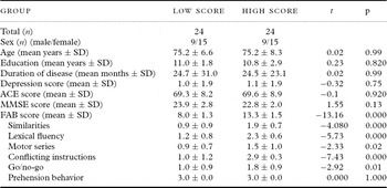
No significant differences were found between high- and low-score groups with respect to age, years of education, or duration of disease by Student's t-test (Table 1). Data on the ApoE genotype were available for 18 low-FAB and 19 high-FAB patients. No ε4 genotype was found in 10 of the 18 low-FAB cases or 9 of the 19 high-FAB, and homozygous E4 was found in 2 of the 18 low-FAB and 3 of the 19 high-FAB cases. The proportion of ε4 carriers in the two groups was similar. With the exception of FAB scores, no significant differences were found between high- and low-score groups with respect to ACE, MMSE, or NPI depression scores (Table 1).
Clinical characteristics and neuropsychological tests

Note: Low score = Alzheimer's disease patients with low FAB score.
High score = Alzheimer's disease patients with high FAB score.
Depression score = Depression score of Neuropsychiatric Inventory.
ACE = Addenbrooke's cognitive examination; FAB = Frontal Assessment Battery; MMSE = Mini-Mental State Examination; SD = standard deviation.
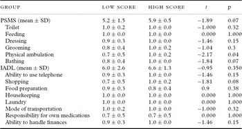
Comparison on PSMS and IADL scales revealed that AD patients with low FAB scores received lower scores than those with high FAB scores on the subscales of physical ambulation on PSMS (Table 2).
Activities of daily living (ADL) and instrumental ADL

Note: Low score = Alzheimer's disease patients with low score on Frontal Assessment Battery test.
High score = Alzheimer's disease patients with high score on Frontal Assessment Battery test.
IADL = Instrumental Activities of Daily Living Scale; PSMS = Physical Self-Maintenance Scale. SD = standard deviation.
Regional cerebral blood flow
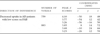
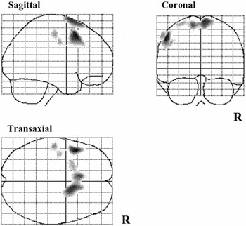
Group comparisons of the SPM results of the high- and low-score groups were performed. Specific voxels with a significantly lower perfusion in the low-score group than in the high-score group are shown in Figure 1 and Table 3. A similar test for specific voxels with a significantly lower perfusion in the high-score group than in the low-score group found no cluster of voxels.
Significant differences in regional uptake between AD patients with low and high scores on FAB

AD = Alzheimer's disease; FAB = Frontal Assessment Battery; MNI = Montreal Neurological Institute.
SPM (z) map of rCBF decrease in AD patients with low scores on the FAB compared with AD patients with high scores. Three-way-glass view of the area of significant hypoperfusion.

Figure 1 shows the z score for each voxel in this cluster superimposed onto a three-way-glass brain view. Figure 1 shows a significant cluster of voxels in the left middle frontal gyrus (MFG), and right superior frontal gyrus (SFG) extending to left SFG. Table 3 shows the probability results of the SPM analysis and the location of peak z scores in terms of the MNI coordinates. These results indicate that AD patients with low FAB scores showed significant hypoperfusion in the left MFG and right SFG compared with patients with high FAB scores.
Discussion
FAB and rCBF
No functional neuroimaging study investigating brain regions in relation to the FAB performance among AD patients has been reported. However, three studies of SPECT imaging on the relationship between the FAB score and rCBF among dementia patients have been reported (Table 4).
Comparison of studies of the relationship between FAB score and decreased rCBF

3D-SSP = three-dimensional stereotactic surface projection; 3D-SRT = three-dimensional stereotactic region-of-interest template; AD = Alzheimer's disease; bil = bilateral; F = frontal lobe; FAB = Frontal Assessment Battery; FTD = frontotemporal dementia; lt = left; MCI = mild cognitive impairment; P = parietal lobe; PaD = Parkinson's disease; rCBF = regional cerebral blood flow; SPECT = single photon emission computed tomography = SPM2, statistical parametric mapping 2.
First, a significant correlation was found between FAB performance and perfusion in the bilateral medial and dorsolateral frontal cortex, independent of age, gender, and MMSE score among 47 patients with a frontal variant of FTD (Guedj et al., Reference Guedj2008). Second, among 117 consecutive patients consulting at a memory clinic, FAB scores were significantly correlated with rCBF in the left callosomarginal and precentral regions independent of age, gender, and MMSE score (Yoshida et al., Reference Yoshida2009). In the same study, a comparison of rCBF between a low FAB group of 20 dementia patients and a high FAB group of 23 dementia patients revealed that the low FAB group showed significantly lower rCBF in the bilateral callosomarginal and left prefrontal regions (Yoshida et al., Reference Yoshida2009). Third, a low FAB group of 18 patients with amnestic mild cognitive impairment (MCI) showed decreased rCBF in some regions of the left lateral frontal and right medial frontal areas compared with a high FAB group of 20 patients with an MCI (Kume et al., Reference Kume2011).
Two correlation studies of FTD and dementia and three comparison studies, including ours, of dementia, MCI, or AD, all showed that the rCBF in the left dorsolateral prefrontal cortex (PFC) was significantly associated with the FAB score (Table 4). In these studies, different software programs (SPM2, 3D-SRT, 3D-SSP) were used to analyze data. However, the results were very similar. In most studies, rCBF in the dorsolateral and/or medial frontal cortex was closely associated with the FAB scores. These results suggest that the dorsolateral and medial PFC play a very important role in the FAB achievement. Only one study, by Matsui et al. (Reference Matsui2006), showed that rCBF in the left inferior parietal lobule was lower in the lower FAB score group of patients with Parkinson's disease than the higher FAB score group. A difference in subjects might have caused the difference in the results.
FAB and ADL
The FAB was developed to evaluate frontal executive function easily and quickly at the bedside. It has been reported that a range of deficits in executive functioning adversely affects the ability of the patients with AD to perform ADL (Feldman et al., Reference Feldman2001; Njegovan et al., Reference Njegovan, Hing, Mitchell and Molnar2001). In this study, AD patients with low FAB scores showed lower scores than those with high FAB scores on the subscale score of physical ambulation on PSMS. Our results suggest that performance on the FAB is closely related to a kind of ADL as well as to rCBF in the SFG and MFG, independent of ACE and MMSE scores. Woodward et al. (Reference Woodward, Brodaty, Boundy, Ames, Blanch and Balshaw2010b) pointed out the possibility that AD patients with low FAB scores may just have more severe AD rather than a discrete frontal variant of AD (FvAD). Obviously, patients with more severe AD showed low FAB scores. However, in our study, the two groups of AD patients with high and low FAB scores were matched for duration of disease, ACE score, and MMSE score. These results suggest that there are AD patients with low FAB scores in the early stage of disease.
Superior and MFGs in AD
Several studies reported that brain activity is increased in the bilateral dorsolateral PFC during executive function tasks (Jonides et al., Reference Jonides, Marshuetz, Smith, Reuter-Lorenenz, Koeppe and Hartley2000; Rypma and D'Esposito, Reference Rypma and D'Esposito2000; Townsend et al., Reference Townsend, Adamo and Haist2006). Performance on the executive-function test was reported to be significantly correlated with regional cerebral glucose metabolism in the dorsolateral PFC in a positron emission tomography (PET) study (Sarazin et al., Reference Sarazin, Pillon, Giannakopoulos, Rancurel, Samson and Dubois1998). Patients with AD had decreased rCBF relative to that in control subjects and MCI patients in the left lateral frontal and left orbitofrontal cortices (Dai et al., Reference Dai, Lopez, Carmichael, Becker, Kuller and Gach2009). We also found a decrease of rCBF in the bilateral dorsolateral PFC among AD patients with low FAB scores.
It has been reported that, in addition to memory impairment, some subjects with mild AD also show frontal dysfunctions (Johnson et al., Reference Johnson, Head, Kim, Starr and Cotman1999). AD patients with greater frontal neuropsychiatric characteristics differ from AD cases with lesser frontal neuropsychiatric characteristics across a range of characteristics including cognition, functioning, and behavior (Johnson et al., Reference Johnson, Head, Kim, Starr and Cotman1999; Woodward et al., Reference Woodward, Jacova, Black, Kertesz, Mackenzie and Feldman2010a). Several studies examined how to distinguish patients with prominent frontal features such as dysexecutive syndrome from typical AD patients by pathology. Patients with AD who presented in the early stages of dementia with disproportionate impairments on tests of frontal lobe function had a greater-than-expected degree of neurofibrillary tangle (NFT) pathology in the frontal lobes (Johnson et al., Reference Johnson, Head, Kim, Starr and Cotman1999; Taylor et al., Reference Taylor, Probst, Miserez, Monsch and Tolnay2008). In addition, several studies reported that 30% to 50% of the patients with AD have TDP-43 pathology (Amador-Ortiz et al., Reference Amador-Ortiz2007; Hu et al., Reference Hu2008; Josephs et al., Reference Josephs2008). However, Bigio et al. (Reference Bigio2010) asserted that the clinical phenotypes of PPA and FTD in cases with pathologic AD are only rarely associated with TDP-43 proteinopathy. Given these previous findings, it is plausible that AD patients with greater frontal neuropsychiatric characteristics can be explained by AD pathology located in the frontal lobes rather than differences in kinds of protein. Our study suggests that some AD patients suffer from frontal neuropsychological deficits and frontal hypoperfusion in the early stage and that those patients might be included in a so-called FvAD. Further studies are needed to explore the differences between groups and to correlate the pathological changes in the early stage using FDG PET or amyloid scanning and autopsy study.
Limitations of this study
Our study has several limitations. The study participants consisted of patients with mild AD who were recruited at a university center. Thus, our results apply only to a clinic-based outpatient population with mild AD. Further study is needed to clarify the neural substrates of the FAB among aged subjects with normal cognitive function or patients with dementias other than AD. Second, we compared rCBF between two groups comprising patients with AD and found a difference between the two groups in the rCBF in the SFG and MFG. It does not mean that these areas are the only important areas for performing the FAB. However, regardless of the above limitations, this is the first report to assess the neural substrates of performance on the FAB among patients with mild AD.
Conflict of interest
None.
Description of authors' roles
E. Oshima and S. Terada designed the study and analyzed the data. E. Oshima wrote the paper. S. Sato assisted in preparing the statistical design and supervised the statistical analysis. E. Oshima, S. Terada, O. Yokota, C. Ikeda, S. Nagao, N. Takeda, and H. Honda collected the data. All authors discussed the results and conclusions. Y. Uchitomi supervised the study design, participated in data analysis, and assisted with writing the paper.
Acknowledgments
We thank Ms. Horiuchi, Ms. Imai, Ms. Yabe, and Ms. Tsuchiyama for their skillful assistance in this study. This work was supported by a grant from the Japanese Ministry of Education, Culture, Sports, Science and Technology (21591517), and the Zikei Institute of Psychiatry.