INTRODUCTION
Bivalves are known as suitable intermediate hosts for many helminths, larval Digenea among them (Lauckner, Reference Lauckner and Kinne1983). Gymnophallid larvae usually parasitize bivalves as first or second intermediate hosts, using shorebirds as definitive hosts for adults (Bartoli, Reference Bartoli1974; Ching, Reference Ching1995). Studies on larval digenean populations affecting bivalves in the south-western Atlantic Ocean are scarce. Szidat (Reference Szidat1962, Reference Szidat1965), described Parvatrema australis Szidat, Reference Szidat1962 (Gymnophallidae) from mytilids; Cremonte (Reference Cremonte1999) reported 7 metacercariae (5 gymnophallids, 1 renicolid and 1 monorchiid) from intertidal bivalve populations. A gymnophallid metacercaria was recorded by Martorelli & Morriconi (Reference Martorelli and Morriconi1998) from patellid gastropods.
Gymnophallid metacercariae occupy and exploit the extrapallial space of their bivalve hosts, a habitat only suitable for a few other parasites. Larvae of Digenea other than those of Gymnophallidae that reach the extrapallial space usually elicit a response in the form of a secretion of calcium carbonate to wall-off the larvae. This reaction often leads to the parasite's death (Bartoli, Reference Bartoli1974; Lauckner, Reference Lauckner and Kinne1983; Ituarte et al., Reference Ituarte, Cremonte and Deferrari2001; Cremonte & Ituarte, Reference Cremonte and Ituarte2003). Contrary to this, gymnophallids have developed various life strategies to overcome the host response. Ituarte et al. (Reference Ituarte, Cremonte and Deferrari2001, Reference Ituarte, Cremonte and Zelaya2005) and Cremonte & Ituarte (Reference Cremonte and Ituarte2003) discussed the wide spectrum of host reactions against infections by gymnophallid larvae, in both modern and Holocene bivalve populations.
In coastal north-western Atlantic Ocean waters, Tagelus plebeius is known from estuarine habitats south north to Cape Cod, Massachusetts, USA (42°N). The parasitic fauna of the northern hemisphere populations of T. plebeius was studied by Holland & Dean (Reference Holland and Dean1977), who described a cestode larva. Wardle (Reference Wardle1983) reported a digenean sporocyst containing trichocercous cercariae and Dungan et al. (Reference Dungan, Hamilton, Hudson, Mc Collough and Reece2002) who found a protozoan of the genus Perkinsus.
In south-western Atlantic coastal waters, T. plebeius (Psammobiidae) is a common clam in sand flats of mixohaline ecosystems south to the San Matías Gulf (41°S), as a commonly important component of benthic infaunal communities (Olivier et al., Reference Olivier, Escofet, Penchaszadeh and Orensanz1972a, Reference Olivier, Escofet, Penchaszadeh and Orensanzb; Botto & Iribarne, Reference Botto and Iribarne1999). The parasites affecting T. plebeius in the south-western Atlantic Ocean have been studied by Cremonte (Reference Cremonte1999) and Vázquez et al. (Reference Váquez, Ituarte, Navone and Cremonte2006). Recently, Lomovasky et al. (Reference Lomovasky, Gutiérrez and Iribarne2005) described an abnormal pigmentation of the inner shell surface, a discoloration previously reported by Cremonte (Reference Cremonte1999) to consist of an orange to brown colour of variable extension affecting the inner shell layer below the valve umbo, and ventrally to the anterior margins of the pallial sinus. This phenomenon was described as concurrent with an irregular calcium carbonate deposition that in some cases results in the formation of localized calcium carbonate concretions. The authors reported this shell alteration as associated with the presence of gymnophallid metacercariae.
In the present paper, the details of the structural characteristics of the bivalve host reaction elicited by metacercariae of Gymnophallidae parasitizing T. plebeius are described.
MATERIALS AND METHODS
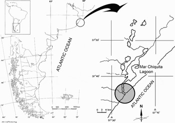
Specimens of Tagelus plebeius from Mar Chiquita lagoon (37° 32′S 57° 19′W) (Figure 1) were collected during low tides from the muddy substrate. Specimens for histological study (N = 19) were fixed in either in Zenker's or Bouin's fixatives and processed according to Gabe (Reference Gabe1968). After dissection, portions of dorsal visceral mass tissues containing parasites were dehydrated in ethanol and embedded in Paraplast®; 5–7 µm thick histological sections were stained with haematoxylin and eosin (H & E) for general purposes. Additionally, sections for specific studies and characterization of particular secretions were stained with Alcian blue/periodic acid Schiff (AB/PAS), tolouidine blue (TB) and Gomori's trichrome (GT) (Gabe, Reference Gabe1968). Some specimens were decalcified by immersion for about 24 hours in a 10% formalin solution with 10% (v/v) acetic acid. Histological sections were examined under a light microscope for presence of parasites. Photomicrographs were taken with a Zeiss Axio Imager microscope equipped with an MRc5 digital camera. Prevalence (percentage of infected specimens in the sample) and intensity of infection (number of parasites per host clam) were calculated following inspection of 50 specimens. Inspection of bivalves was performed under a stereoscopic microscope dissecting the sacs containing gymnophallid larvae and counting all of them. Data were analysed after grouping them into four shell-length groups, to determine if the intensity of infection was related to size (=age) of host bivalves. Data on intensity of parasitism were not normally distributed, so a non-parametric Spearman rank-correlation analysis was performed to assess the existence of a linear association between these two variables (Morales & Pino, Reference Morales and Pino1987).
Location map, shadow circle indicates the collecting site.

RESULTS
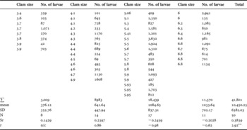
Fifty specimens of Tagelus plebeius were collected between July 2005 and March 2006, their sizes (maximum shell length) ranged between 3.4 to 6.8 cm length. The prevalence of infection was 100%; bivalves were infected with metacercariae of an unknown species of the family Gymnophallidae (Digenea). The intensity of infection varied from 42 to 3632 larvae per bivalve (Table 1). Metacercariae occupied exclusively the extrapallial space, they were never found affecting organs within the visceral mass; none of the 19 specimens examined histologically showed alterations in the gametogenesis or in the hepatic tissues.
Values of intensity of infection grouped according to host size (maximum shell length, cm) (N, number of specimens; SD, standard deviation; Sr, Spearman rank; t, t-statistic value: statistically significant values marked with **).

Intensity of infection and its relationship with size of hosts
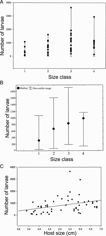
The number of gymnophallid larvae per host individual was plotted against host shell length (SL). Mean and standard deviation of values of intensity of infection (number of larvae per host specimen) for each size-class are shown in Table 1. Figure 2A shows the distribution of values of intensity of infection according to four hosts size-classes (3.0–3.99, 4.0–4.99, 5.0–5.99 and 6.0–7.0 cm SL). Median values of intensity corresponding to each size-class (Figure 2B) showed a progressive increase throughout the analysed host size-range, however, dispersion of values of intensity within each class was high and consequently, high values of standard deviation of means for each size-class were observed (Table 1). Results of the Spearman rank-order correlation test are presented in Figure 2C and Table 1. When pooled data of intensity of infection versus size were plotted, the correlation value obtained was statistically significant (N = 50; rs = 0.3889; t = 2.92) (Table 1), i.e. higher values of infection intensity in larger (=aged) bivalve hosts differed significantly from values expected if data were not correlated. However, when the test was performed for each of the four shell length-classes, test results were not statistically significant within shell-length groups (Table 1).
Intensity of infection of Tagelus plebeius by gymnophallid metacercariae and its relationship with size (=age) of hosts. (A) Number of larvae per individual distributed according to the 4 selected host size-classes; (B) median and non-outlier range values of intensity of infection for each size-class; (C) Spearman rank-order correlation of number of larvae against host size.

General anatomy and histopathology
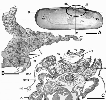
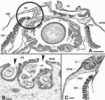
The main site of infection is the extrapallial space adjacent to the mantle isthmus, immediately below the hinge and ligament (Figure 3A–C) this area corresponds to the subarticular extrapallial space (Bartoli, Reference Bartoli1973).
General characteristics of infection in Tagelus plebeius by gymnophallid metacercariae. (A) Inner view of a shell of T. plebeius showing the position in the visceral mass and the typical site of infection (encircled oval area), as well as levels of sections depicted in 3B (horizontal line) and 3C (parallel line); (B) frontal section of the dorsal visceral mass showing the extension of infection by metacercariae (GT); (C) transverse section (TB). Scale bars A, 1 cm; B, 1 mm; C, 250 µm (aa, anterior adductor muscle; ct, cardinal teeth; ime, inner mantle epithelium; li, ligament; m, metacercaria; mtl, mantle; mi, mantle isthmus; n, nephridium; od, outer gill demibranch; ome, outer mantle epithelium; pa, posterior adductor muscle; ps, pallial sinus; r, rectum; s, sacs containing metacercariae; sct, space corresponding cardinal teeth).

HOST REACTION
The gymnophallid metacercariae found in T. plebeius had colonized the extrapallial space, i.e. the space between the outer mantle epithelium and the inner shell surface. The mantle epithelium consisted of two one-cell thick sheets of mantle epithelia separated by a loose connective tissue layer; the outer epithelium was formed of cuboidal or low-columnar cells (about 13–14 µm high, 11–12 µm wide) and the inner layer was composed of slender, fusiform cells (6 µm height, 12 µm length) (Figure 4A–C). The site at which cercariae enter the extrapallial space of the bivalve host is still unknown. The host reaction to colonization of its extrapallial space by gymnophallid metacercariae consisted of the hyperplasia and metaplasia of the outer mantle epithelium adjacent to larvae (Figure 4B, C). According to the site in the extrapallial space the larvae occupy, some differences in the extent, intensity, and final result of host tissue responses were observed.
Details of the host reaction. (A) Transverse section of clam visceral mass showing the normal histology of mantle epithelium, and the initial site of gymnophallid infections (circle) (H & E); (B) detail of a portion of outer mantle epithelium showing the initial phase of infection by metacercariae (arrows indicate the sacs still opened to the extrapallial space) (H & E); (C) detail of the mantle site where infection starts (approximately equivalent to the area encircled in Figure 4A) of a specimen with incipient reaction (GT). Scale bars, A, C, 200 µm; B, 100 µm (ct, connective tissue; es, extrapallial space; h, haemocytes; ime, inner mantle epithelium; m, metacercaria; mi, mantle isthmus; mtl, mantle; od, outer gill demibranch; ome, outer mantle epithelium; r, rectum; s, sacs containing metacercariae; v, ventricle).

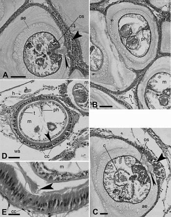
At the initial stages of infection, the cells of the outer mantle epithelium adjacent to a metacercaria change their normal shape, becoming tall-columnar (metaplasia) (up to 27 µm high). Progressively the cells multiply and invaginate, forming a shallow depression that surrounds a single larva (Figure 4B). Usually, additional larvae are added and the sac enlarges, progressively sinking into the dorsal part of the visceral mass above the kidney (Figures 3C & 4B, C). As the infection progresses, larvae that subsequently reach the infection zone incorporate into existing sacs, or stimulate the formation of new ones (Figures 3C & 4B). The dorsal portion of the visceral mass above the pericardium and kidney is expanded laterally and filled by the multiple sacs generated by the host tissues reaction (Figure 3B, C). The modified outer mantle epithelium forming the one-cell-thick wall of sacs showed a variable height, 16–27 µm high according to the degree of distension caused by the number of metacercariae contained (Figures 5A–E & 7A–C). The layer of columnar cells forming the sac wall, which showed a PAS positive reaction (Figure 5C), is supported by a thin layer of flattened conjunctive cells (Figure 5D, E). At the initial phase of reaction, the cells of the sac wall showed apical cytoplasmic regions filled with secretory granules (Figure 5D, E). The secretions of these cells accumulate as a thin layer covering the inner surface of the sac walls (Figures 5E & 6A–C) forming an envelope surrounding larvae. The secretion is acidophilic, PAS negative, Alcian blue positive (Figure 6B, C), indicating its acid mucopolysaccharide nature.
Details of the tissue reaction of Tagelus plebeius elicited by gymnophallid metacercariae. (A) Transverse section of a metacercaria at the level of oral sucker; note the alveolar envelope, the details of the sac wall (wide arrow indicates the proliferative process just in front of the oral sucker) and the host secretion entering into the oral sucker (narrow arrow) (GT); (B) detail of four contiguous sacs (three sections show metacercariae), with scarce haemocytes in the point of junction (GT); (C) transverse section of a metacercaria at the level of caeca; note the enlargement of the epithelium of the wall sac (arrow) facing the oral sucker of a metacercaria (GT); (D) transverse section of a recently settled metacercaria still without alveolar envelope; the apical zone of sac wall cells shows the apical portion of cytoplasm filled with secretion material (arrow) (H & E); (E) detail of sac wall epithelium cells showing secretory granules at the apical portion of cytoplasm and the beginning of secretion to form the alveolar envelope of a metacercaria (arrow) (H & E). Scale bars: A, B, D, 100 µm; C, E, 50 µm (ae, alveolar envelope; c, caecum; cc, connective cell of the sac wall; ct, connective tissue; h, haemocytes; m, metacercaria; os, oral sucker; ph, pharynx; t, tegument of larva; ws, epithelium of sac wall).

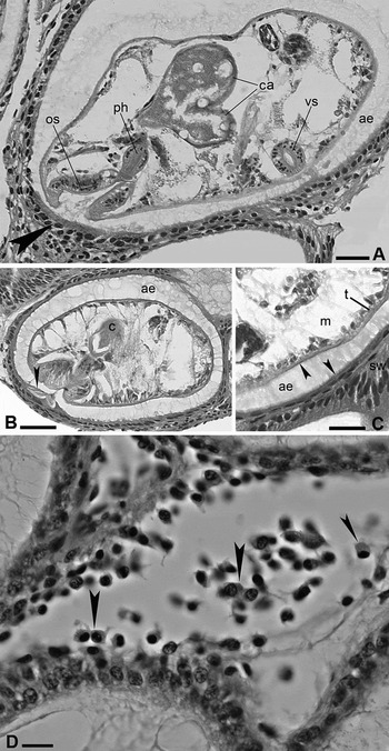
Details of the tissue reaction of Tagelus plebeius elicited by gymnophallid metacercariae. (A) Sagittal section of a metacercaria showing an advanced stage of secretion of the alveolar envelope (note the difference in epithelium height near the oral sucker (arrow) and secreted material seeming to be ingested by the larvae) (H & E); (B) sagittal section of a metacercaria showing detail of the epithelium of the sac wall. Arrow indicates secretion in front of oral sucker showing tinctorial affinity different to that of alveolar envelope (compare with that in the caecum) (AB/PAS); (C) detail of 6B showing the flattened epithelium and its secretion Alcian blue positive (AB/PAS); (D) point of junction of 4 sacs with haemocytes (arrows) (H & E). Scale bars: A, C, 50 µm; B, 100 µm; D,10 µm (ae, alveolar envelope; c, caecum; ca caeca; m, metacercaria; os, oral sucker; ph, pharynx; sw, sac wall; t, tegument of larva; vs, ventral sucker).

Each larva is individually surrounded by an acellular multilayered envelope with alveolar appearance, 44–67 µm thick, secreted by the epithelium of the sac wall (Figure 5A–C). The envelope entirely surrounds the body of the metacercaria except the region immediately in front of the oral sucker that remains in direct contact with the sac wall and its secretions (Figures 5A, C & 6A, B). The material forming the envelope was negative to PAS and GT and Alcian blue positive, a reaction compatible with that of a non-sulphated acid mucopolysaccharide substance. At the point where various sacs containing metacercariae join, a moderate number of haemocytes appear (Figures 5B & 6D).
Regardless of the degree of distension of the sac containing metacercariae, the height of the epithelium facing the oral sucker of the larvae was usually higher than other portions of the sacs walls (Figures 5A, C & 6A, B). The secreting activity was in cases more intense at these points, showing a β metachromatic reaction (toluidine blue coloration), differing from the secretions at other portions of the sac wall (Figures 5A & 7B). The digestive cecae of metacercariae were usually found filled with a material showing the same affinities (γ metachromatic, Alcian blue positive and PAS negative) as secretions from the mantle sac epithelium near the gymnophallid oral sucker (Figures 5A & 6B), suggesting ingestion of those host secretions by metacercariae.
In older infections, as determined by the size (=age) of the bivalve hosts, an amorphous yellow-brown material was frequently found within sacs. Sacs in bivalves larger than 6 cm length sometimes contained small calcium carbonate aggregations (about 0.67 µm diameter) in the form of pearls loose within sacs.
When the intensity of infection in the subarticular space was very high, metacercariae were also found settled up to the pallial line (the insertion of the orbicular mantle muscle), and at the deepest (anterior) portion of the pallial sinus, just below the area of insertion of the siphon retractor muscles (Figure 3A). In older specimens with high intensities of infection, metacercariae occurred on the anterodorsal part of the pallial sinus, eliciting the same reaction described in the subarticular extrapallial space by forming a sac containing metacercariae which are smaller than those developed in the subarticular space remaining between the mantle and shell. Frequently in these cases, the host reaction leads to a calcium carbonate deposition surrounding the larvae in the form of a blister which was accompanied by a discoloration of the inner shell surface.
DISCUSSION AND CONCLUSIONS
Cremonte (Reference Cremonte1999) and Vázquez et al. (2006) described for the first time the infection of Tagelus plebeius from Mar Chiquita lagoon by larval gymnophallid Digenea, reporting a prevalence of 100%, as was also found in the present study.
The reactions of T. plebeius against gymnophallid larvae that are described here, resembled those elicited by Bartolius pierrei (Gymnophallidae) in Darina solenoides (Bivalvia: Mactridae) (Cremonte & Ituarte, Reference Cremonte and Ituarte2003), and Meiogymnophallus strigatus (Gymnophallidae) in Donax trunculus (Bivalvia: Donacidae) (Bartoli, Reference Bartoli1974). The reaction includes the alteration of the outer mantle epithelium (hyperplasia and metaplasia) to form a sac surrounding a variable number of metacercariae. However, there are great differences in the host load capacity (i.e. the number of larvae a host is able to accumulate) and this fact seems to determine some of the particular characteristics of the host reaction. As in D. solenoides, in T. plebeius the subarticular extrapallial space, the site where larvae settle to start the reaction, remains permanently free of metacercariae due to the fact that the larvae that arrive to the extrapallial space are either incorporated to an existing sac or immediately elicit a host reaction forming a new sac. In this way, the extrapallial space is always receptive to new infective larvae. This, explains the correlation between the number of infecting larvae and size (= age) of the bivalve host. Lomovasky et al. (Reference Lomovasky, Gutiérrez and Iribarne2005) examined 610 specimens of T. plebeius from Mar Chiquita lagoon with sizes from 6.57 to 73.78 mm L (mean length = 60.24 mm) and also found that the intensity of parasitism was related to size (= age) of specimens. Higher intensities of parasitism (more than 800 metacercariae per specimen) were found in specimens larger than 60 mm shell length, and medium intensities (200–800 larvae/specimen) were common in specimens from 47–72 mm in shell length. Results from the present study generally agree with those of Lomovasky et al. (Reference Lomovasky, Gutiérrez and Iribarne2005), however, the maximum number of metacercariae found was markedly higher in the present study. Vázquez et al. (2006) reported a mean intensity of infection of 662 in a sample of 104 specimens of T. plebeius of lengths ranging from 11 to 62 mm.
In D. solenoides, the metacercariae of B. pierrei (Gymnophallidae) enter the bivalve through the mantle border, reach the dorsolateral portion of the extrapallial space, and elicit the outer mantle epithelium reaction to form large sacs that sink into the visceral mass. Although in low- or moderate-intensity infections, Cremonte & Ituarte (Reference Cremonte and Ituarte2003) considered that the process of incorporation of larvae would be unlimited; in the case of high infection intensities it was observed that a limitation of space available for sacs surrounding larvae does exist. It was observed, particularly in cases of high intensities of infection, that newly formed sacs do not have space for sinking into the visceral mass, and remain as extended lateral depressions in the outer mantle epithelium.
In T. plebeius, the epithelium of the sacs where larvae are enclosed secretes an Alcian blue positive substance, which is also PAS negative, confirming it to be an acid muco-polysaccharide. This secretion progressively forms a complete envelope surrounding each metacercaria as it was described for B. pierrei in D. solenoides. However, in the latter, an additional acellular secondary envelope (Cremonte & Ituarte, Reference Cremonte and Ituarte2003) surrounds several larvae, unlike in T. plebeius.
Lomovasky et al. (Reference Lomovasky, Gutiérrez and Iribarne2005) reported abnormal calcifications associated with an intense coloration in the inner surface of the shell in aged specimens, which represented about 5.9% of the studied sample. In the present study, abnormal calcifications seemed to be related to extremely high intensities of parasitism elicited by metacercariae settled in the extrapallial space areas other than the subarticular space; particularly in the central area of inner shell surface and the deepest portion of the pallial sinus. The calcium secretion observed in some larger (=aged) specimens in the present study never formed massive calcareous structures like those described in Gaimardia trapesina, Neolepton sp., Cyamiomactra sp. and Yoldia sp. (Ituarte et al., Reference Ituarte, Cremonte and Deferrari2001, Reference Ituarte, Cremonte and Zelaya2005), where host reactions to gymnophallid metacercariae formed igloo-like structures surrounding each larva.
Gymnophallid metacercariae do not encyst as most Digenea do at this life stage. The host-produced envelope is used by larvae as a ‘cyst’ that protects them during passage through the avian host's digestive system (Bowers & James, Reference Bowers and James1967). The metacercaria in this family does not represent a resting stage; on the contrary, the growth and development that they undergo are so spectacular that a simple temperature increase is enough to trigger egg production in a few hours (Bartoli, Reference Bartoli1974; Pekkarinen, Reference Pekkarinnen1984; Cremonte, Reference Cremonte2001; Cremonte et al., Reference Cremonte, Vázquez and Ituarte2008). It is clear, then, that gymnophallid metacercariae feed on extrapallial fluids that are rich in nutrients, avoiding in some unknown way the precipitation of calcium carbonate around them. Gymnophallid metacercariae have several cephalic glands whose functions are unknown, but whose probable function may be to prevent defensive calcium deposition by host molluscs (Ituarte et al., Reference Ituarte, Cremonte and Deferrari2001).
The case of G. trapesina is a good example of the quite different alternatives in the relationship established between a bivalve host and different species of larval digeneans, even belonging to the same family. Two different gymnophallid species simultaneously parasitize the general extrapallial space (Ituarte et al., Reference Ituarte, Cremonte and Deferrari2001); however, each elicits a quite different host reaction. Larvae of one species, a relatively small metacercaria (386–458 µm length) that lives ‘solitary’, elicit a host response leading to the formation of igloo-like calcareous coverings surrounding single larvae. Metacercariae of the second species (a minute larva of 190–230 µm length) lives in ‘aggregates’ of up to 30 individuals, elicit a reaction of the mantle adjacent to larvae, to form a wide sac delimited by the outer mantle epithelium and the inner shell surface, where calcium deposition never takes place. In the latter case, the metacercariae contained within the sac never showed any kind of hyaline envelope, as larvae did in the cases of D. solenoides and T. plebeius.
The wide spectrum of reactions elicited by metacercariae of different gymnophallid species (for a review see Ituarte et al., Reference Ituarte, Cremonte and Zelaya2005) are directed toward avoiding the walling-off of larvae by the bivalve host, specifically by calcium deposition. The factors by which different host reactions are elicited or modulated by different gymnophallid species, are still unknown.
ACKNOWLEDGEMENTS
The collection of specimens of Tagelus plebeius from Laguna Mar Chiquita, a natural reserve area, was authorized by the Secretaría de Recursos Naturales, Buenos Aires Province. The authors warmly acknowledge Pablo Penchaszadeh for allowing the use of the Zeiss Axiovision microscope to obtain photographs that illustrate this paper. This work was funded by PIP 5990 from CONICET and PICT 38015 from ANPCy T to C.I. C.I. and F.C. are members of the CONICET and A.S. received a scholarship from Facultad de Ciencias Naturales y Museo, Universidad Nacional de La Plata.