





Introduction
Measles is a severe, vaccine-preventable disease that continues to cause extensive morbidity and mortality in large parts of the world, despite the widespread use of measles vaccine, either as a single antigen vaccine or as a multi-component antigen vaccine: measles and rubella or measles, mumps, and rubella. In 2017, 172,939 measles cases were reported globally, with an estimated 122,000 deaths from measles [Reference Patel and Orenstein1]. Measles outbreaks increased and peaked in 2019. Subsequently, the COVID-19 pandemic led to reduced reports of measles cases and had a severe impact on routine vaccination programs, leading to an increasing number and scale of measles outbreaks in 2022 [Reference Akindele2–Reference Atim4]. Vaccine coverage is highly variable between World Health Organization (WHO) regions, and measles continues to be endemic in the African and South-East Asia regions, where vaccine coverage is less than 80% in many areas [Reference Dixon5]. These regions account for approximately 94% of all global measles deaths. This was the epidemiological situation in Uganda at the time of this study, with widespread measles outbreaks [Reference Walekhwa6, Reference Mensah and Gyasi7]. In the African region, WHO has set a goal of measles elimination in 80% of countries by 2030 based on the maintenance of high vaccination coverage, for which ongoing political commitment and sustainable surveillance will be required [Reference Masresha8, 9].
A key component of elimination plans is surveillance to monitor impact. Laboratory confirmation of measles cases is a vital aspect of surveillance at all stages of control programmes because clinical diagnosis is unreliable. The mainstay of laboratory confirmation is the detection of measles-specific immunoglobulin M (IgM) antibodies in serum samples. More recently, alternative samples such as dried capillary blood (CAP) spots and oral fluids (OFs) have been used for measles diagnosis by antibody detection [Reference Helfand10–Reference Uzicanin12]. OF samples can also be used to detect viral ribonucleic acid (RNA), which is important for tracking transmission of measles in the community and for documenting elimination [Reference Hutse11, Reference Jin, Vyse and Brown13]. The use of OF and CAP samples is becoming increasingly common because samples can be obtained safely and non-invasively, without the risks associated with venous blood collection. This improves patient compliance with specimen collection, as the procedures are simple and painless. Rapid diagnostic tests (RDTs) are increasingly used for the diagnosis of infections [Reference Aidoo and Incardona14, Reference Bouzid15]. They can be performed in a single incubation step at ambient temperature without complex electrical equipment, and their results can be read visually. By increasing diagnostic capacity and facilitating rapid diagnosis in resource-poor countries, they have the potential to improve the timeliness of measles surveillance and the response of health authorities to possible outbreaks [Reference Brown16]. In the African Region, many countries continue with endemic measles, and surveillance may be compromised by poor infrastructure, challenges with timely transport, and limited laboratory capacity. Thus, surveillance in remote or poorly served areas remains a logistical challenge. Use of a measles RDT by health workers at the first contact has the potential to facilitate rapid case confirmation and a timely public health response, even in hard-to-reach areas [Reference Brown16].
A lateral flow RDT based on an antibody capture format for measles IgM has been described, which enables the detection of measles-specific IgM antibodies in both serum and OF specimens in <30 min [Reference Warrener17]. The RDT had high sensitivity and specificity (>90%) with serum and OF compared to a reference enzyme immunoassay with serum in a laboratory setting [Reference Warrener17, Reference Warrener18]. Measles virus RNA was detected by reverse transcriptase polymerase chain reaction (RT-PCR) and genotyped from RDTs used for OF testing, after dry storage for 5 weeks at 20°C–25°C [Reference Warrener17].
This prototype RDT was subsequently redesigned into a cassette-housed format for ease of handling and robustness in field settings [Reference Warrener18]. As part of a project funded by the Bill and Melinda Gates Foundation, this RDT was modified by the incorporation of a blood filter pad to enable both OF and capillary (finger prick) blood samples to be tested. A pilot batch of these RDTs was manufactured commercially and used in this study. However, the role of alternative samples and the RDT has not been evaluated for use in the global measles control programmes, and this study was one of the first to investigate performance and usability in an outbreak programmatic setting.
The introduction of measles IgM RDT into the measles surveillance programme has the potential to provide important information on real-time measles case confirmation, improve timeliness of case confirmation where transport of clinical specimens from remote areas to the central laboratory is challenging, to reduce delays in initiating appropriate control measures, to reduce laboratory-based turnaround times and to rapidly enable scale-up of testing services.
To realize these opportunities, studies are required to evaluate the RDT performance and to define the feasibility and needs to support its introduction into surveillance.
This study aimed to determine the following: (1) the performance sensitivity and specificity, of a measles IgM RDT to confirm measles infection using OFs, collected using the Oralight Device, and CAP specimens performed in field conditions, compared to laboratory-based IgM detection in serum using the Siemens anti-measles IgM enzyme-linked immunosorbent assay (ELISA) as the gold standard; (2) to assess the accuracy and consistency of on-site visual RDT interpretation; (3) assess the rate of detection of measles virus by qRT-PCR in measles IgM-positive RDTs; and (4) to survey the perception of health workers, patients, and guardians on the use, potential problems and benefits of the Measles IgM RDT for measles diagnosis.
Methods
Ethical approval for the study
The study received approval from Uganda Virus Research Institute Science and Ethics Committee (GC/127/18/02/632) and the Uganda National Council for Science and Technology (HS 2372).
Rapid diagnostic test (RDT) for measles IgM
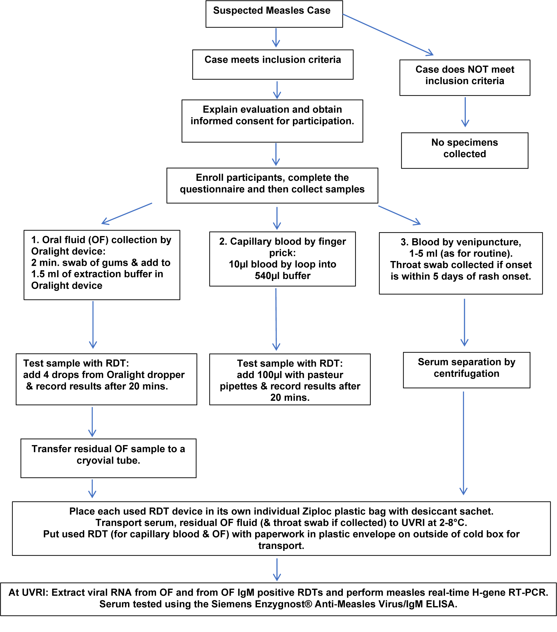
The measles IgM RDTs were prepared under contract manufacture, according to specifications of the UK Health Security Agency (UKHSA). The measles IgM RDT is an immunochromatographic assay enclosed in a plastic cassette. Components and principle of the assay are as described previously [Reference Warrener17], modified by replacement of the glass fibre sample pad with a blood separation matrix. The procedure for performing the measles IgM RDT on OFs and CAP specimens is outlined in Figure 1. Specimen processing time was minimal, and results were available following a 20-min incubation at ambient temperature by interpreting the presence or absence of the control and test lines that appear on the RDT. The RDT was required to have a visible control line to be considered a valid test. If, in addition to the control line, the test line was visible, the test was interpreted as positive. If the control line was visible, but the test line was not, the test was interpreted as negative.
Procedural steps involved in the evaluation of the measles IgM RDT within the Ugandan national surveillance system, 2018–2019.

Figure 1. Long description
The flowchart begins at the top with Suspected Measles Case.
Two paths emerge from the top. To the right, Case does NOT meet inclusion criteria leads to No specimens collected. To the center, Case meets inclusion criteria leads to Explain evaluation and obtain informed consent for participation, followed by Enroll participants, complete the questionnaire and then collect samples.
From the enrollment step, three parallel sample collection methods are shown from left to right.
1. Oral fluid O F collection by Oralight device. 2 minute swab of gums and add to 1.5 ml of extraction buffer. This leads to Test sample with R D T by adding 4 drops and recording results after 20 minutes, then Transfer residual O F sample to a cryovial tube.
2. Capillary blood by finger prick. 10 microliters blood by loop into 540 microliters buffer. This leads to Test sample with R D T by adding 100 microliters with pasteur pipettes and recording results after 20 minutes.
3. Blood by venipuncture, 1 to 5 ml as for routine. Throat swab collected if onset is within 5 days of rash onset. This leads to Serum separation by centrifugation.
All three paths converge at a step to Place each used R D T device in its own individual Ziploc plastic bag with desiccant sachet. Transport serum, residual O F fluid and throat swab if collected to U V R I at 2 to 8 degrees Celsius. Put used R D T for capillary blood and O F with paperwork in plastic envelope on outside of cold box for transport.
The final step at the bottom states At U V R I. Extract viral R N A from O F and from O F I g M positive R D T s and perform measles real-time H-gene R T-P C R. Serum tested using the Siemens Enzygnost Anti-Measles Virus/I g M E L I S A.
The presence of the control line confirmed the reactivity between the measles antigen conjugate and the anti-measles antibody.
Staff training
The Uganda Virus Research Institute (UVRI) and Ministry of Health (MoH) team who led the outbreak investigations attended a 2-day practical training course in study design, sample collection, the use, result interpretation and troubleshooting of the measles IgM RDTs and transportation of residual OFs, used RDTs and serum specimens to the UVRI laboratory.
Study design and recruitment of participants
This was a community-based prospective study nested within the ongoing outbreak-based measles surveillance in Uganda from May 2018 to May 2019. Cases were recruited from a population which included all suspected measles cases and outbreaks investigated that met the following criteria: Any person with a history of fever and maculopapular rash (onset within 30 days), with any one of cough, coryza, or conjunctivitis in outbreaks identified by healthcare workers or surveillance officers anywhere in Uganda. When the National Expanded Programme on Immunisation (EPI) laboratory at UVRI learnt of a suspected outbreak through surveillance reports to the MoH, UVRI staff visited the district and worked with district officials, including district surveillance focal persons and health workers, to investigate the outbreak. In a few instances, cases in districts with laboratory-confirmed outbreaks were also recruited.
The district officer guided the team to the area with confirmed cases, and the team searched for other suspected cases of measles.
On arriving at the household of a suspected measles case, the surveillance team informed the family about the purpose of the visit. The team established whether the case met the eligibility criteria.
Participants were recruited following a verbal explanation of the study and reading of the information sheets that were provided. The participants (18 years old and above) or guardians of participants less than 18 years old provided written consent, and assent was received from children ≥7 years before inclusion in the study.
Participants with any contraindication to venepuncture, such as bleeding disorders, and for OF collection, such as sore mouth from oral ulcers or infection, inflammation, or dehydration, or any other major illness were excluded from the study.
After consent was obtained, the subject was enrolled in the study and provided a unique subject identification number. A short study questionnaire was administered to the subject/guardian, containing questions found in the case investigation form used in the measles surveillance guidelines in Uganda. District or health sub-district surveillance staff then collected an OF sample using the Oralight device (UKHSA (formerly Public Health England), London, United Kingdom), designed to collect samples and then dispense them in the same setting, and a CAP sample collected by finger prick from suspected measles cases. The specimens were tested immediately with the measles IgM RDTs on site by district or health sub-district surveillance staff, following cascade training delivered by, and under instruction from, UVRI staff.
Visual result interpretation of RDTs was performed independently by two field staff, and results were recorded using a five-point WHO reading scale based on colour intensity of the test line; 0: negative, 1: indeterminate, 2: weak, 3: moderate, and 4: strong positive [Reference Warrener18]. The results were recorded on the study questionnaire.
For data analysis, indeterminate results were interpreted as positive, as a faint visible test line was present. If there was doubt about an individual RDT result, the lines were photographed for records or audit, and for real-time referral for a second opinion, using a mobile phone. Throughout the study, the normal surveillance samples, approximately 4 mL venous blood, were collected according to standard practice. The used RDTs, together with the separated serum sample and residual OF sample, were returned to UVRI in a specimen carrier with ice packs. Residual OF specimens and sera were stored at −20°C and 2°–8°C in the laboratory, respectively, until tested. Used RDTs were stored in Ziploc plastic bags with desiccant sachets at room temperature (approx. 25°C) prior to molecular testing at UVRI. A flow diagram outlining the procedural steps followed within the study is provided in Figure 1.
Sample size
At the start of the study, it was hypothesized that the RDT would have a specificity of 85% and sensitivity of at least 90% compared to the Siemens anti-Measles IgM ELISA performed using serum. The study was designed to show this sensitivity with 80% power at a 5% significance level. Thus, 100 IgM-positive measles cases should yield a 95% confidence interval (95% CI) of 81%–95% and for 150 cases, 84%–94% (95% CI). The aim was to recruit up to 400 suspected measles cases, but closed recruitment at 320 suspected cases of measles as the study period approached 1 year. The prevalence of confirmed measles in this cohort was 55%, which gave us sufficient power to estimate sensitivity and specificity with a precision of 5.4%.
Laboratory testing
Enzyme-linked immunosorbent assay (ELISA)
Sera were tested using the Enzygnost® Anti-Measles Virus/IgM ELISA (Siemens Healthcare GmbH, Marburg, Germany) with a reported sensitivity of 87.9% and specificity of 98.7% [Reference Stambos19]. Testing and result interpretation followed the manufacturer’s instructions. An optical density (O.D., 450 nm/620 nm) measurement ≥0.2 was interpreted as positive, an O.D. of 0.1 to <0.2 was interpreted as equivocal, and an O.D. of <0.1 was interpreted as negative.
RNA extraction
Viral RNA was extracted from residual OF samples and from the blood separation pad (BSP) of RDTs used for OF testing only. Extraction was performed using the QIAamp Viral RNA Mini Kit (QIAGEN GmbH, Hilden, Germany) following the manufacturer’s instructions essentially as described, with the following modification for BSPs from RDTs. Briefly, the BSP was removed from the RDT, placed in an extraction tube, 140 μL phosphate-buffered saline was added, followed by 560 μL Buffer AVL–carrier RNA prepared according to the manufacturer’s instructions, and incubated at room temperature for 20 min. Tubes were centrifuged at 11000 rpm for 2 min, and the supernatant was transferred to a clean tube to prevent BSP fibres from interfering with subsequent steps, which were performed as described by the manufacturer. Negative controls: OF elution buffer only and OF from healthy individuals, and positive controls: measles vaccine diluted 10−1 and 10−2 in nuclease-free water, were included in each extraction.
Measles N-gene, H-gene and RNase P gene amplification and detection by real-time RT-PCR
Extracted RNA was tested for the presence of a 67-nucleotide fragment of the measles haemagglutinin gene (H-gene) [Reference Thomas20], for the presence of a 75-nucleotide fragment of the measles nucleoprotein gene (N-gene) for residual OF extracts only and the human ribonuclease P (RNase P) [Reference Hummel21] gene-specific target as an endogenous control, in separate real-time RT-PCRs (qRT-PCR). Amplification of the above genes was performed using the SuperScript™ III platinum™ one-step qRT-PCR kit (Invitrogen, Carlsbad, USA) and gene-specific primers and probes; the sequences of which are provided in Supplementary Table 1, using an ABI 7500 thermocycler (Applied Biosystems, Singapore). Measles-specific primers and probe were obtained from Thermo Fisher Scientific, UK and RNase P-specific primers and probe were obtained from the US Centers for Disease Control and Prevention International Reagent Resource.
For the H gene qRT-PCR target, the reaction mixture contained the following reagents per sample to be amplified; 6.3 μL of nuclease free water, 12.5 μL of 2X reaction buffer, 0.4 μL of primer/probe mix, 0.25 μL of RNase inhibitor, 0.05 μL of ROX reference and 0.5 μL of Superscript III Platinum Taq enzyme mix to give final concentrations of 0.9 μM each primer, 0.25 μM probe, 10 U/μL of RNase inhibitor and 2.5 U/μL of Superscript III Platinum Taq enzyme mix. Five microliters of the extracted RNA were then added to give a final reaction volume of 25 μL. The thermal cycling conditions consisted of a reverse transcription reaction at 50°C for 30 min, an activation step of 95°C for 10 min, followed by amplification cycling of 95°C for 15 s and 60°C for 1 min, for 40 cycles.
For the N gene qRT-PCR target, the reaction mixture contained the following reagents per sample to be amplified; 7.2 μL of nuclease free water, 12.5 μL of 2X reaction buffer, 2 μL of primer/probe mix, 0.25 μL of RNase inhibitor, 0.05 μL of ROX reference and 0.5 μL of Superscript III Platinum Taq enzyme mix to give final concentrations of 0.3 μM MeV primer, 0.1 μM RNase P primer, 0.25 μM probe, 10 U/μL of RNase inhibitor, and 2.5 U/μL of Superscript III Platinum Taq enzyme mix. Two point five microlitres of the extracted RNA were then added to give a final reaction volume of 25 μL. The thermal cycling conditions consisted of a reverse transcription reaction at 48°C for 30 min, an activation step of 95°C for 5 min, followed by amplification cycling of 95°C for 15 s and 60°C for 1 min, for 40 cycles.
For quality control purposes, the extraction controls and a non-template control were included in each qRT-PCR run, and for the N-gene PCR run only, RNase P primer kit positive controls were also included.
Usability of RDTs in field setting
A questionnaire on the usability of the Oralight OF collection device and the Measles IgM RDT was completed by the health workers who participated in the study. After completion of the field testing, responses were analysed to inform future use of OF collection and of the Measles IgM RDT using OF and CAP samples.
Data analysis
Data were double-entered in EPI INFO version 3.5.4 (Centers for Disease Control and Prevention, GA, USA) and was exported to Microsoft Office, EXCEL workbook and analysed in STATA version 13.0 (StataCorp LP, College Station, TX, USA).
The study population demographics were summarized. The RDT readings made using CAP and OF samples by two health workers in the district were compared using Kappa statistics [Reference McHugh22].
Sensitivity and specificity of the RDTs were compared with: (1) the serum measles IgM ELISA result as the gold standard and (2) Serum measles IgM ELISA and/or qRT-PCR positive as the gold standard. The proportion command in STATA 13.0 and confidence intervals produced with the Wilson score method were used to determine the sensitivity and specificity of the RDTs [Reference McHugh22, Reference Newcombe23].
In the performance analysis of IgM detection in OF and CAP specimens by RDT, only RDTs with concordant test results, negative or positive, between the two readers were included, and those with invalid results were excluded.
Results
Three hundred and twenty participants were recruited for the study from 43 districts in Uganda (Figure 2). The participants’ age ranged from 3 months to 41 years 6 months, with a median age of 6 years. Not all participants gave all 3 samples; 313 gave serum samples, 319 CAP samples, and 320 OF samples (Supplementary Figures S1 and S2). Specimens were collected within 7 days of rash onset from the majority (62.5%) of study participants (Supplementary Table 2).
Distribution of measles cases included in the study by district.

Figure 2. Long description
A map of Uganda divided into administrative districts. A legend in the bottom right indicates that light blue shading represents Districts visited, while white areas represent Districts not visited. A north arrow is located in the top right corner.
* In the Northwest, visited districts include Yumbe, Maracha, Arua, and Madi-Okollo.
* In the North and Northeast, visited districts include Amuru, Gulu, Pader, Omoro, Otuke, Alebtong, and Katakwi.
* In the Central and Western regions, visited districts include Masindi, Nakasongola, Nakaseke, Luwero, Kiboga, Kikuube, Kagadi, Kibaale, Kyegegwa, and Bundibugyo.
* In the Southwest, visited districts include Kasese, Lyantonde, Kiruhura, Lwengo, Masaka, and Kyotera.
* In the South and Southeast, visited districts include Wakiso, Mpigi, Kampala, Mukono, Buikwe, Buvuma, Jinja, Kamuli, Kaliro, Iganga, Mayuge, Namutumba, and Butaleja.
* The Eastern border districts and the far Northern districts like Kaabong and Karenga are primarily white, indicating they were not visited.
Of the 313 serum samples collected, 303 were tested by measles IgM ELISA.
Three hundred and twelve cases were included in the analysis of RDT IgM results compared to the ‘true measles case’ classification, with true cases designated as those for which the Siemens anti-measles IgM ELISA and/or the qRT-PCR gave a positive result. Eight cases were not able to be assigned a final classification as they had neither qRT-PCR nor serum IgM ELISA results (Supplementary Table 2).
To identify ‘true measles cases’, measles H-gene qRT-PCR was performed on 311 OF specimens. Measles N-gene qRT-PCR and human RNase P qRT-PCR were performed on a subset of 136 specimens to enable comparison with H-gene qRT-PCR and to ascertain OF sample quality, respectively. Out of the 136 OFs tested by N-gene and H-gene qRT-PCRs, 9 were confirmed by N-gene only, 87 by both H-gene and N-gene and none by H-gene only (Supplementary Table 3).
Of the 136 OF specimens tested, 134/136 (98.5%) were RNase P positive and this was split between H-gene positive and negative specimens: 88/89 (98.9%) H-gene qRT-PCR positive OFs were RNase P positive and 46/47 (97.9%) H-gene negative OFs were RNase P positive. This showed that OF sample quality was high.
Measles IgM RDT result interpretation
Analysis of the measles IgM RDT result interpretation revealed that 300 OF and 288 CAP samples tested had valid control lines recorded following visual interpretation by both readers. For most of the invalid results, the readers disagreed on whether the control line was visible: In 6/14 (42.9%) samples, there was agreement between the two readers of CAP RDT in the interpretation of invalid control line results, while for OF RDTs, the agreement between the two readers in interpretation of an invalid control line was only 1/8 (12.5%). The overall agreement of direct visual result interpretation of the RDT device as positive or negative between two staff in valid tests was 92.9% for OF specimens (Kappa = 0.86), and 92.4% for CAP (Kappa = 0.85).
Measles case designation based on laboratory testing
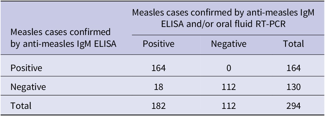
Table 1 demonstrates the impact of using both the serum anti-measles IgM ELISA and the qRT-PCR results for case classification, compared to serum IgM ELISA alone. There was excellent agreement (276/294, 93.9%) overall; however, 18 additional measles cases were confirmed by qRT-PCR. Specimens from 14 of these 18 (77.8%) cases were collected within 7 days of rash onset. Two of the qRT-PCR-positive, serum IgM ELISA-negative cases were measles IgM RDT positive with both CAP and OF samples.
Measles cases confirmed by serum anti-measles IgM ELISA compared to those confirmed using the combined results of serum measles IgM ELISA and RT-PCR testing of oral fluid specimens

Table 1. Long description
The table presents data for 294 participants. The top header defines the columns as Measles cases confirmed by anti-measles I g M E L I S A and or oral fluid R T - P C R, subdivided into Positive, Negative, and Total. The left-most column defines the rows as Measles cases confirmed by anti-measles I g M E L I S A, subdivided into Positive, Negative, and Total.
* Row 1 (Positive E L I S A): 164 cases are Positive by the combined method, 0 are Negative, totaling 164.
* Row 2 (Negative E L I S A): 18 cases are Positive by the combined method, 112 are Negative, totaling 130.
* Row 3 (Total): The vertical totals are 182 Positive, 112 Negative, and a grand total of 294.
The data shows that the combined method identified 18 additional positive cases that were negative by serum E L I S A alone.
Note: Summary of results obtained for 294 study participants, out of a total of 320, for whom laboratory-based measles serum IgM ELISA and RT-PCR results were available. Samples with equivocal measles IgM results in ELISA were excluded from this analysis. Measles serum IgM ELISA used throughout was Siemens Enzygnost® Anti-Measles Virus/IgM ELISA.
RDT performance using CAP and OF samples compared to the Siemens IgM ELISA as the reference test
Following application of the inclusion criteria described above (Supplementary Figure S1), IgM results from 255 CAP RDTs and 267 OF RDTs were analysed compared to Siemens IgM ELISA and summarized in Table 2. The overall sensitivity of the RDT with CAP samples was 97.9% (95% CI: 93.7%–99.3%), and this did not differ by time from rash onset to specimen collection. The specificity of CAP RDT was 91.7% (95% CI: 84.8%–95.6%). The overall sensitivity for OF RDT was 96.7% (95% CI: 92.2%–98.6%) and specificity was 88.0% (95% CI: 80.1%–92.8%).
Performance of the measles IgM RDT using capillary blood and oral fluid specimens compared to anti-measles IgM ELISA on sera

Table 2. Long description
The table is organized into six columns: Specimen used for measles I g M R D T testing, Measles I g M R D T results, Anti-measles virus/I g M E L I S A Positive, Anti-measles virus/I g M E L I S A Negative, R D T sensitivity percentage with 95 percent C I, and R D T specificity percentage with 95 percent C I.
* Oral fluid specimen row:
- Positive R D T results: 145 E L I S A positive, 14 E L I S A negative.
- Negative R D T results: 5 E L I S A positive, 103 E L I S A negative.
- Performance: Sensitivity is 96.7 percent (92.2–98.6); Specificity is 88.0 percent (80.7–92.8).
* Capillary blood specimen row:
- Positive R D T results: 143 E L I S A positive, 9 E L I S A negative.
- Negative R D T results: 3 E L I S A positive, 100 E L I S A negative.
- Performance: Sensitivity is 97.9 percent (93.7–99.3); Specificity is 91.7 percent (84.7–95.7).
Abbreviations used: R D T stands for rapid diagnostic test and C I stands for confidence interval.
Abbreviations: RDT, rapid diagnostic test; CI, confidence interval.
Note: Measles IgM ELISA results were available for 294 sera. Specimens for which concordant RDT results interpretations were obtained between the two observers were included. Results of concordant invalid RDT test results between the two observers were excluded in the analysis. Results for 267 oral fluid RDTs and 255 capillary blood RDTs were included in this analysis.
RDT performance using CAP and OF samples compared to the Siemens IgM ELISA and/or RT-PCR as the reference ‘true case’ criteria
Following exclusion of invalid and discordant RDTs (Supplementary Figure S2), the number of CAP and OF RDTs included in analyses compared to the measles true case criteria was 269 and 282, respectively. The overall sensitivity of the RDT using CAP was 90.4% (95% CI: 84.9%–94.1%) (Table 3). Sensitivity of IgM detection by RDT was lower in participants with rash onset of 7 days or less: 89.4% (95% CI: 83.6%–95.1%), compared to those for whom specimens were collected more than 7 days after rash onset: 94.0% (95% CI: 87.2%–100.0%). The specificity of the CAP RDT was 89.2% (95% CI: 81.5%–93.9%).
Performance of capillary blood and oral fluid measles IgM RDT compared to true measles cases

Table 3. Long description
The table is organized into six columns: Specimen used for measles I g M R D T testing, R D T results, True measles cases (subdivided into Detected and Not detected), R D T sensitivity percent with 95 percent C I, and R D T specificity percent with 95 percent C I.
* Oral fluid specimen row:
- Positive results: 155 detected, 14 not detected.
- Negative results: 16 detected, 97 not detected.
- Sensitivity: 88.5 (82.8 to 92.5).
- Specificity: 86.1 (78.1 to 91.5).
* Capillary blood specimen row:
- Positive results: 151 detected, 11 not detected.
- Negative results: 16 detected, 91 not detected.
- Sensitivity: 90.4 (84.9 to 94.1).
- Specificity: 89.2 (81.5 to 94.0).
True measles cases are defined as positive anti-measles I g M E L I S A on serum or H-gene or N-gene R T-P C R positive result on oral fluid.
a True measles cases in this study were defined as any suspected case which gave a positive anti-measles IgM ELISA result on serum and/or an H-gene or N-gene RT-PCR positive result on residual oral fluid liquid tested in the laboratory. Results for 282 oral fluid RDTs and 269 capillary blood RDTs were included in this analysis.
Abbreviations: RDT, rapid diagnostic test; CI, confidence interval.
The overall sensitivity of IgM detection in OFs by RDT compared to the true case criteria was 88.5% (95% CI: 82.8%–92.5%) and specificity was 86.1% (95% CI: 78.1%–91.5%) (Table 3). Sensitivity of IgM detection in OFs by RDT was higher in the participants who gave samples more than 7 days from rash onset: 92.3% (95% CI: 84.8%–99.7%) compared to those whose specimens were collected less than 7 days from rash onset: 87.4% (95% CI: 81.3%–93.4%).
Detection of measles virus RNA from used OF RDTs
Measles H-gene qRT-PCR results were available for 307 RDTs used for OF testing and their matching residual OF specimens. Results are summarized in Table 4. High concordance, 91.5% (281/307), was observed between the two results.
Comparison of measles H-gene RT-PCR results generated from RNA extracted from RDTs used for oral fluid testing with RT-PCR results for matched liquid oral fluid specimens

Table 4. Long description
The table is organized into five columns. The first column lists the R T - P C R results for O F R D T s. The second and third columns show the results for residual liquid oral fluid specimens, categorized as Detected and Not detected. The fourth and fifth columns provide performance characteristics.
Row 1: Detected on R D T. Liquid oral fluid Detected: 146. Liquid oral fluid Not detected: 6.
Row 2: Not detected on R D T. Liquid oral fluid Detected: 20. Liquid oral fluid Not detected: 135.
Row 3: Total. Liquid oral fluid Detected: 166. Liquid oral fluid Not detected: 141.
Performance characteristics spanning these rows:
- Sensitivity percentage with 95 percent C I: 88.0 (82.0 to 92.1).
- Specificity percentage with 95 percent C I: 95.7 (90.8 to 98.1).
Abbreviations: H-gene, measles haemagglutinin gene; RDT, rapid diagnostic test; OF, oral fluid; RNA, ribonucleic acid; CI, confidence interval; RT-PCR, one-step, real-time, reverse transcription polymerase chain reaction.
Note: The total number of suspected cases for which the OF RDT and the matched residual liquid oral fluid were tested by RT-PCR was 307. RDT used for OF testing and that gave an invalid control line result for the antibody test were not excluded from this analysis.
Results were then stratified by specimen collection timing post-symptom onset, in Table 5. For both the liquid OF specimen and the matched OF RDT, the highest detection rate of measles H-gene by qRT-PCR occurred in specimens collected in the first week following onset of rash, 63.4% and 59.1%, respectively. The measles qRT-PCR detection rate declined progressively in subsequent weeks post-rash onset. Nevertheless, measles virus was detected by qRT-PCR from some of the OF RDTs within the first 4 weeks of sample collection after rash onset. No significant difference in qRT-PCR detection rate by week post-rash onset was observed between liquid OF specimens and matched OF RDTs.
Detection of measles haemagglutinin gene by RT-PCR from measles RDTs used for oral fluid testing compared to RT-PCR results obtained from residual, liquid oral fluids by timing of specimen collection post rash onset

Table 5. Long description
The table consists of four columns and six data rows including a total.
Column headers are:
1. Timing of specimen collection post rash onset by week (days).
2. Total number of cases collected per week.
3. Measles H-gene R T - P C R detection in liquid oral fluid specimens (percent; 95 percent C I).
4. Measles H-gene R T - P C R detection from O F R D T s (percent; 95 percent C I).
Data rows:
- Week 1 (0 to 7 days): 200 cases (62.5 percent). Liquid oral fluid: 123/194 (63.4 percent; 56.4 to 69.9). O F R D T s: 114/193 (59.1 percent; 52.0 to 65.8).
- Week 2 (8 to 14 days): 79 cases (24.7 percent). Liquid oral fluid: 32/77 (41.5 percent; 31.0 to 53.0). O F R D T s: 31/76 (40.8 percent; 30.2 to 52.3).
- Week 3 (15 to 21 days): 25 cases (7.8 percent). Liquid oral fluid: 10/24 (41.7 percent; 23.3 to 62.7). O F R D T s: 7/23 (30.4 percent; 14.6 to 52.8).
- Week 4 (22 to 28 days): 12 cases (3.8 percent). Liquid oral fluid: 3/12 (25.0 percent; 7.0 to 59.1). O F R D T s: 1/12 (8.3 percent; 0.9 to 47.5).
- Greater than Week 4 (29 to 35 days): 4 cases (1.2 percent). Liquid oral fluid: 0/4 (0 percent). O F R D T s: 0/4 (0 percent).
- Total: 320 cases. Liquid oral fluid: 168/311 (54.0 percent; 49.3 to 60.2). O F R D T s: 153/308 (49.7 percent; 44.1 to 55.2).
Abbreviations: H-gene, measles haemagglutinin gene; RDT, rapid diagnostic test; OF, oral fluid; RT-PCR, one-step, real-time, reverse transcription polymerase chain reaction.
Usability of RDT within study
Table 6 summarizes the findings from the completed questionnaires from 320 participants, their parents or guardians and health workers involved in the study. Collection of OF was well accepted and used successfully within the study. Questionnaire responses indicate the ease of OF collection, elution from the swab and use of the RDT at the point of patient contact were all rated very highly (>97%). Both OF sampling and finger prick blood collection were preferred to standard serum collection by colleagues involved in the field.
Summary of observational findings of oral fluid quality, responses to questionnaire on sample collection methodology for RDT use and staff perspectives on the use of Measles IgM RDTs in Ugandan clinics

Table 6. Long description
The table is divided into two primary sections across six columns: Issues for collecting oral fluids (left) and Measles I g M R D T: Issues for surveillance (right). Each section includes columns for Number and Frequency (%).
Section 1: Issues for collecting oral fluids
* O F easy to collect? Yes: 313 (97.8%), No: 5 (1.6%), Missing: 2 (0.6%).
* O F easy to extract from collection device? Yes: 311 (97.2%), No: 1 (0.3%), Missing: 8 (2.5%).
* R D T easy to perform? Yes: 319 (99.7%), No: 1 (0.3%).
* Blood stains in the sample? Yes: 53 (16.7%), No: 265 (82.8%), Missing: 2 (0.6%).
Section 2: Measles I g M R D T: Issues for surveillance
* Food particles in oral fluid sample? Yes: 20 (6.3%), No: 298 (93.1%), Missing: 2 (0.6%).
* Sample type preferred by patient? Finger prick: 111 (34.7%), Oral fluid: 164 (51.3%), Serum: 38 (11.9%), Missing: 7 (2.2%).
* R D T will improve sampling compliance? Yes: 310 (96.9%), No: 3 (0.9%), Missing: 7 (2.2%).
* R D T will improve measles management? Yes: 309 (96.6%), No: 4 (1.3%), Missing: 7 (2.1%).
Abbreviations: RDT, rapid diagnostic test; OF, oral fluid specimen.
Discussion
The study demonstrated successfully that the measles IgM RDT has sufficient accuracy for use in this setting, where outbreaks are characterized based on confirming three measles cases in a district or health unit catchment area [24]. The sensitivity and specificity of the RDT compared well to the IgM ELISA alone, and measles cases confirmed by serum IgM and /or qRT-PCR for both CAP and OF testing were assessed. Measles IgM RDT performance was more sensitive with CAP samples than OF for measles IgM detection. In this case series, the sensitivity of the serum measles IgM ELISA was comparable to the RDT at 90% of confirmed cases, which illustrates how additional cases are detected using RT-PCR in an outbreak setting. These findings support the wider use of measles detection by RT-PCR in the measles-rubella surveillance programme. It should be noted that the Siemens Enzygnost IgM assay used as the reference comparator in this study is no longer commercially available. While this represents a limitation, it may also be viewed as an advantage, as currently available commercial measles IgM kits generally demonstrate inferior sensitivity and specificity, suggesting the RDT may compare even more favourably against the alternatives now accessible to programmes.
The ability to consistently detect measles virus RNA extracted from used IgM-positive OF RDTs, following ambient temperature storage in this study, suggests it may also be possible to amplify the 450-nucleotide region at the carboxy terminal of the measles N-gene used for measles sequence analysis and genotyping, as demonstrated previously [Reference Warrener17]. Measles sequence analysis has become an important surveillance tool, enabling tracking of transmission chains and strain diversity, contributing supporting data to document progress towards measles elimination for national or regional verification [Reference Masresha8]. The use of nucleic acid extracts from IgM-positive RDTs could increase the number and representativeness of viral genotypes and strains to enhance current surveillance data, particularly from hard-to-reach, low-resourced, or previously underserved settings. Further studies to establish the rate of successful genotyping directly from used OF RDTs are needed.
The study was conducted in the context of Ugandan measles outbreak surveillance and demonstrates the successful use of both CAP and OF samples with the measles IgM RDT in the field. This followed a 2-day practical and theoretical training course for a team of core staff, who successfully trained and supervised clinic staff in the use of the RDTs based on simple visual guides during outbreak investigations. Many of the clinic and surveillance staff involved in the study already had experience with the use of HIV and malaria RDTs, which aided its introduction. The study also demonstrated that the measles RDTs gave accurate results in the field based on visual reading (Kappa >0.8) with two independent readers. However, the agreement between the readers for both OF RDT and CAP RDT was lower than what was observed in another study in Malaysia [Reference Senin25]. In the Uganda study, approximately 3.2% of OF RDTs and 6.6% of CAP RDTs results were invalid. This is higher than in other studies and is likely to improve with simplified interpretation of the RDT results as positive or negative only, and increased familiarity with the assay [Reference Senin25]. In a programme, invalid test results would be repeated using the original sample, which was not performed here because of limited availability of the trial batch of RDTs.
There are several potential models for introducing the RDT into surveillance, and its use in this study for outbreak confirmation by a central surveillance team is one. The results of the study provide support for the use of RDTs for confirming measles outbreaks, as indicated in previous studies [Reference Warrener17, Reference Warrener18]. This study recommends either CAP or OF samples for RDT testing. The choice of sample type should be guided by in-country resourcing and requirements for downstream molecular testing of the RDT to meet surveillance objectives. Residual measles N-gene nucleic acid present in antigen used in the RDT may be detected by N-gene qRT-PCR and by the endpoint PCR used to generate amplicons for molecular sequence analysis [Reference Warrener17]. Due to the expected higher viral load in OFs relative to the diluted CAP specimens tested in the RDTs, it is possible that OFs may be less affected by this than the CAP specimens; hence, the use of OFs may be preferable if molecular investigation using the RDTs is planned. In a previous evaluation conducted in a low-incidence setting, of the six measles cases confirmed by qRT-PCR on OF, measles H-gene was amplified successfully from one of the matching RDTs tested with CAP and four of the matching RDTs used for OF testing [Reference Senin25]. While the number of qRT-PCR-positive OFs and matched CAP and OF RDTs in that study was small, it suggests amplification of the measles H-gene from dried CAP RDTs is possible. Even if sensitivity of genotyping of end-point PCR amplicons generated from CAP RDTs is lower than for OF RDTs, identifying a small number of representative circulating measles strains can be beneficial.
Wider-scale use for measles surveillance should be considered once other key programme issues such as where to place the RDTs, either centrally or dispersed in clinics, how to arrange logistics, and ensuring both positive and negative results are reported to the surveillance programme are resolved.
The simple survey of staff and guardians’ perspectives in this study suggests there is likely to be improved compliance using either OF or CAP samples for measles surveillance in children, but this needs further investigation.
Supplementary material
The supplementary material for this article can be found at http://doi.org/10.1017/S0950268826102052.
Data availability statement
The raw data supporting the conclusions of this article will be made available by the corresponding author on request.
Author contribution
Conceptualization: D.W.B., D.F., L.W.; Data Curation: J.B., EPI Laboratory Data Group, H.B.; Formal Analysis: J.B., T.L., L.W.; Funding acquisition: D.W.B.; Investigation: T.K., P.T., I.T., J.P.E., H.B., P.N.; Methodology: L.W., D.F., D.W.B., J.B.; Project administration: J.B.; Resources: L.W.; Supervision: J.B., D.W.B., D.F., T.K.; Visualization: J.B., P.T., L.W., P.N.; Writing – original draft preparation: J.B., D.W.B., P.T., I.T.; Writing – review, editing, redrafting: J.B., L.W., D.F., P.T.
Funding statement
This work was funded by the Bill & Melinda Gates Foundation, Seattle, WA (Grant No. OPP1066619). In-country surveillance activities were also supported by the World Health Organization.
Competing interests
L.W., D.F., and D.W.B. were supported in part by the Bill & Melinda Gates Foundation grant number OPP1066619 to develop the measles IgM RDT and Oralight oral fluid collection device. The measles IgM rapid diagnostic test has been licensed nonexclusively from UKHSA to Global Access Diagnostics (GADx) Ltd., England. A modified version of this device is being developed for regulatory approval and commercialization by GADx and Diatropix, a not-for-profit initiative of Institut Pasteur de Dakar, Senegal, with funding from the Bill & Melinda Gates Foundation, Seattle, WA (Grant No. INV-003213). L.W. is employed by UKHSA and has been subcontracted by Institut Pasteur de Dakar to support the technical transfer of measles RDT technology from UKHSA to GADx, and D.W.B. held a fellowship at LVRE, FIOCRUZ, Brazil, funded with this grant support. The remaining authors declare no competing interests.



 Open access

