Introduction
Diffusion-weighted magnetic resonance imaging (MRI), specifically non-echo-planar imaging, has an evolving role in the assessment of patients with cholesteatoma. The predominant clinical application for diffusion-weighted MRI in the assessment of cholesteatoma is the detection of residual disease after canal wall preserving surgery.Reference Jindal, Riskalla, Jiang, Connor and O'Connor 1
A number of series have reported on the positive and negative predictive values of diffusion-weighted MRI in this context.Reference De Foer, Vercruysse, Bernaerts, Deckers, Pouillon and Somers 2 – Reference Dubrulle, Souillard, Chechin, Vaneecloo, Desaulty and Vincent 4 There are also series that include clinically diagnosed primary cholesteatomas in their analysis.Reference Pizzini, Barbieri, Beltramello, Alessandrini and Fiorino 5 – Reference Ilıca, Hıdır, Bulakbaşı, Satar, Güvenç and Arslan 7 In these series, diffusion-weighted MRI is used to assess ears with clinically evident cholesteatoma and those patients awaiting ‘second-look’ surgery, in order to determine the diagnostic accuracy of the imaging technique. Diffusion-weighted MRI has been shown to localise cholesteatoma,Reference Khemani, Lingam, Kalan and Singh 8 and has consequently been used to plan microscopicReference Majithia, Lingam, Nash, Khemani, Kalan and Singh 9 or endoscopicReference Migirov, Wolf, Greenberg and Eyal 10 surgical approaches.
The role of diffusion-weighted MRI in the assessment of patients with suspected cholesteatoma, however, is controversial, and little has been published on this topic. One series reported its use in the diagnosis of ‘high-risk’ retraction pockets.Reference Alvo, Garrido, Salas, Miranda, Stott and Delano 11 In this series from 2015, 15 patients with retraction pockets that were either collecting skin or were not fully visualised with a microscope underwent operative management. A pre-operative diffusion-weighted MRI was compared with the operative findings, and there was a high level of concordance: 9 out of 10 cholesteatomas were identified, and there were no false positives. Retraction pockets that are not clearly cholesteatomatous are common, and they present otologists with a challenging clinical situation. Assistance in making a decision on whether to operate or not is clearly particularly helpful.
However, within the spectrum of chronic ear disease, there are a number of other settings in which a clinician can be aided by imaging to guide a patient's diagnosis and management. Persistent granulation or polyposis of the tympanic membrane may be due to underlying cholesteatoma. Inflammatory tissue may obstruct a view of the tympanic membrane defect. Whilst in many cases this may be amenable to topical treatment, underlying cholesteatoma may cause granulation to persist, preventing a clear assessment of the tympanic membrane. Aural polyps can be removed, but this may require general anaesthesia. Furthermore, abnormal appearances of a tympanic membrane may arise as a result of mesotympanic pathologies, such as congenital or implantation cholesteatoma, but may be due to tympanosclerosis, or misplaced cartilage grafts from prior tympanoplasty. Imaging to determine the presence or absence of cholesteatoma may have a role in these and other similar scenarios.
This article aimed to report our experience of using diffusion-weighted MRI to aid the diagnosis of patients with chronic ear disease.
Materials and methods
Study design and setting
We searched a prospectively updated database of patients undergoing diffusion-weighted MRI for cholesteatoma assessment at our institution. The data were collected from 2009 to 2014 (six years). All patients undergoing imaging to aid the clinical diagnosis of chronic middle-ear disease were included. All patients undergoing imaging for other indications were excluded. Diffusion-weighted MRI findings were correlated with the clinical record, computed tomography (CT) findings and a prospectively updated database of operative findings.
Subjects
Seventy-eight investigations in 74 patients were identified. Four patients had bilateral middle-ear disease. Forty-four ears underwent operative management. This was considered the ‘gold standard’ for determining the presence or absence of cholesteatoma. The patients’ age range was 5.7–79.2 years (mean, 41.7 years). Forty-four patients (59 per cent) were female, and 30 (41 per cent) were male. Forty-five ears were left ears and 33 were right ears.
Classification
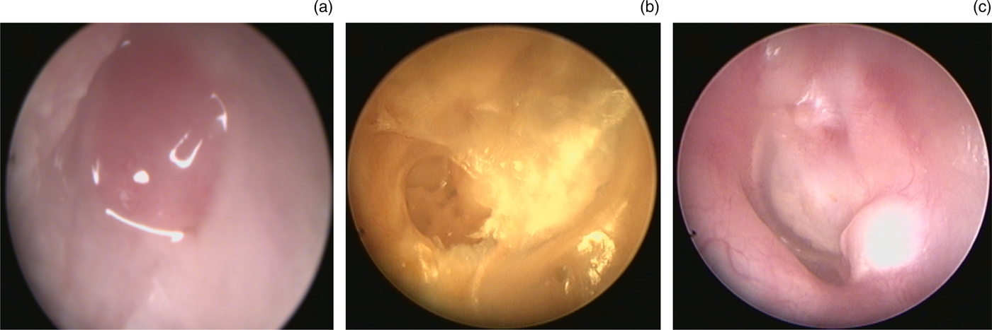
The 78 investigations were categorised into 4 groups, as described below, based on the stated reason for cholesteatoma being suspected, but not diagnosed (Figure 1). In one group, the patients had persistent granulation of the tympanic membrane, or polyposis of the tympanic membrane or external auditory canal, with suspected underlying cholesteatoma. A second group had tympanic membrane retraction with suspected cholesteatoma not visible on otomicroscopy or otoendoscopy. A third group had atypical tympanic membrane appearances, with possible implantation or congenital cholesteatoma. Finally, there was a miscellaneous group that did not fit the above classifications. This latter group included patients with concomitant external ear canal stenosis, those not compliant with microsuction clearance of ear canal debris, patients with incidental imaging findings thought to be suggestive of cholesteatoma, and those with persistently symptomatic ears with no evident disease.
Indications for requesting diffusion-weighted magnetic resonance imaging to aid diagnosis: (a) persistent granulation or, as in this case, aural polyps; (b) a deep focal retraction pocket; and (c) abnormal appearing tympanic membrane.

Imaging technique
Non-echo-planar diffusion-weighted imaging was performed using a half-Fourier single-shot turbo-spin echo (‘HASTE’) sequence. Magnetic resonance imaging was performed on a 1.5-T superconductive unit (Magnetom Avanto; Siemens Medical Solutions, Erlangen, Germany) using a standard Head Matrix coil. Coronal, 2 mm thick, turbo-spin echo, T2-weighted images (repetition time = 4640 ms; echo time = 103 ms; matrix = 245 × 384; field of view = 150 × 200 mm), and coronal, 2 mm thick, half-Fourier single-shot turbo-spin echo, diffusion-weighted images (repetition time = 1600 ms; echo time = 113 ms; matrix = 134 × 192; field of view = 220 × 220 mm; b-factors = 0 and 1000 seconds ⁄mm2) were acquired in all patients. An apparent diffusion co-efficient map was calculated using the diffusion scan raw data. All scans were reviewed by an experienced head and neck radiologist with an interest in middle-ear imaging (RKL).
Statistical analysis
The accuracy of diffusion-weighted MRI in aiding diagnosis was compared to its diagnostic performance in the context of planning surgery in patients with clinically evident primary cholesteatoma (72 cases). Fisher's exact test was applied using GraphPad statistical software (GraphPad Software, La Jolla, California, USA). This analysis was conducted to see if there was a significant difference in the accuracy of diffusion-weighted MRI between these clinical contexts. Accuracy was defined as (true positives + true negatives)/total number of patients with operative confirmation.
Results
Overall results
In the 44 cases that underwent operative confirmation, the accuracy of diffusion-weighted MRI was 63.6 per cent. Sensitivity was 55 per cent, specificity was 71 per cent, the positive predictive value was 61 per cent and the negative predictive value was 65 per cent.
Comparison with evident cholesteatoma
Our database included 72 cases of clinically evident primary cholesteatoma where diffusion-weighted MRI was requested to plan surgery. The demographics for this group were similar to those of the suspected cholesteatoma group; patient age ranged from 4.9 to 74.2 years (mean, 33.6 years). Cholesteatoma was evident in 68 cases, and there were 4 false negatives. It should be noted that the control sample was weighted towards more advanced disease, as the predominant role for diffusion-weighted MRI in these patients was staging disease in cases with total or subtotal opacification of the mastoid on CT. These data were nevertheless used to compare the diagnostic accuracy and sensitivity of diffusion-weighted MRI between suspected cholesteatoma and clinically evident cholesteatoma.
There was a significant difference in the diagnostic performance (sensitivity) of diffusion-weighted MRI between suspected and clinically evident cholesteatoma cases (p < 0.0001).
Results by indication
Diffusion-weighted MRI was most reliable in the context of persistent inflammatory tissue, where the accuracy was 80 per cent in 10 patients who underwent operative management. Diffusion-weighted MRI performed least well in tympanic membrane retractions, in which the accuracy was 45 per cent in 22 patients who underwent operative management. The diagnostic performance is shown by indication in Table I.
Diagnostic performance of diffusion-weighted MRI in assessment of suspected cholesteatoma

Data represent numbers, unless indicated otherwise. MRI = magnetic resonance imaging
Patients not undergoing surgery
Our series included a significant number of patients who had not undergone surgery (and are therefore not included in the statistical analysis). Whilst 26 of these patients’ scans had not shown cholesteatoma, 8 did demonstrate cholesteatoma. A proportion of these patients were, at the time of writing, awaiting surgical treatment, or had moved to a different area for treatment at another centre. There were, however, a small number of patients (n = 3) who were not offered operative management despite a scan demonstrating restricted diffusion. This was because the appearances and symptoms that had raised the suspicion of cholesteatoma resolved.
Discussion
Diagnostic performance
This series demonstrates a statistically significant difference between the sensitivity of diffusion-weighted MRI in the assessment of suspected and clinically evident cholesteatoma. This distinction is highly relevant. It is likely that a number of the factors that make it difficult to assess cholesteatoma in an out-patient setting also interfere with imaging assessment.
A greater bulk of cholesteatoma arising within a retraction may make it more likely for a cholesteatoma to be clinically evident and radiologically detectable. Cholesteatomatous, metaplastic retractions in their early stage will be limited by the resolution of diffusion-weighted MRI, which cannot detect lesions less than 2 mm in size.Reference Khemani, Singh, Lingam and Kalan 12 Furthermore, factors that can conceal the tympanic membrane and prevent clinical diagnosis may cause false positive signals. These include wax, and pus released from inflammatory tissue.
In addition, patients with retractions generally underwent CT imaging prior to diffusion-weighted MRI. It may be that those cases with large volumes of disease were detected with CT, and that diffusion-weighted MRI was reserved for those more indeterminate cases in which the CT did not provide a clear diagnosis.
Indeterminate results
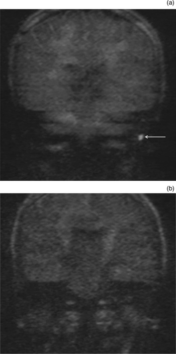
There are cases in which diffusion-weighted MRI results can be considered indeterminate; however, in our series, all scans were categorised as positive or negative. This allowed a clearer assessment of diagnostic performance. Nevertheless, we acknowledge that there are cases in which a radiologist may have to make a decision aided by clinical acumen. One case that demonstrates this point is shown in Figure 2. A patient with an epitympanic retraction had a solitary focus of restricted diffusion in the mastoid tip (Figure 2a). This was therefore ascribed to proteinaceous fluid, and was considered to be negative. Subsequent imaging showed resolution of this fluid (Figure 2b). Interpretation of the imaging and clinical context makes it clear that the signal does not represent primary cholesteatoma.
Coronal, b1000 diffusion-weighted magnetic resonance imaging requested to assess a retraction shows solitary restricted diffusion in the mastoid tip (a) (marked with arrow). Follow-up imaging demonstrates resolution of likely proteinaceous fluid (b).

Role of computed tomography and clinical judgement
Computed tomography is generally considered to be first line in the assessment of chronic ear disease. The limited diagnostic performance of diffusion-weighted MRI in this study supports this position, although there are cases when the ability of diffusion-weighted MRI to characterise soft tissue may be useful (Figure 3). There are cases in which these modalities are complementary. Computed tomography has high definition, and may demonstrate bony middle-ear anatomy very clearly. Diffusion-weighted MRI may then help characterise soft tissue. However, neither modality is infallible, and clinical assessment of the ear is crucial in determining management. There may also be a role for the fusion of diffusion-weighted MRI and CT images, as previously described.Reference Plouin-Gaudon, Bossard, Ayari-Khalfallah and Froehlich 13
Soft tissue in Prussak's space indicates cholesteatoma on computed tomography (CT) (a) (marked with arrow); however, diffusion-weighted magnetic resonance imaging (MRI) (correctly) excludes the condition (b) (coronal CT reformat shown with b1000 diffusion-weighted coronal MRI).

It is important to note that a retraction pocket which causes episodic otorrhoea may be appropriate for operative management, whether or not it is cholesteatomatous. The clinical context is crucial in both the interpretation of this imaging and the planning of treatment.
Implications of false positives and negatives
When assessing residual disease after canal wall preserving surgery, it is thought that the false negative findings is particularly relevant. This is because a ‘second-look’ operation is commonly considered both the gold standard management and a part of routine practice. Thus, a false positive finding would not mean that the patient has been harmed as a result of imaging, as the patient would receive ‘standard’ management, whilst a false negative finding may mean that the patient develops extensive residual disease. When diagnosing cholesteatoma pre-operatively, however, both false positive and false negative signals can be highly problematic. They may lead patients to undergo primary mastoid surgery unnecessarily, or falsely reassure patients and clinicians. Furthermore, it is likely that ‘missed’ primary cholesteatoma may be more problematic than focal ‘pearls’ of residual disease, the clinical significance of which has not been formally established. In this series, one patient with a retraction had their primary surgery delayed by almost a year as a result of diffusion-weighted MRI that did not initially show cholesteatoma (although CT was suggestive), but repeated imaging subsequently detected keratin accumulation (Figure 4).
In a patient with retraction, computed tomography (CT) suggests cholesteatoma (a). Initial diffusion-weighted magnetic resonance imaging (MRI) does not show restricted diffusion (b). At an interval of one year, follow-up diffusion-weighted MRI does show cholesteatoma (c) (marked with arrow) (coronal CT reformat shown with b1000 diffusion-weighted coronal MRI).

Awareness of the possibility of false positives is important in light of the literature published on the use of diffusion-weighted MRI in cholesteatoma assessment. As prior series have focused on evident disease, or possible residual disease or recurrence, only six patients who never had cholesteatoma have been previously reported.Reference Alvo, Garrido, Salas, Miranda, Stott and Delano 11 Our series highlights the presence of false positives in this patient group (Figure 5).
A patient with persistent discharging tympanic membrane granulation despite topical treatment had computed tomography (CT) findings which were reassuring (a), but diffusion-weighted magnetic resonance imaging (MRI) (b) indicated cholesteatoma. No keratin was found at the time of operation. Retained pus was the likely source of restricted diffusion signal (coronal CT reformat shown with b1000 diffusion-weighted coronal MRI).

Categorising indications
The division of the imaging indications into four groups could be considered subjective. For example, granulation and retractions commonly co-exist. However, the reason for suspecting the presence of cholesteatoma is usually clear. This paper specifies a number of distinct clinical scenarios in which diffusion-weighted MRI may play a role. The ‘miscellaneous’ group included a wide variety of patients, and should therefore be interpreted with caution. Assessment of retractions appears to rely on adequate pre-operative microsuction and a sufficient bulk of disease to allow detection. Persistent granulation may cause false positives if retained pus is present, as pus restricts diffusion. Whilst diffusion-weighted MRI may play a role in the rare patient with ear canal stenosis or aberrant imaging findings, its limited accuracy means that it should nevertheless be used with caution, and its use is best reserved to experienced clinicians.
Comparison with previous series
Our series shows significantly inferior diagnostic accuracy when compared to the previously published series, which included only retraction pockets (p < 0.01, Fisher's exact test).Reference Alvo, Garrido, Salas, Miranda, Stott and Delano 11 The reasons for this are not entirely clear. It may be that our patients had a smaller volume of disease, making them less amenable to detection by diffusion-weighted MRI.
-
• Diffusion-weighted magnetic resonance imaging (MRI) is highly diagnostic in detecting residual cholesteatoma after canal wall preserving surgery
-
• It is effective at staging the extent of clinically evident cholesteatoma when planning surgery
-
• Diffusion-weighted MRI is being more widely used in cholesteatoma assessment
-
• It has lower diagnostic accuracy at detecting cholesteatoma when the disease is not clinically evident
-
• Diagnostic performance was lowest in patients with retraction pockets that could not be fully visualised
-
• Care should be taken when using diffusion-weighted MRI to diagnose patients in these settings
An ear with a ball of keratin, with a minimum width of 3–4 mm, in the depths of an otherwise clean retraction is an uncommon phenomenon. It is clear there is a spectrum between the deep retraction pocket which collects wax or small amounts of keratin, inflamed or metaplastic epithelium, and the established ball of cholesteatoma. It may be that the classification of these as cholesteatoma or otherwise varied between our institutions.
Conclusion
Diffusion-weighted MRI has a significantly lower diagnostic accuracy in the assessment of suspected cholesteatoma than clinically evident disease. Computed tomography and diffusion-weighted MRI may be complementary in this context, but should be used with caution, and clinical judgement is paramount.