Interrupted aortic arch is a very rare congenital cardiovascular anomaly characterised by the presence of discontinuous segment between the ascending and descending aorta.Reference Reardon, Hallman and Cooley 1 It has been classified into three types depending on the level of interruption and is commonly associated with other cardiovascular abnormalities (seen in about 97% cases).Reference Reardon, Hallman and Cooley 1 , Reference Celoria and Patton 2 It is usually identified and treated in newborns. Rarely, interrupted aortic arch may go undiagnosed into adulthood.Reference Shirani and Soleymanzadeh 3 Although echocardiography, supplemented with MRI is routine in view of radiation exposure to child, electrocardiography-gated multidetector CT angiography can help in further delineating the anatomical details.Reference Shirani and Soleymanzadeh 3 Here, we report an unusual case of type A interrupted cervical aortic arch associated with long segment coarctation of the descending thoracic aorta diagnosed with the help of electrocardiography-gated multidetector CT angiography in a neonate.
Case report
A full-term female baby of 3 kg was delivered to a primiparous mother by emergency caesarean section due to fetal distress. The baby cried immediately after birth. On day 2 of life, the baby was found to have a systolic grade 3 murmur. Echocardiography revealed post-ductal long segment coarctation of the aorta, small mid-septum ventricular septal defect with left to right shunt, dilated right atrium and ventricle, and severe pulmonary arterial hypertension. Baby was on 100% moist oxygen support; however, on day 9 of life, she was referred to the tertiary care unit in gasping condition requiring intubation and mechanical ventilation. She had features of both cardiac and respiratory failure at that time, hepatomegaly and poor perfusion evidenced by mottling, delayed capillary filling, and metabolic acidosis. Differential pulses were noted between the two upper limbs.
Loss of pulsatility in the abdominal aorta was found on Doppler study, suggestive of proximal aortic occlusion. In view of deranged renal parameters, contrast magnetic resonance angiography was not carried out. Non-contrast cardiac MRI showed long segment interruption involving the distal part of the arch of the aorta and descending thoracic aorta. However, the detailed anatomy was not ascertained as it was a non-contrast scan.
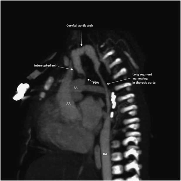
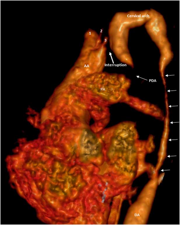
Finally, electrocardiography-gated multidetector CT angiography was done under light sedation for detailed vascular anatomy and revealed interruption of the aortic arch distal to the origin of the left subclavian artery (type A interrupted aortic arch) (Figs 1 and 2). The descending thoracic aorta was reconstituted by a narrow communication from the main pulmonary artery, suggestive of patent ductus arteriosus. Reconstituted arch, distal to the interrupted segment, was high up in location (cervical arch). A long segment narrowing was found in the descending thoracic aorta. The brachiocephalic artery was hypertrophied and the right subclavian artery was excessively tortuous. The left common carotid and left subclavian arteries were atretic at their origin and distal left subclavian artery was reconstituted by collateral from branches of right subclavian artery. The distal left common carotid artery was possibly filled retrograde from the Circle of Willis. A small ventricular septal defect was also noted. The abdominal aorta and its branches were normal.
Sagittal oblique view of multidetector computed tomographic angiography (MDCTA) image showing type A interrupted arch. Distal part of the arch is reconstituted by narrow PDA. A long segment narrowing is also seen in the descending thoracic aorta after a well developed cervical aortic arch. AA=ascending aorta; DA=descending aorta; PA=pulmonary artery; PDA=patent ductus arteriosus.

Volume-rendered multidetector computed tomographic angiography (MDCTA) image showing type A interrupted aortic arch along with the long segment narrowing in descending thoracic aorta (multiple arrows). Note the enlarged brachiocephalic trunk (1) along with atretic left common carotid artery (2) and left subclavian artery (3). Aortic arch is of cervical type and is reconstituted by PDA. AA=ascending aorta; DA=descending aorta; PA=pulmonary artery; PDA=patent ductus arteriosus.

The child was considered for surgical repair, which was refused by her parents. She was then managed with supportive care, inotropes, and prostaglandin infusion for maintaining ductal patency. Subsequently, she developed sepsis with deranged coagulation profile and renal failure. On day 26, her condition deteriorated with increasing ventilator requirement due to pulmonary oedema. Saturation and haemodynamics continued to deteriorate despite maximum inotropic support. Cardiopulmonary failure ensued and she expired on the next day.
Discussion
Interrupted aortic arch is a very rare congenital cardiovascular anomaly and is usually diagnosed by electrocardiography supplemented with MRI, in order to reduce the radiation dose. Anatomy differs in different cases and determines the surgical approach. Our case was an unusual case of type A interrupted aortic arch (cervical arch) and was associated with long segment coarctation of the descending thoracic aorta. To the best of our knowledge this combination has not been described in the literature. Another unique feature of our case was that, detailed anatomy was ascertained with the help of electrocardiography-gated multidetector CT angiography under light sedation. Multidetector CT angiography not only identified the type of aortic arch interruption but also delineated the exact anatomical details, which was not well delineated in echocardiography and non-contrast cardiac magnetic resonance.
Interrupted aortic arch was first described in year 1778. Celoria and PattonReference Celoria and Patton 2 classified interrupted aortic arch into three types (A, B, and C).Interrupted aortic arch is commonly associated with ventricular septal defect (90%) and patent ductus arteriosus (98%).Reference VanPraagh, Bernhard, Rosenthal, Parisi and Fyler 4 , Reference Ho, Wilcox, Anderson and Lincoln 5 Our case was a triad of interrupted arch, ventricular septal defect, and patent ductus arteriosus, as already described by Everts-Saucey and Carson;Reference Everts-Suacey and Carson 6 however, our case was unique having interrupted cervical aortic arch associated with long segment coarctation of the descending thoracic aorta.
Embryologically, a normal thoracic aorta develops from six aortic arches and seventh intersegmental arteries. The two dorsal aortae fuse in the midline to form a single descending aorta.Reference Reardon, Hallman and Cooley 1 Type A interrupted aortic arch develops due to delayed abnormal regression of left fourth arch after cranial migration of the left subclavian artery.Reference Reardon, Hallman and Cooley 1 Abnormal involution of the left fourth arch may be associated with failure of descent of the same, giving rise to association of multilevel coarctation and cervical aortic arch.Reference Kumar, Mandalam, Unni, Roy, Gupta and Rao 7
Electrocardiography-gated multidetector CT angiography not only identified the type of aortic arch interruption but also delineated the exact anatomical details. Our case demonstrates that electrocardiography-gated multidetector CT angiography can help in delineating complicated anatomy in an interrupted aortic arch in difficult to do situations. It does not require deep sedation and has better temporal and spatial resolution, less scan time, and is compatible with electrocardiographic gating; however, in most cases, echocardiography followed by magnetic resonance angiography is sufficient to make the diagnosis.
Conclusions
Cases of interrupted aortic arch have variable anatomy. Here, we describe a case of interrupted cervical aortic arch associated with long segment coarctation of the descending thoracic aorta. Electrocardiography-gated multidetector CT angiography can help in delineating complicated anatomy in interrupted aortic arch in difficult to do situations, even in neonates.
Acknowledgements
None.
Financial Support
This research received no specific grant from any funding agency, commercial, or not-for-profit sector.
Conflicts of Interest
None.
Ethical Standards
The authors assert that all procedures contributing to this work comply with the ethical standards of the relevant national guidelines on human experimentation ICMR and with the Helsinki Declaration of 1975, as revised in 2008, and has been approved by the Institutional ethics committee, SGPGIMS, Lucknow.
Supplementary material
To view supplementary material for this article, please visit https://doi.org/10.1017/S1047951117001913