Introduction
Callous-unemotional traits and the brain
Recent years have seen recognition and formalization of the idea that the presentation of callous-unemotional (CU) traits, or “limited prosocial emotions” as denoted in the fifth edition of Diagnostic and Statistical Manual of Mental Disorders (DSM-5; American Psychiatric Association, 2013), may delineate a specific subtype of youth prone to serious, persistent, and stable antisocial behavior (Frick, Cornell, Barry, Doug Bodin, & Dane, Reference Frick, Cornell, Barry, Doug Bodin and Dane2003; Frick, Ray, Thornton, & Kahn, Reference Frick, Ray, Thornton and Kahn2014; Frick, Stickle, Dandreaux, Farrell, & Kimonis, Reference Frick, Stickle, Dandreaux, Farrell and Kimonis2005; Kimonis, Kennealy, & Goulter, Reference Kimonis, Kennealy and Goulter2016). Clinically, CU traits include lack of remorse or guilt, callousness and lack of empathy, and lack of concern about performance in important activities (American Psychiatric Association, 2013). Evidence has pointed to both genetic and environmental (e.g., early deprivation, negative parenting) etiologies of CU traits (Humphreys et al., Reference Humphreys, McGoron, Sheridan, McLaughlin, Fox, Nelson and Zeanah2015; Hyde et al., Reference Hyde, Waller, Trentacosta, Shaw, Neiderhiser, Ganiban and Leve2016; Waller, Gardner, & Hyde, Reference Waller, Gardner and Hyde2013). A review of the literature covering treatment for disruptive behavior disorders (DBDs) has suggested that while CU traits may be associated with reduced treatment response, certain interventions in youth with CU traits can indeed lessen behavioral problems (Humphreys et al., Reference Humphreys, McGoron, Sheridan, McLaughlin, Fox, Nelson and Zeanah2015; Waller et al., Reference Waller, Gardner and Hyde2013; Wilkinson, Waller, & Viding, Reference Wilkinson, Waller and Viding2016). Scholars have suggested that improved understanding of these mechanisms may support early diagnosis and superior treatment efficacy, as specific interventions such as social-learning based parent training (Hawes, Price, & Dadds, Reference Hawes, Price and Dadds2014) could be tailored to address characteristic deficiencies.
A growing body of research (Crowe & Blair, Reference Crowe and Blair2008; Herpers, Scheepers, Bons, Buitelaar, & Rommelse, Reference Herpers, Scheepers, Bons, Buitelaar and Rommelse2014; Umbach, Berryessa, & Raine, Reference Umbach, Berryessa and Raine2015) has connected high levels of CU traits and its adult counterpart, psychopathy (Barry et al., Reference Barry, Frick, DeShazo, McCoy, Ellis and Loney2000), to specific behavioral and neurological abnormalities. Convergent findings from different methodologies (e.g., task-based functional magnetic resonance imaging [fMRI] studies, neurocognitive tasks, MRI) have most consistently implicated frontal, temporal, and limbic areas associated with emotion, moral, and reward processing (Baskin-Sommers, Neumann, Cope, & Kiehl, Reference Baskin-Sommers, Neumann, Cope and Kiehl2016; Cohn et al., Reference Cohn, Veltman, Pape, van Lith, Vermeiren, van den Brink and Popma2015b; Herpers et al., Reference Herpers, Scheepers, Bons, Buitelaar and Rommelse2014; Jones, Laurens, Herba, Barker, & Viding, Reference Jones, Laurens, Herba, Barker and Viding2009; Lozier, Cardinale, VanMeter, & Marsh, Reference Lozier, Cardinale, VanMeter and Marsh2014; Marsh et al., Reference Marsh, Finger, Fowler, Adalio, Jurkowitz, Schechter and Blair2013, Reference Marsh, Finger, Mitchell, Reid, Sims, Kosson and Blair2008; Pujol et al., Reference Pujol, Batalla, Contreras-Rodríguez, Harrison, Pera, Hernández-Ribas and Cardoner2012; Sebastian et al., Reference Sebastian, McCrory, Cecil, Lockwood, De Brito, Fontaine and Viding2012; Veroude et al., Reference Veroude, von Rhein, Chauvin, van Dongen, Mennes, Franke and Buitelaar2016; Viding et al., Reference Viding, Sebastian, Dadds, Lockwood, Cecil, De Brito and McCrory2012; White et al., Reference White, Marsh, Fowler, Schechter, Adalio, Pope and Blair2012). Additionally, there is some evidence of an association between gray matter volume differences in those same areas (e.g., frontal cortex, amygdala, insula, striatum) and CU traits in children and adolescents (Bolhuis et al., Reference Bolhuis, Viding, Muetzel, El Marroun, Kocevska, White and Cecil2019; Cardinale et al., Reference Cardinale, O'Connell, Robertson, Meena, Breeden, Lozier and Marsh2019; Cohn et al., Reference Cohn, Veltman, Pape, van Lith, Vermeiren, van den Brink and Popma2015b; Cohn et al., Reference Cohn, Viding, McCrory, Pape, van den Brink, Doreleijers and Popma2016; Raschle et al., Reference Raschle, Menks, Fehlbaum, Steppan, Smaragdi, Gonzalez-Madruga and Stadler2018; Rogers & De Brito, Reference Rogers and De Brito2016; Sebastian et al., Reference Sebastian, De Brito, McCrory, Hyde, Lockwood, Cecil and Viding2016; Wallace et al., Reference Wallace, White, Robustelli, Sinclair, Hwang, Martin and Blair2014). From a developmental standpoint, it is hypothesized that these neurobiological profiles may result in deficient learning of empathy and morality, which in turn leads to higher rates of externalizing behavior and aggression, particularly proactive or instrumental aggression (Blair, Reference Blair2017; Fanti, Frick, & Georgiou, Reference Fanti, Frick and Georgiou2009; Kochanska, Gross, Lin, & Nichols, Reference Kochanska, Gross, Lin and Nichols2002; Waller & Hyde, Reference Waller and Hyde2018). These studies have primarily been conducted in clinical and forensic populations, but considering that some degree of CU behaviors exist in many healthy persons, it seems plausible that the neurobiological characteristics of these behaviors can also be found (albeit at lower levels) in healthy populations. In recent years, and with the adoption of the research domain criteria (RDoC) framework, which integrates multiple levels of information to investigate behaviors, psychiatric symptoms are increasingly conceptualized as disorders of neural circuits that exist on a continuum from typical to pathological (Cuthbert & Insel, Reference Cuthbert and Insel2013).
Resting state network connectivity
From this RDoC perspective, this study acknowledges that psychopathologies exist on a spectrum of functionality, and many disparate cognitive processes can be implicated in symptomatology. This “psychopathology as a spectrum” perspective is supported by a growing body of research finding associations between a variety of psychopathologies and abnormal resting state connectivity in specific networks, even within this healthy sample, including anhedonia (Pornpattananangkul, Leibenluft, Pine, & Stringaris, Reference Pornpattananangkul, Leibenluft, Pine and Stringaris2019) and psychotic-like experiences (Karcher, O'Brien, Kandala, & Barch, Reference Karcher, O'Brien, Kandala and Barch2019). To date, it is an open question whether this is the case with CU traits. There is a dearth of literature surrounding potential associations between resting state connectivity and CU traits in community-based children. Nevertheless, adult literature and a few adolescent studies provide reasons to hypothesize a priori that reduced activation in the default mode network (DMN) specifically, may be associated with higher levels of CU traits. Empirical support for this hypothesis would contribute to the growing support for the hypothesis that CU traits also fit into the RDoCs continuum framework.
Activation in the DMN, which consists primarily of the precuneus, posterior cingulate cortex, inferior parietal cortices, and the medial prefrontal cortex (Wade et al., Reference Wade, Prime, Jenkins, Yeates, Williams and Lee2018), is more active during rest periods than when a subject is engaged in goal-directed behavior that demand attention to external stimuli. The DMN has been linked to Theory of Mind (Saxe, Carey, & Kanwisher, Reference Saxe, Carey and Kanwisher2004), moral decision-making (Reniers et al., Reference Reniers, Corcoran, Völlm, Mashru, Howard and Liddle2012), personal introspection, self-referential thinking, perspective taking, and emotional processing (Andrews-Hanna, Reference Andrews-Hanna2012; Buckner & Carroll, Reference Buckner and Carroll2007; Harrison et al., Reference Harrison, Pujol, Lopez-Sola, Hernandez-Ribas, Deus, Ortiz and Cardoner2008; Mak et al., Reference Mak, Minuzzi, MacQueen, Hall, Kennedy and Milev2017; Pujol et al., Reference Pujol, Batalla, Contreras-Rodríguez, Harrison, Pera, Hernández-Ribas and Cardoner2012; Raichle, Reference Raichle2015). These functions make the DMN a good candidate for the neural substrates of the CU traits. Longitudinal and developmental literature have identified the DMN in children as young as five years old (de Bie et al., Reference de Bie, Boersma, Adriaanse, Veltman, Wink, Roosendaal and Sanz-Arigita2012), and suggested that the functional architecture of the DMN resembles that of mature adults by the age of 10 (Sherman et al., Reference Sherman, Rudie, Pfeifer, Masten, McNealy and Dapretto2014; Supekar et al., Reference Supekar, Uddin, Prater, Amin, Greicius and Menon2010). The connectivity, however, appears to strengthen internally with age, particularly between the medial prefrontal cortex and posterior cingulate cortex (Sherman et al., Reference Sherman, Rudie, Pfeifer, Masten, McNealy and Dapretto2014; Supekar et al., Reference Supekar, Uddin, Prater, Amin, Greicius and Menon2010). In clinical and forensic samples, hypoactivity in this network has been found to be negatively associated with antisocial psychopathologies, including CU traits (Philippi et al., Reference Philippi, Pujara, Motzkin, Newman, Kiehl and Koenigs2015; Thijssen & Kiehl, Reference Thijssen and Kiehl2017), as well as with conduct disorder (CD) more generally (Broulidakis et al., Reference Broulidakis, Fairchild, Sully, Blumensath, Darekar and Sonuga-Barke2016; Lu et al., Reference Lu, Zhou, Zhang, Xiang, Zhang, Liu and Yuan2015; Zhou et al., Reference Zhou, Yao, Fairchild, Cao, Zhang, Xiang and Wang2016).
Default mode network and CU traits
Prior studies have used clinical and forensic samples of antisocial and CU adolescents to identify the DMN as a likely network of interest. In adolescents, Broulidakis et al. (Reference Broulidakis, Fairchild, Sully, Blumensath, Darekar and Sonuga-Barke2016) examined 56 male adolescents aged 13–18 years, half of whom presented with CD. They found lower connectivity between specific areas implicated in the DMN in adolescents diagnosed with CD as compared to control adolescents after controlling for attention-deficit hyperactivity disorder (ADHD). Within the CD group, there was no association between scores on the Youth Psychopathic Inventory (YPI) and connectivity in the DMN. Other studies have found the same, but without accounting for potential confounding effects of CU traits (Lu et al., Reference Lu, Zhou, Zhang, Xiang, Zhang, Liu and Yuan2015; Zhou et al., Reference Zhou, Yao, Fairchild, Cao, Zhang, Xiang and Wang2016). Within an antisocial incarcerated adolescent sample, Thijssen and Kiehl (Reference Thijssen and Kiehl2017) found a negative relationship between DMN connectivity and psychopathic traits. One contradictory finding by Cohn et al. (Reference Cohn, Pape, Schmaal, van den Brink, van Wingen, Vermeiren and Popma2015a) is a positive association between connectivity in the anterior DMN in the lateral frontopolar cortex and scores on the CU subscale of the YPI, in a juvenile arrestee cohort aged 12–20 years. They hypothesize that this may be a result of hyperconnectivity within two localized areas involved in the DMN, as opposed to more widespread hyperconnectivity. Importantly, these findings have been based on studies using relatively small (typically male) samples drawn from clinical or forensic populations that tend to be limited in their ability to control for co-occurring externalizing problems and CD, symptomology which may also contribute to abnormal resting state network (RSN) connectivity.
Findings in studies of adults involving psychopathy broadly parallel those of the adolescent literature. Studies typically examine three outcomes with regard to psychopathy: Factor 1 scores, which capture interpersonal affective deficits; Factor 2 scores, which include antisocial lifestyle characteristics; and an overall total score combining the two scores. Consistent with the CU literature, Factor 1 scores are most commonly negatively associated with connectivity between parts of the DMN (Motzkin, Newman, Kiehl, & Koenigs, Reference Motzkin, Newman, Kiehl and Koenigs2011; Philippi et al., Reference Philippi, Pujara, Motzkin, Newman, Kiehl and Koenigs2015; Pujol et al., Reference Pujol, Batalla, Contreras-Rodríguez, Harrison, Pera, Hernández-Ribas and Cardoner2012; Reniers et al., Reference Reniers, Corcoran, Völlm, Mashru, Howard and Liddle2012). One notably large study (n = 985; Espinoza et al., Reference Espinoza, Vergara, Reyes, Anderson, Harenski, Decety and Calhoun2018) of incarcerated men found associations between Factor 1 scores and abnormal connectivity patterns in a number of networks, including reduced within-network connectivity in the DMN.
It is important to note that this research often recruits individuals with prior criminal justice system involvement, or participants diagnosed with DBDs Because of the cost and logistical demands of neuroimaging studies, many studies are relatively small in size. Thus, detection of effects may necessitate studying relatively homogenous samples of significantly antisocial participants (e.g., clinically referred or individuals involved in criminal justice). Unfortunately, these participants are also more likely than community-based healthy populations to have experienced childhood trauma (Cuomo, Sarchiapone, Di Giannantonio, Mancini, & Roy, Reference Cuomo, Sarchiapone, Di Giannantonio, Mancini and Roy2008; Weeks & Widom, Reference Weeks and Widom1998), traumatic brain injury (Farrer & Hedges, Reference Farrer and Hedges2011; Shiroma, Ferguson, & Pickelsimer, Reference Shiroma, Ferguson and Pickelsimer2010), and other negative environmental risk factors. All of these may also influence brain activity and integrity, providing a number of complicating limitations and potential confounding variables. Because maladaptive behaviors exist on a spectrum, it would be beneficial to explore whether cognitive or neural substrates of CU behavior can also be identified in healthy individuals recruited from the community who have not been selected for problem behaviors. Doing so may reduce some of the uncertainty and endogeneity around causes of both the behavior and the associated brain correlates.
This study
The gap in the literature that this study aims to address is whether an association exists between nonclinical levels of CU traits and resting state brain network connectivity in community-based children. Few previous studies have examined the association between callous unemotional traits and resting state MRI (rs-fMRI). Those that have been completed have used adult and/or clinical or forensic samples (e.g., Broulidakis et al., Reference Broulidakis, Fairchild, Sully, Blumensath, Darekar and Sonuga-Barke2016; Cohn et al., Reference Cohn, Pape, Schmaal, van den Brink, van Wingen, Vermeiren and Popma2015a). To date, there are no data about CU traits in children and resting state connectivity, which in young children has been shown to have high test–retest reliability compared to task-based fMRI (Song et al., Reference Song, Desphande, Meier, Tudorascu, Vergun, Nair and Prabhakaran2012; Thomason et al., Reference Thomason, Dennis, Joshi, Joshi, Dinov, Chang and Gotlib2011). Because CU traits are thought to be relatively stable even in nonreferred youth (Frick, Kimonis, Dandreaux, & Farell, Reference Frick, Kimonis, Dandreaux and Farell2003; Loney, Taylor, Butler, & Iacono, Reference Loney, Taylor, Butler and Iacono2007), this study will test whether the findings in older samples can be extended downward. This study is of particular note because of the age of the participants, community-based nature of the recruitment process, and the size of the sample, which allows for more stringent sensitivity checks and to detect even small brain-behavior relationships. Depending on the results, we can provide more evidence for potential neural substrates of CU behavior, and findings are less limited by potential confounders often found in more rarified populations.
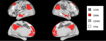
Moreover, this study aims to provide a map of considerations for future researchers to consider when looking at the relationship between these traits and resting state connectivity in community children. The previous literature provides reason to hypothesize a negative association between CU traits and connectivity of the DMN specifically, as shown in Figure 1, but this study will take a conservative approach in assessing all eight networks that could plausibly be involved. This exploratory approach is also in acknowledgement of previous studies that have implicated additional networks in emotional processing deficits (Malhi, Das, Outhred, Bryant, & Calhoun, Reference Malhi, Das, Outhred, Bryant and Calhoun2019).
Default mode network (DMN) connectivity in the ABCD study. Adapted from Biological Psychiatry, 86/1, Karcher, N. R., O'Brien, K. J., Kandala, S., & Barch, D. M. Resting State Functional Connectivity and Psychotic-Like Experiences in Childhood: Results from the Adolescent Brain Cognitive Development Study, Pages No. 7–15, Copyright (2019), with permission from Elsevier. Note: Other major networks are depicted in gray, in order of the legend: cingulo-opercular network (CON), cingulo-parietal network (CPAR), fronto-parietal network (FPN).

Materials and methods
Data
This study used preprocessed, precomputed data from the Adolescent Brain Cognitive Development (ABCD) study curated annual release 2.0, containing baseline data from 11,875 children aged 9–11 years (47.8% F/52.2% M). Goals of this longitudinal National Institute of Health (NIH)-funded study include the recruitment and retention of a representative national sample of 12,000 healthy children; full recruitment details can be found in Garavan et al. (Reference Garavan, Bartsch, Conway, Decastro, Goldstein, Heeringa and Zahs2018). The long-term aim is to understand the impact of genes and environment on brain development, structure, and function, as well as their role in substance use. Data were collected at 22Footnote 1 sites across the United States; sites were chosen based on the requirement that selected sites have the research expertise and neuroimaging equipment necessary for adherence to the protocol. The human research protection programs and institutional review boards at all universities participating in the ABCD project approved all experimental and consenting procedures. While recruitment is delegated to the individual sites, coordination of the study and storage and analysis of the data are centralized at the University of California, San Diego. This centralization helps ensure consistent quality checks across the data. Despite maintaining equivalent protocols, there are correlations between resting state connectivity data and the scanner type, thus we follow the precedent set by earlier papers (e.g., Karcher et al., Reference Karcher, O'Brien, Kandala and Barch2019) using these resting state data and include scanner as a covariate.
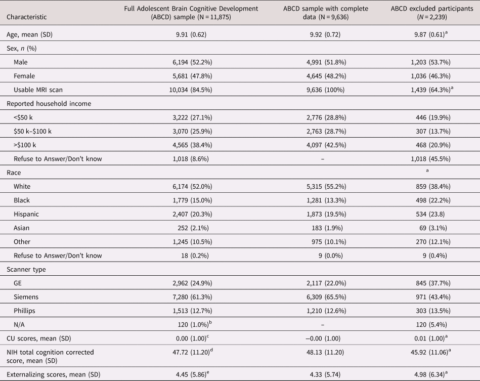
In keeping with prior studies using this sample (Karcher et al., Reference Karcher, O'Brien, Kandala and Barch2019; Paulus et al., Reference Paulus, Squeglia, Bagot, Jacobus, Kuplicki, Breslin and Tapert2019), participants missing data on any of the variables of interest were excluded. As an example, in the primary analysis, of the 11,875 children, participants were eliminated from analyses in the following order: 63 were missing data on questionnaire items related to CU traits, an additional 116 participants never underwent an MRI, 678 participants did not pass ABCD's overall MRI quality control check (Hagler et al., Reference Hagler, Hatton, Makowski, Cornejo, Fair, Steven Dick and Xue2018), and 919 participants were eliminated because income was not reported by parents. An additional 463 participants were eliminated because they were missing a mean motion calculation for the rs-fMRI data. After this elimination process, the final sample size was 9,636. In each individual analysis, additional participants were eliminated based on presenting as outliers in that particular network (described in more detail in the analysis plan section) or if they were missing data on the added covariates (e.g., externalizing problems, NIH toolbox score). Demographics of the full sample, the excluded participants, and the main analysis sample after elimination for missing data can be found in Table 1. Independent sample t tests and chi-squared tests with Pearson residuals were conducted for appropriate variables to compare excluded and included participants. Included participants were more likely to be white, not be black, be slightly older, have higher cognition scores, and have lower externalizing scores than excluded participants. However, we do note a consequence of the large sample sizes are small standard errors, hence even small differences register as significant.
Demographics

Notes: aIndicates significant differences between included and excluded participants. Analyses not conducted for variables where missing data acted as an exclusionary variable. Fisher's exact test was conducted on the race/ethnicity proportions, and indicated a significant difference.
b Only those subjects who did not undergo the MRI (n = 120) are missing data for scanner type.
c Excluding the 63 individuals with missing data for this score.
d Excluding the 1,026 individuals with missing data for this score.
e Excluding the 11 individuals with missing data for this score.
Resting state functional magnetic resonance imaging data
All neuroimaging data were collected on a 3 T scanner (either Siemens, General Electric, or Phillips) with a 32-channel head coil and completed T1-weighted (T1w) and T2-weighted (T2w) structural scans (0.7 mm isotropic). Full details of the acquisition and preprocessing were described elsewhere (Casey et al., Reference Casey, Cannonier, Conley, Cohen, Barch, Heitzeg and Dale2018; Hagler et al., Reference Hagler, Hatton, Makowski, Cornejo, Fair, Steven Dick and Xue2018) but briefly, over the course of multiple scans, the ABCD Study collected T1w and T2w structural MRI (sMRI), diffusion MRI (dMRI), and functional MRI, including both resting-state fMRI and task-fMRI (Casey et al., Reference Casey, Cannonier, Conley, Cohen, Barch, Heitzeg and Dale2018). Because of the age of the participants and the length of the scan, real-time motion correction and monitoring is used, which allows the operator to know when the required amount of frames with the ABCD movement criterion (>12.5 min of data with framewise displacement (FD (Power et al., Reference Power, Mitra, Laumann, Snyder, Schlaggar and Petersen2014) <0.2 mm)) has been achieved. Despite these state-of-the-art techniques, the age of the subjects and the length of the scan resulted in some scans failing the quality control assessment. Those subjects were excluded from this analysis.
During rs-fMRI data collection the participants viewed a crosshair for 20 min total (four 5-min sessions with breaks in between). Resting state images were obtained in the axial plane using an echo planar imaging (EPI) sequence; the oblique EPI prescriptions are not recommended or supported by General Electric and Phillips. Connectivity strength during rs-fMRI was calculated through a seed-based correlational approach on parcellated regions. Correlations were calculated within and between pre-defined cortical networks (n = 13, default mode, auditory, cingulo-opercular, retrosplenial-temporal, salience, somatomotor hand, somatomotor mouth, ventral attention, dorsal attention, fronto-parietal, visual, cingulo-parietal, and “none”, which includes any remaining regions) between each network and each subcortical region of interest (ROI), and within each subcortical ROIs. Fisher r to z transformation of the mean correlations between pairs of regions within each large-scale network were conducted, resulting in 13 measures of with-network connectivity. Five networks were excluded a priori (e.g., somatomotor hand, visual). While the DMN was identified as the most likely network candidate, considering the relatively dearth of literature in this space, this study took an exploratory approach and examined within-network connectivity of the remaining eight networks. Visual depictions of four (cingulo-opercular, default mode, fronto-parietal, and cingulo-parietal) of the eight networks examined in this sample can be found in Figure 1, with an emphasis on the DMN specifically.
Measures
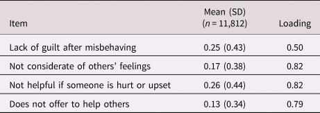
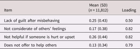
This study utilized a CU measure previously derived in this sample by Hawes et al. (Reference Hawes, Waller, Thompson, Hyde, Byrd, Burt and Gonzalez2020). Full details can be found therein, but briefly, items from the Child Behavior Checklist (CBCL; Achenbach, Reference Achenbach1991) and the Strengths and Difficulties Questionnaire (SDQ; Goodman, Reference Goodman1997) were initially selected based on their connection to theoretical conceptualizations of CU traits and similarity to items from other scales (e.g., Inventory of Callous-Unemotional Traits; Kimonis et al., Reference Kimonis, Frick, Skeem, Marsee, Cruise, Munoz and Morris2008). Both instruments ask parents to answer questions about their child according to a three-level Likert scale. The SDQ items were reverse coded such that higher scores represented more CU traits. Due to the distribution of the data, all items were dichotomously recoded so that any endorsement (1 or 2) was recoded to 1. After screening items out based on low internal consistency and low rates of endorsement, the final four item scale (consisting of one item from the CBCL and three from the SDQ) had good psychometric properties, was validated in an independent sample (youth from the on-going Michigan Twin Neurogenetics Study) through comparisons to self-report and parent scores on the Inventory of Callous-Unemotional traits, and had appropriate associations with oft-comorbid outcomes as measured by the CBCL (e.g., Conduct Problems, ADHD behaviors). The four items identified by Hawes et al. (Reference Hawes, Waller, Thompson, Hyde, Byrd, Burt and Gonzalez2020) were submitted to a principal component analysis. Only one component had an eigenvalue >1 (2.21). Item loadings for the single resulting component were between .49 and .83, and accounted for 55% of the variance (see Table 2). Participant scores for this component were saved and retained as a measure of CU scores. As the majority of subject scores on the initial scale were 0 (max = 4), the CU variable was highly skewed (1.33), and the kurtosis was 3.48. Principal components analysis requires complete data, thus participants missing data for any of the four items were eliminated from the main analyses.
Loadings and descriptive statistics of callous-unemotional items
Covariates
Mean-centered age, sex, mean motion during the rs-fMRI sessions, parental reported household income (categorized as <50 k, >50 k and <100 k, and >100 k), and scanner type were included as covariates in all analyses.
Robustness analyses
To further test this hypothesis, several robustness checks were conducted. The externalizing subscale from the CBCL (Achenbach, Reference Achenbach1991) was included as a covariate in one analysis, to test whether any found association was the result of a more generalized relationship between externalizing behavior and the DMN, or was specific to CU traits.
The participants were administered the NIH toolbox cognitive function battery, which provides a number of individual scores, as well as three composite scores (i.e., a fluid cognition composite, a crystallized cognition composite, and a total composite score). The fully corrected t scores for the total composite score were used as an additional control in another robustness check, to test whether findings remain after controlling for total cognition.
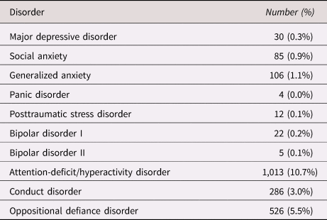
Parents were also administered the Kiddie Schedule for Affective Disorders and Schizophrenia (KSADS-5) through an interview. The KSADS-5 provides present and past diagnoses of a range of psychiatric disorders. A robustness analysis excluded any participants whose parents had not completed the entire KSADS-5 (n = 142) as well as participants with a current diagnosis of a DBD, bipolar disorder, generalized anxiety disorder, social anxiety disorder, major depressive disorder, posttraumatic stress disorder, panic disorder, or attention deficit hyperactivity disorder according to the KSADS. The final sample size for this robustness analysis was 8,001, before exclusion based on MRI quality control or outlier status. Distribution of these disorders can be found in Table 3.
Kiddie Schedule for Affective Disorders and Schizophrenia (KSADS) disorders (n = 9,494)

Analysis plan
Of the 13 networks, five were eliminated a priori as networks of no interest: somatomotor mouth, somatomotor hand, auditory, visual, and none. Mixed effects models were conducted in R lme4 package using Sattherthwaite approximate degrees of freedom (Bates, Mächler, Bolker, & Walker, Reference Bates, Mächler, Bolker and Walker2015). Family unit (to account for sibling participants) and the 22 research sites were modeled as random intercepts. Age, sex, motion, scanner type, and household income included as fixed effect covariates. Network connectivity score was the dependent variable in all analyses. The R package lmerTest (Kuznetsova, Brockhoff, & Christensen, Reference Kuznetsova, Brockhoff and Christensen2017) was used to derive the appropriate p values, which were then corrected for multiple comparisons using false discovery rate (FDR), and 95% confidence intervals were calculated using bootstrapping resampling techniques (via the percentile interval with 5,000 resamples). Following convention, an effect was considered significant if the 95% CI did not include zero. As in prior studies using these data (Pornpattananangkul et al., Reference Pornpattananangkul, Leibenluft, Pine and Stringaris2019), before each analysis, the distribution of correlation values for the network of interest were inspected and values that deviated over 1.5 interquartile ranges above the upper quartile or below the lower quartile were eliminated as outliers.
In addition to these primary analyses, significant models were run a second time controlling for externalizing behavior as measured by the CBCL, to determine whether relationships between network connectivity and CU traits were an artifact of comorbid externalizing behavior, or were independently related. Similarly, models were rerun controlling for the NIH toolbox total cognition score. These secondary analyses were also subjected to FDR p value corrections, which are reported in the text where relevant.
A final robustness analysis involved rerunning the DMN model using a subset of children absent of a current diagnosis of specific common psychiatric disorders based on the KSADS interview of the parents. As one goal of this study was to test whether relationships found in clinical/forensic samples can be detected in community-based samples, this comparison is very strict in restricting the analysis to children both community-based and free of psychopathology.
Results
Descriptive statistics and correlations
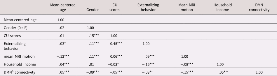
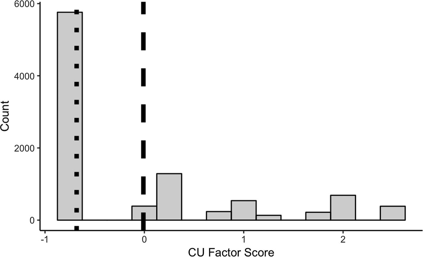
Of the participants with complete data (n = 9,639), the sample was relatively evenly split on gender (52.2% M/ 47.8% F). The mean age of the participants was 9.92 (SD = 0.62). Consistent with the representative goals of recruitment, the majority of participants were Caucasian (55.2%), followed by Hispanic (19.5%). Of those reporting, the majority of families reported a yearly income of less than $100,000. As seen in Table 4, bivariate correlations were performed on the demographic variables. As necessary, factor variables (i.e., household income) were recoded to continuous variables. DMN connectivity was positively associated with age, and higher socioeconomic status (SES), and negatively associated with being male, externalizing behavior, CU traits, and motion within the scanner. Higher CU scores were positively associated with being male, higher externalizing behavior, less DMN connectivity, and more average motion within the scanner. Of the children with CU factor scores (n = 9,636), 4.0% had the highest possible score (2.62), and 59.8% had the lowest possible score (−0.68). The frequency of these disorders can be seen in Figure 2.
Correlations of covariates and variables of interest

Notes: aDefault mode network.
* p < .05, ** p < .01, ***p < .001.
Distribution of callous-unemotional (CU) factor scores in the sample with complete data (n = 9,636). Note: The dotted line represents the median of the scores, the dashed line is the mean of the scores.

Primary analyses
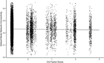
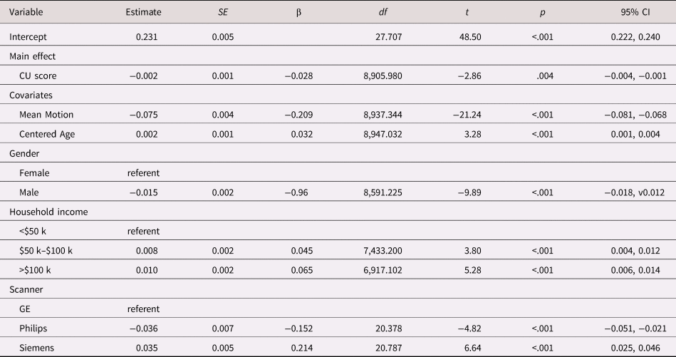
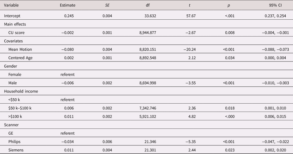
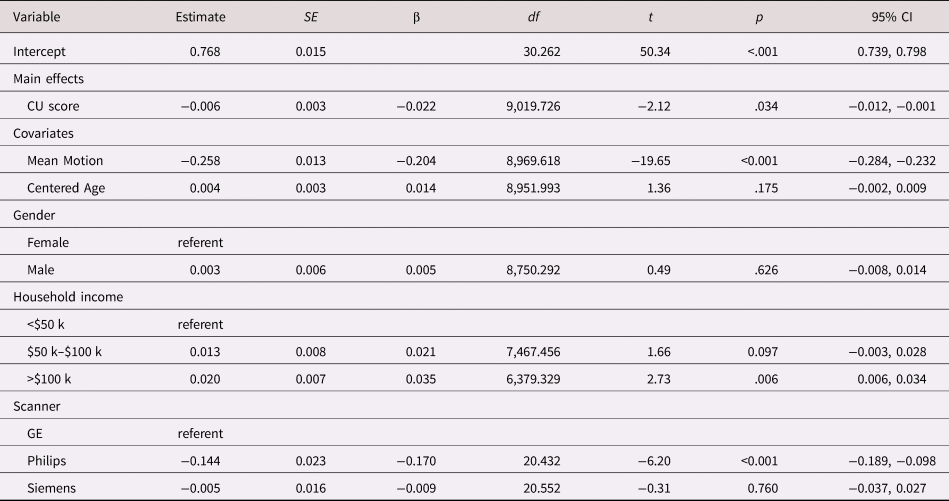
Of the eight brain networks tested, three were significantly associated with CU scores (model coefficients show in Tables 5–7). CU scores were negatively associated with DMN connectivity (b = −0.002 p = .004, 95% CI [−.004, −.001]; seen in Figure 3), dorsal attention network connectivity (b = −0.002, p = .008, 95% CI [−.004, −.001]), and cingulo-parietal network connectivity (b = −0.006, p = .034, 95% CI [−0.012, −0.001]). These significant associations were mildly attenuated after FDR corrections for eight analyses (DMN and dorsal attention network ps = .031), with the cingulo-parietal network association being reduced to trending significance (p = .090). CU scores were not associated with any of the other five networks of interest, even before FDR corrections (all ps > .497, uncorrected). Results of the regressions for the insignificant networks can be found in Supplementary Tables 1–5.
Default mode connectivity as a function of callous-unemotional (CU) factor scores after controlling for covariates (n = 8,984). Note: rs-fMRI DMN connectivity was calculated using Fisher r to z transformation of the mean correlations between pairs of regions within the default mode network (DmN). The gray dotted line is the slope of the regression of CU scores on DMN connectivity values. The gray area surrounding the line is the 95% confidence interval.

Default mode connectivity model (n = 8,984)

Note: Estimate unstandardized beta, SE standard error, β standardized beta, df Satterthwaite approximate degrees of freedom.
Dorsal attention network model (n = 8,996)

Note: Estimate Unstandardized beta; SE standard error, df Satterthwaite approximate degrees of freedom.
Cingulo-parietal network model (n = 9,059)

Note: Estimate unstandardized beta, SE standard error, β standardized beta, df Satterthwaite approximate degrees of freedom.
Externalizing behavior
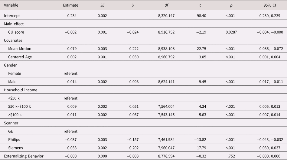
The two models that survived p value correction were tested further to see whether the associations remained even after controlling for scores on the externalizing subscale of the CBCL. The association between DMN and CU traits remained, although the strength was weakened to trending significance after p value corrections for two tests (b = −0.002, p = .029, 95% CI [−.004, −.000], p corrected = .057), and externalizing behavior was no longer significantly related to DMN connectivity (b = −0.000, p = .752, 95% CI [−.000, .000]). Full results of the model can be found in Table 8. In contrast, the addition of externalizing behavior completely eliminated the association between CU and dorsal attention network connectivity (b = −0.001, p = .282, 95% CI [−.003, .001]), while externalizing behavior was significantly and negatively associated with dorsal attention network connectivity (b = −0.001, p = .002, 95% CI [−.001, −.000]). Results of this model can be found in Supplementary Table 6.
Default mode network model with externalizing behavior covariate (n = 8,980)

Note: Estimate unstandardized beta, SE standard error, β standardized beta, df Satterthwaite approximate degrees of freedom.
Total cognition
The two models that survived p value correction were rerun controlling for fully corrected t scores on the NIH toolbox total composite score of overall cognition, which was the ABCD study's measure of intelligence. The association between DMN and CU traits remained (b = −0.002, p = .025, 95% CI [−.003, −.000], p corrected = .026), and cognition was not significantly related to DMN connectivity (b = 0.000, p = .406, 95% CI [−.000, .000]). Full results of the model can be found in Supplementary Table 7. Similarly, cognition had no effect on the relationship between CU and dorsal attention network connectivity (b = −0.002, p = .027, 95% CI [−.004, −.001], ], p corrected = .026), and it was not negatively associated with dorsal attention network connectivity (b = 0.000, p = .127, 95% CI [−.001, −.000]). Results of this model can be found in Supplementary Table 8.
Moderators
Interactions between connectivity and sex and age were tested in the default model. There was no moderating effect of sex (b = −0.000, p = .901, 95% CI[−.003, .003]), or age (b = −0.001, p = .230, 95% CI [−.002, .001]). FDR corrections were not performed on these insignificant results, model coefficients are shown in Supplementary Tables 9 and 10.
Clinical diagnoses
As an additional analysis, children with complete data but who presented with a current diagnosis were excluded (n = 1,635). Participants were also excluded if their parents had not completed the KSADS (n = 142). The analysis for the DMN was then rerun in this healthy sample. Participants continued to be excluded based on outlier status in each analysis. In this smaller sample (n = 7, 506), CU traits were no longer predictive of resting state connectivity (b = 0.001, p = .135, 95% CI [−0.000, 0.003]), and indeed went in the opposite direction. Full results of this model can be found in Supplementary Table 11. This additional analysis and the overlapping nature of symptomologies within psychopathology suggests that the DMN–CU trait relationship may also be dependent on comorbid psychopathology within this sample.
Discussion
Higher levels of CU traits were associated with lower connectivity in the DMN, and this finding seems to be specific to CU and not a result of comorbid externalizing behavior. Externalizing behavior, not CU, was related to reduced connectivity in the dorsal attention network; a relationship that was eliminated after exclusion of participants with active psychopathologies. The association between the DMN and CU was unaffected by age (albeit in a very narrow age range), or sex, but was eliminated after the removal of children with a current psychiatric disorder from the analysis. This could indicate that children with significant CU traits may also exhibit comorbid psychopathologies that complicate this DMN–CU relationship; this is in keeping with research that has connected CU symptoms with conduct problems, hyperactivity, and emotional problems associated with more internalizing problems (e.g., Saunders et al., Reference Saunders, Anckarsäter, Lundström, Hellner, Lichtenstein and Fontaine2019).
The findings in the DMN are consistent with prior literature in clinical and forensic adolescents and with adults with psychopathy, which suggests that the relationship between deficient affective processing and the DMN is not a false finding or an artifact of sampling bias. The relatively small size of the effect also suggests that this relationship likely exists on a spectrum. It seems likely that the exceptionally large sample allowed us to detect an effect that has presented more strongly in clinical, forensic samples, and older samples. This idea is also supported by the null findings in the clinical diagnoses secondary analyses. Future research on CU traits may consider how CU traits may play a role in disorders other than DBD.
Identification of the default network as a potential neural substrate of CU traits in childhood, even if in the normative range, supports prior neural findings suggesting that CU traits may characterize youth with impaired experiences of guilt, remorse, and empathy; all constructs necessary for the development of interpersonal prosocial behavior. The presence of these neural correlates in healthy, community-based children speaks to the importance of incorporating an RDoC perspective in antisocial behavior research, and further supports the idea of CU traits as a continuum, rather than a set of present or absent symptoms. The results are consistent with a true association between CU traits and the DMN, not one confounded by comorbid psychopathology. However, in keeping with the RDoC perspective, it is possible that decreased DMN connectivity during resting state is not unique to CU traits. In fact, ADHD, often comorbid with CU traits, has been associated with weaker connectivity between some of the regions existing within the DMN (Castellanos et al., Reference Castellanos, Margulies, Kelly, Uddin, Ghaffari, Kirsch and Milham2008; Sidlauskaite, Sonuga-Barke, Roeyers, & Wiersema, Reference Sidlauskaite, Sonuga-Barke, Roeyers and Wiersema2016).
There were also findings in the dorsal attention network. However, the inclusion of externalizing behaviors, typically found comorbidly with CU traits, eliminated the relationship, and indeed higher scores on the externalizing subscale were themselves significant predictors of reduced dorsal attention network connectivity. These two paired findings suggest specificity of the DMN connectivity to CU traits, and dorsal attention network connectivity as a more generalized correlate of externalizing behavior. The dorsal attention network includes the dorsolateral prefrontal cortext(dlPFC), frontal eye fields, inferior precentral sulcus, superior occipital gyrus, middle temporal motion complex, and superior parietal lobule (Spreng, Sepulcre, Turner, Stevens, & Schacter, Reference Spreng, Sepulcre, Turner, Stevens and Schacter2013), and is thought to subserve goal-directed, top-down processing (Farrant & Uddin, Reference Farrant and Uddin2015). This finding is relatively novel with regard to resting state network connectivity and externalizing problems and requires further replication, although deficiencies (both structural and functional) in the prefrontal cortex, including the dlPFC, have been strongly implicated in antisocial behavior (see Yang & Raine, Reference Yang and Raine2009 for a meta-analysis).
Preliminary findings also identified the cingulo-parietal network as a potential neural substrate of CU traits; however, the inclusion of externalizing behavior eliminated the effect. It is possible that the relatively low number of nodes included in that network resulted in a false positive, as this network has not been implicated in any prior literature. Indeed, prior work using this parcellation has folded the cingulo-parietal network into the fronto-parietal network based on similarity in patterns. Regardless, it does provide a potential exploratory finding for future follow-up research.
Importantly, genetically informed adoption studies (e.g. Hyde et al., Reference Hyde, Waller, Trentacosta, Shaw, Neiderhiser, Ganiban and Leve2016) and research involving interventions have come to the same conclusion that positive environmental interventions (e.g., parental warmth and positive reinforcement, which can be facilitated by parent-child interventions) can be effective in treating CU traits, particularly if introduced early in the life course (Hawes et al., Reference Hawes, Price and Dadds2014; Waller et al., Reference Waller, Gardner and Hyde2013). The presence and extension of the DMN–CU trait association into childhood is in line with other studies that have used complementary methods to identify relationships between CU traits/Factor 1 scores and reduced reactivity to negative stimuli (Marsh et al., Reference Marsh, Finger, Mitchell, Reid, Sims, Kosson and Blair2008). In short, both the behaviors and the neurocognitive/neurological traits associated with CU traits appear to be relatively stable (barring intervention). This study adds support to the growing belief that early intervention is key for these behaviors. Neurobiology is still highly plastic and parents can still exert influence over behaviors (Pardini, Lochman, & Powell, Reference Pardini, Lochman and Powell2007; Waller et al., Reference Waller, Gardner and Hyde2013). In time, the ABCD study will provide a unique opportunity to study both the stability of the CU behaviors and the relationship between DMN and CU traits in a large, community-based sample as the participants transition into adulthood.
There are a few important limitations to this study. The measure of CU traits is newly validated, with relatively low variability by virtue of the limited number of items contributing to the scale and due to the community-based nature of the sample (Hawes et al., Reference Hawes, Waller, Thompson, Hyde, Byrd, Burt and Gonzalez2020). It would be expected that the relationships we found may be stronger if the measure used had better discriminatory capabilities. As this study is cross-sectional, we cannot establish directionality of the relationship between DMN connectivity and CU traits. Because ABCD is a longitudinal study, however, future studies will be well-positioned to test directional hypotheses. Additionally, resting state network data limit the conclusions that can be drawn with regard to cognitive mechanisms, as they are explicitly nontask related. Thus, in this study, DMN connectivity was not connected to performance on an emotion recognition task, for example. Regardless, this study provides a target area and network for future studies that can use task paradigms designed to test the behaviors thought to be implicated in the DMN. This study examined CU traits specifically. There is a body of literature surrounding the relationship between DMN activity and depression (Hamilton et al., Reference Hamilton, Furman, Chang, Thomason, Dennis and Gotlib2011; Sheline et al., Reference Sheline, Barch, Price, Rundle, Vaishnavi, Snyder and Raichle2009), which has typically identified an increase in the DMN activity, rather than the decrease seen here. Although that is outside the scope of this study, future research may test for a relationship between depressive symptoms and DMN connectivity in this healthy community sample. This is particularly important given the neutralizing effect of excluding clinically diagnosed children from the analysis. It seems likely that comorbid psychopathologies may help drive some of these findings.
The characteristics of this study provided both strengths and limitations. As a strength, because these children were recruited from the community and this study was not designed for children with DBDs, it is less likely that parents were biased in their reporting. Parents were asked to report on a number of behaviors and demographic variables, not just CU traits or externalizing behaviors. As a limitation, because CU traits were not an a priori interest of the ABCD study, the measure of CU that was used in this study is not one of the traditionally used measures, and was reliant on only four questions, resulting in low variability. Despite the limitations of this measure, the size of the sample still permitted the detection of a brain-behavior effect. This finding could be supported if future waves of ABCD make use of the Inventory of Callous-Unemotional Traits (Kimonis et al., Reference Kimonis, Frick, Skeem, Marsee, Cruise, Munoz and Morris2008) or another well-validated CU trait instrument.
Future studies of this sample can work toward determining causality by using future waves of this longitudinal data. CU traits are thought to be both genetically and environmentally informed, so future studies can incorporate environmental variables to probe the importance of each. Additionally, while participants are currently community-based, future waves of data can be used to construct predictive models in an attempt to pinpoint early identifiers of serious externalizing and offending behavior later in life.
Conclusion
Practically speaking, these findings are epidemiological in nature. Such findings support the conceptualization of CU traits as a continuous spectrum, where associations in forensic and clinical samples can also be found in community-based children (albeit on a smaller scale, and likely with large sample demands). Constructing a profile of CU traits that includes neural substrates is in keeping with the RDoC perspective. Not only is this relationship found in children—an extension of the previous literature—but it is also found in community-based children therefore addressing some of the potential endogeneity problems extant in prior literature. This brain-behavior relationship provides more evidence for a dimensional aspect to CU-traits, and adds to existing literature that suggests the importance of early identification and intervention for youth with high levels of CU traits.
Supplementary Material
The supplementary material for this article can be found at https://doi.org/10.1017/S0954579420000401.
Financial support
Data used in the preparation of this article were obtained from the Adolescent Brain Cognitive Development (ABCD) Study (http://abcdstudy.org), held in the NIMH Data Archive (NDA). This is a multi-site, longitudinal study designed to recruit more than 10,000 children age 9–10 and follow them over 10 years into early adulthood. The ABCD Study is supported by the National Institute of Health (and additional federal partners) under award numbers U01DA041022, U01DA041028, U0ik1DA041048, U01DA041089, U01DA041106, U01DA041117, U01DA041120, U01DA041134, U01DA041148, U01DA041156, U01DA041174, U24DA041123, U24DA041147, U01DA041093, and U01DA041025. A listing of participating sites and a complete listing of the study investigators can be found at https://abcdstudy.org/study-sites/. ABCD consortium investigators designed and implemented the study and/or provided data but did not necessarily participate in analysis or writing of this report. This manuscript reflects the reviews of the authors and may not reflect the opinions or views of the NIH or ABCD consortium investigators.