Introduction
There is increasing interest in the use of two-dimensional speckle tracking echocardiography to assess cardiac pathology. This technology provides an angle-independent measure of myocardial deformation, allowing direct quantification of regional and global myocardial mechanics.Reference Dandel and Hetzer1, Reference Clemmensen, Eiskjaer, Kofoed-Nielsen, Hoyer and Poulsen2 Myocardial strain imaging has gained traction as a possible non-invasive technique to assess graft function in heart transplant recipients, and may be beneficial in the diagnosis of rejectionReference Podrouzkova, Bedanova and Tretina3–Reference Lisi, Cameli, Tacchini, Ballo, Maccherini and Mondillo5 and cardiac allograft vasculopathy.Reference Zoeller, Miyamoto, Younoszai and Landeck6, Reference Clemmensen, Logstrup, Eiskjaer and Poulsen7
Multiple reports have suggested that strain and strain rate imaging may allow for earlier detection of sub-clinical cardiac pathology.Reference Dandel and Hetzer1, Reference Barbosa, Mota, Simoes, Nunes Mdo and Barbosa8–Reference Marcus, Barends, Morava-Kozicz, Feuth, de Korte and Kapusta11 This provides clear incentive to evaluate the use of this technology in heart transplant recipients, for whom early detection of rejection is of critical importance. However, paediatric data assessing the temporal changes in strain associated with the development of rejection are lacking. The aim of this project is to assess the changes over time in strain, strain rate, and diastolic strain before a diagnosis of rejection in paediatric heart transplant recipients, and to compare these results to more traditional measures of ventricular performance.
Methods
This was a retrospective study and was approved by the Vanderbilt University Institutional Review Board. All children who underwent heart transplant at Vanderbilt University Medical Center with at least one episode of treated rejection (2004–2015) were included. Rejection was defined as an event that prompted augmentation of the immunosuppression regimen. This includes patients with a clinical diagnosis of rejection with or without endomyocardial biopsy evidence of rejection. A positive endomyocardial biopsy at our centre is defined as International Society of Heart and Lung Transplantation grade ≥2R (2004 grading systemReference Stewart, Winters and Fishbein12), grade 1B (1990 grading systemReference Billingham, Cary and Hammond13), or ≥pathologic antibody-mediated rejection-2.Reference Berry, Angelini and Burke14
At our centre, 1 to 4 cardiac catheterisations with surveillance endomyocardial biopsy are performed in the first post-transplant year depending on patient age, and annually thereafter. Biopsies are also performed when there is clinical suspicion for rejection. C4d immunofluorescence is routinely performed on all specimens. Complete echocardiograms are obtained at the time of endomyocardial biopsy, whether for surveillance or suspected rejection. Echocardiograms performed in conjunction with rejection episodes were identified for inclusion in the study. All preceding echocardiograms were also identified for inclusion, up to the most recent negative endomyocardial biopsy prior to the rejection event. Echocardiograms were excluded if image quality was inadequate for accurate assessment using two-dimensional speckle tracking echocardiography.
Echocardiography
Images were obtained on Siemens Acuson Sequoia (Siemens Medical Solutions, Erlangen, Germany), Philips IE33, or Philips EPIQ7 ultrasound machines (Philips Medical Systems, Best, The Netherlands). A standard protocol was utilised for all echocardiograms including multiple acquisitions of the short axis at the level of the papillary muscles and the apical 4-chamber view to ensure adequate visualisation of the endocardial border. All echocardiograms were obtained by a core group of 6 sonographers with additional training in the heart transplant protocol. Although the heart transplant protocol includes assessment at the base and apex in the short axis and the 2-chamber and 3-chamber apical views, due to variable image quality, these images were not analysed.
Post-processing
All strain measurements were performed using vendor independent software (Cardiac Performance Analysis, TomTec Imaging Systems, Unterschleissheim, Germany). Images were imported in standard DICOM format with a frame rate of 30 Hz. Measurements included global longitudinal strain, global circumferential strain, longitudinal strain rate, circumferential strain rate, peak longitudinal early diastolic strain rate, and peak circumferential early diastolic strain rate. Circumferential strain and strain rate measurements were obtained from a parasternal short axis image at the level of the papillary muscles and longitudinal strain and strain rate measurements were obtained from an apical 4-chamber view. Measurements were completed for one cardiac cycle, from end-diastole to end-diastole. Measurements were performed by two readers with expertise in speckle tracking echocardiography (MS and CH), blinded to rejection history. One set of measurements was performed for each echocardiogram. To ensure consistency in tracking and measurement technique, all measurements were reviewed and validated by an additional reader with expertise in speckle tracking echocardiography who was also blinded to patient history (JG). Left ventricular ejection fraction was independently measured for each study using either Simpson’s biplane technique or 5/6 area length method.
Statistical analysis
Patient demographics were described using standard descriptive statistics. For each variable, baseline measurements for each patient were defined as the average across all echocardiograms performed ≥180 days preceding the rejection event. Baseline values were compared to measurements at the time of rejection using the Wilcoxon signed rank test. Smoothing technique (LOESS) was used to visualise the pattern of each variable over time from 180 days prior to the time of rejection until the rejection event. Changes in ejection fraction and strain measurements were modelled using separate regression models of the form Y ij = f i (t ij | β) + e ij for rejection i in a subject at time j. Ejection fraction and strain (Y ij) were allowed to change smoothly over time before rejection, specified by function f i (t ij | β) with parameters β. The time of rejection (t ij) is 0, such that t ij < 0 is pre-rejection. Since repeated measurements are taken on the same subject over time and we expected the residuals, e ij, may be correlated, error structure of AR-1 was used in the model. Restricted cubic splines with four knots were used in each model on the time variable. Predicted values versus time with 95% confidence intervals were plotted to present the overall time effect. The timing of the inflection for each variable prior to rejection was estimated to assess the timing of alterations preceding rejection events.Reference Laird and Ware15
All statistical analyses were performed in R version 3.3.3 (R core team (2017), R: A language and environment for statistical computing. R Foundation for Statistical Computing, Vienna, Austria) or STATA version 15 (StataCorp LLC; College Station, TX, USA) with p<0.05 considered statistically significant. Study data were collected and managed using REDCap (Research Electronic Data Capture) electronic data capture tools hosted at Vanderbilt.Reference Harris, Taylor, Thielke, Payne, Gonzalez and Conde16
Results
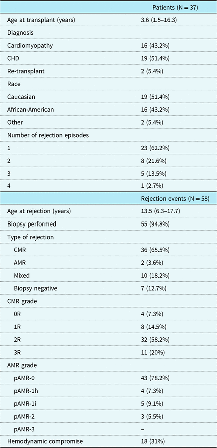
A total of 58 rejection episodes were identified in 37 unique patients (Table 1). A single episode of rejection was present in 23 (62.2%) patients and 14 (37.8%) patients had multiple included rejection events. Of the rejection events, 45 (77.6%) were >1-year post-transplant. An endomyocardial biopsy was performed for 55 (94.8%) rejection episodes. Cellular rejection was confirmed in 46 (83.6%) and evidence of antibody mediated rejection was documented in 12 (21.8%) biopsy specimens. A total of 10 biopsy specimens had features of both cellular and antibody mediated rejection. Endomyocardial biopsy was negative for evidence of either cellular or antibody mediated rejection for seven (12.7%) of the treated rejection events. A total of four patients (accounting for five rejection events) had evidence of cardiac allograft vasculopathy at the time of the included rejection. Two patients were diagnosed with allograft vasculopathy preceding the rejection event and two were diagnosed concurrent with rejection.
Demographics based on patients and rejection events.

AMR = antibody mediated rejection; CHD = congenital heart disease; CMR = cellular mediated rejection.
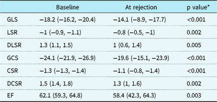
A total of 325 echocardiograms were identified for inclusion and 34 (10.5%) studies were excluded due to suboptimal image quality. A total of 291 echocardiograms were analysed with a median of 5 (interquartile range 4–8) studies analysed per rejection event. Baseline strain values compared to measurements at the time of rejection are shown in Table 2. A significant decrement from baseline was noted in all strain measurements at the time of rejection. Ejection fraction also declined from baseline measurements at the time of rejection.
Baseline strain measurements compared to those obtained at the time of rejection.

p values from the Wilcoxon signed rank test.
CSR = circumferential strain rate; DCSR = peak circumferential early diastolic strain rate; DLSR = peak longitudinal early diastolic strain rate; EF = ejection fraction; GCS = global circumferential strain; GLS = global longitudinal strain; LSR = longitudinal strain rate.
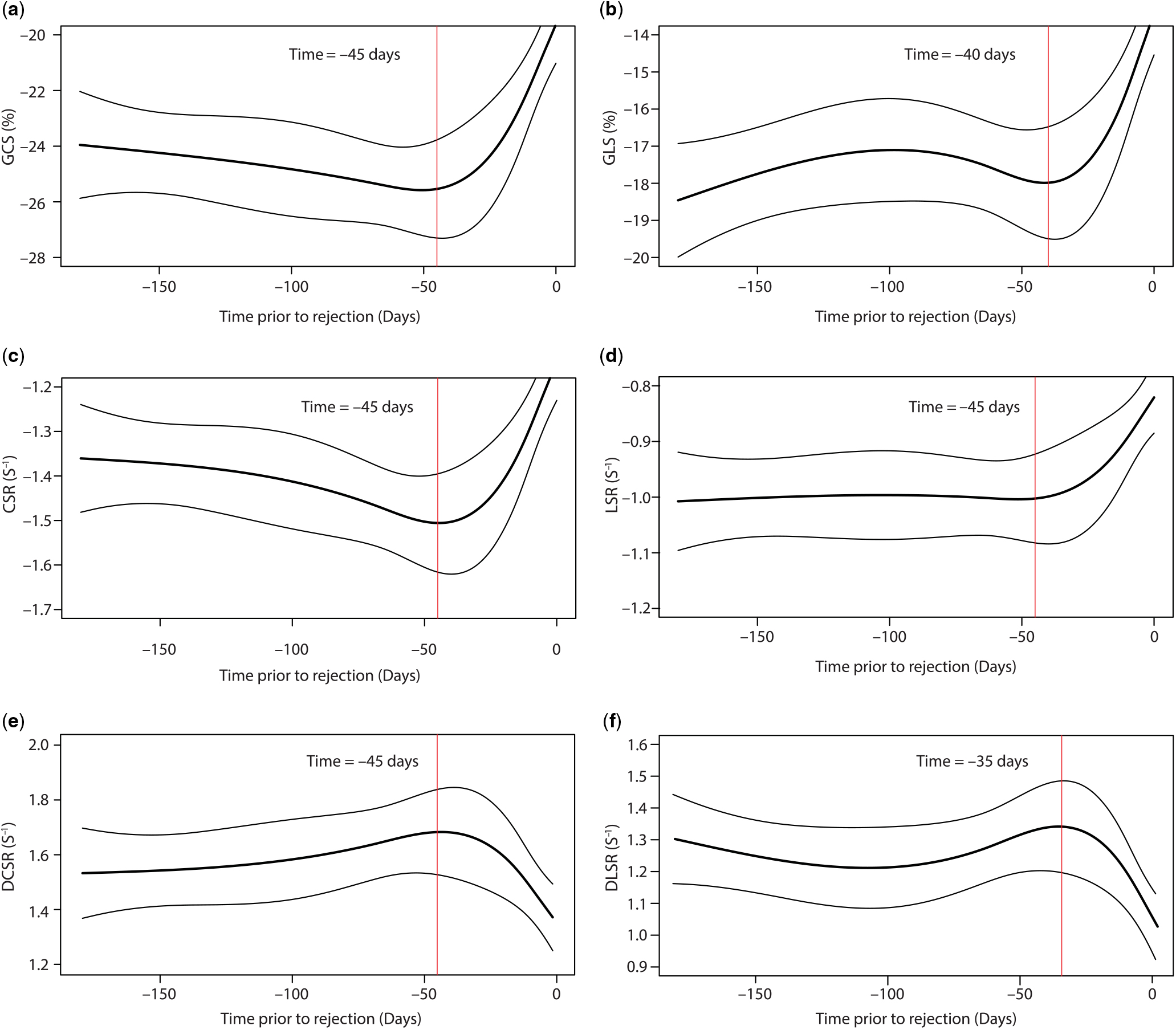
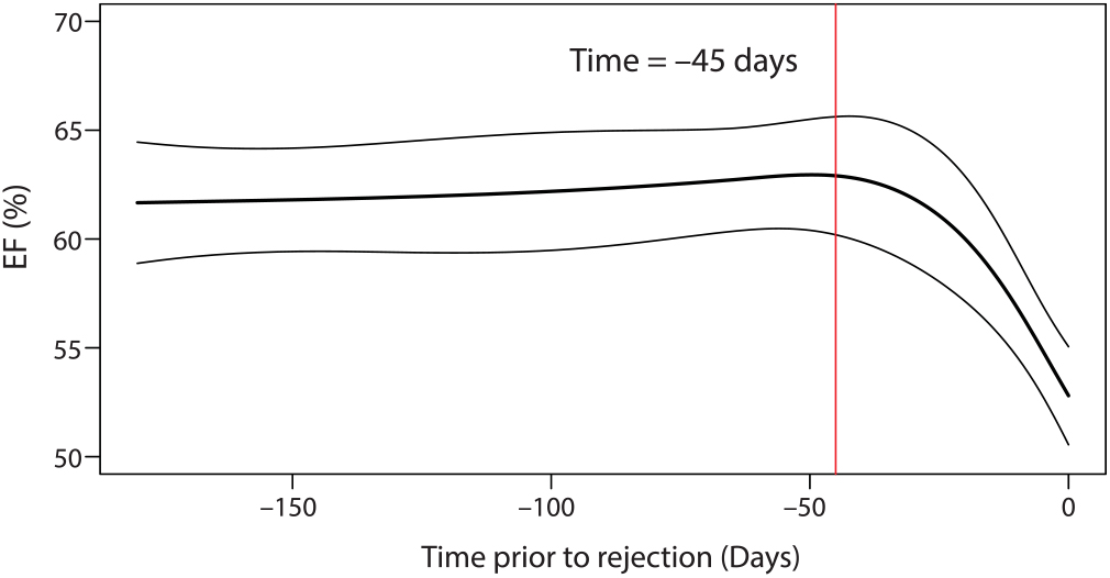
Regression models demonstrating the temporal association of strain measurements with rejection are shown in Figure 1 (a–f) and ejection fraction shown in Figure 2. The earliest changes prior to rejection are estimated to occur 45 days prior to the event. Measurements noted to change at this time were ejection fraction, global circumferential strain, circumferential strain rate, peak circumferential early diastolic strain rate, and longitudinal strain rate. Global longitudinal strain and peak longitudinal early diastolic strain rate demonstrated changes later in the time course, at 40 and 35 days prior to the rejection event, respectively.
Regression models depicting the changes prior to a diagnosis of rejection in (a) GCS – global circumferential strain, (b) GLS – global longitudinal strain, (c) CSR – circumferential strain rate, (d) LSR – longitudinal strain rate, (e) DCSR – peak circumferential early diastolic strain rate and (f) peak longitudinal early diastolic strain rate.

Regression model depicting the changes in ejection fraction (EF) prior to a diagnosis of rejection.

Discussion
Our analysis demonstrates the temporal association of strain measurements leading up to a diagnosis of rejection in paediatric heart transplant recipients. Significant alterations in strain occur with the development of rejection; however, these changes occur concurrently with changes in ejection fraction. Therefore, the use of strain measurements in isolation may not predict rejection earlier compared to more traditional measures of ventricular performance. However, serial changes in strain measures may be useful as an adjunct to standard echocardiographic measures for non-invasive assessment of graft function, as ejection fraction in our study remained within the normal range despite the development of rejection.
Changes in strain measurements have been previously documented in the setting of rejection in both paediatric and adult heart transplant recipients.Reference Podrouzkova, Bedanova and Tretina3–Reference Lisi, Cameli, Tacchini, Ballo, Maccherini and Mondillo5, Reference Kato, Oda and Hashimura10, Reference Gursu, Varan, Sade, Erdogan, Sezgin and Aslamaci17–Reference Pieper, Shah, Harmann, Cooley, Ionova and Migrino20 While the results of our analysis confirm this association, our study does not support the assertion that strain and strain rate imaging diagnose allograft rejection earlier compared to standard measurements. Multiple studies have suggested that strain is more sensitive to sub-clinical changes in ventricular performance and therefore may provide a non-invasive method for early rejection diagnosis.Reference Kato, Oda and Hashimura10, Reference Marciniak, Eroglu and Marciniak19, Reference Syeda, Hofer and Pichler21 The contradictory findings in our study may reflect variation in the evolution of strain versus more standard echocardiographic measures with the development of rejection or may simply be secondary to differences in thresholds to define normal and abnormal between measurements.
While our analysis suggests that strain measurements in isolation may have limited ability to predict rejection early, utilisation of strain in conjunction with standard echocardiographic measures may prove clinically useful. Previously developed models utilising multiple echocardiographic measurements have proven useful in the non-invasive detection of rejection.Reference Dodd, Brady, Carden, Frist, Boucek and Boucek22 The addition of strain measurements into such a model may be advantageous and help to improve the sensitivity and specificity for rejection diagnosis. Other analyses have incorporated strain into similar models and demonstrated value in the prediction of all-cause mortality following heart transplantation.Reference Clemmensen, Eiskjaer, Logstrup, Ilkjaer and Poulsen23
It is important to consider that changes in strain occur following transplantation during the normal remodelling process. Abnormal strain has been documented in the early post-transplant period in both childrenReference Godown, Dodd and Stanley24, Reference Kailin, Miyamoto, Younoszai and Landeck25 and adults.Reference Saleh, Villarraga and Kane26 This may limit the utility of strain to detect rejection in the first year post-transplant, a time when the risk of rejection is greatest.Reference Gossett, Canter and Zheng27, Reference Kirklin, Naftel and Bourge28 Strain measurements normalise by 1-year post-transplant and remain stable in the absence of rejection or cardiac allograft vasculopathy to at least 5 years.Reference Godown, Dodd and Stanley24, Reference Pichler, Binder and Hofer29 Therefore, strain may be better suited to detect rejection beyond 1-year, following initial stabilisation in the early post-transplant period. At least one study suggests that there may be significant intra-patient variability in strain measurements,Reference Godown, Dodd and Stanley24 which may further impact the utility of strain in the assessment of rejection. However, others have reported that strain measurements are reliable and reproducible in the paediatric heart transplant population.Reference Wisotzkey, Jorgensen and Albers30
Limitations
Our study has inherent limitations. Given the variable quality of echocardiographic views, segmental analysis was not possible and therefore our study focused on global strain assessment. Additionally, 10.5% of echocardiograms identified for inclusion were excluded due to suboptimal image quality, potentially impacting the results of our analysis. The number and timing of echocardiograms included varied by patient and rejection event, an inherent limitation of a retrospective analysis. Unfortunately, prospective assessment is not feasible given the limited number of paediatric heart transplant recipients and relative infrequency of treated rejection. The statistical modelling utilised in this analysis attempts to minimise this limitation. Given lack of robust standardisation, there may be variation in strain measurements based on the software package used.Reference Koopman, Slorach and Hui31, Reference Koopman, Slorach and Manlhiot32 However, the use of third-party strain analysis software improves the generalizability of this study as images from multiple vendors can be analysed with Cardiac Performance Analysis Software. The events in our cohort consisted primarily of cellular rejection, but antibody-mediated, mixed, and biopsy negative rejection also occurred. Given the relatively small number of rejection events that were not cellular rejection, comparison among these groups was not feasible. It is also possible that subclinical rejection episodes occurred prior to the formal diagnosis of rejection, as our centre utilises surveillance biopsy sparingly. Lastly, our analysis did not focus on the changes in strain following an episode of rejection and therefore the evolution of strain parameters following a rejection event cannot be assessed.
Conclusions
In the presence of acute allograft rejection, there are detectable alterations in myocardial strain in paediatric heart transplant recipients. These changes occur simultaneously with a decline in ejection fraction. Therefore, the assessment of strain in isolation may not facilitate earlier detection of rejection. However, strain may prove to be a useful non-invasive adjunct in the assessment of graft performance and may aid in the identification of rejection when taken into consideration with other non-invasive measures.
Author ORCIDs
Justin Godown 0000-0002-7068-0625; Jonathan H. Soslow 0000-0001-9194-5330
Acknowledgements
None.
Financial Support
This project was generously supported by the Enduring Hearts Foundation and the Thrasher Research Fund (JG). Research reported in this publication was supported by the National Heart, Lung, and Blood Institute of the National Institutes of Health under Award Number K23HL123938 (Bethesda, MD) (JHS). The project was also supported by the National Center for Research Resources, Grant UL1 RR024975-01, and is now at the National Center for Advancing Translational Sciences, Grant 2 UL1 TR000445-06 (Bethesda, MD). The content is solely the responsibility of the authors and does not necessarily represent the official views of the NIH.
Conflicts of Interest
None.
Ethical Standards
The authors assert that all procedures contributing to this work comply with the ethical standards of the relevant national guidelines on human experimentation Belmont Report and with the Helsinki Declaration of 1975, as revised in 2008, and has been approved by the institutional committee. The Institutional Review Board of Vanderbilt University Medical Center approved this study.