Anomalous origin of a pulmonary artery (AOPA) branch from the aorta is a rare congenital heart disease, in which one of the pulmonary branches arises from the ascending aorta. It is well known that when occurring as an isolated lesion, the right PA (RPA) branch is usually involved; and when associated with tetralogy of Fallot (TOF), the left branch is more commonly affected.Reference Kutsche and Van Mierop 1 The condition is included in the group of aortic arch abnormalities. Mostly this condition is reported to be associated with the right aortic arch. Our patient had double aortic arch with additional coronary arteriovenous fistula, a rare association that made our case a first of its kind. Most patients of AOPA and TOF do not survive infancy without surgical treatment.Reference Morgan 2
Case study
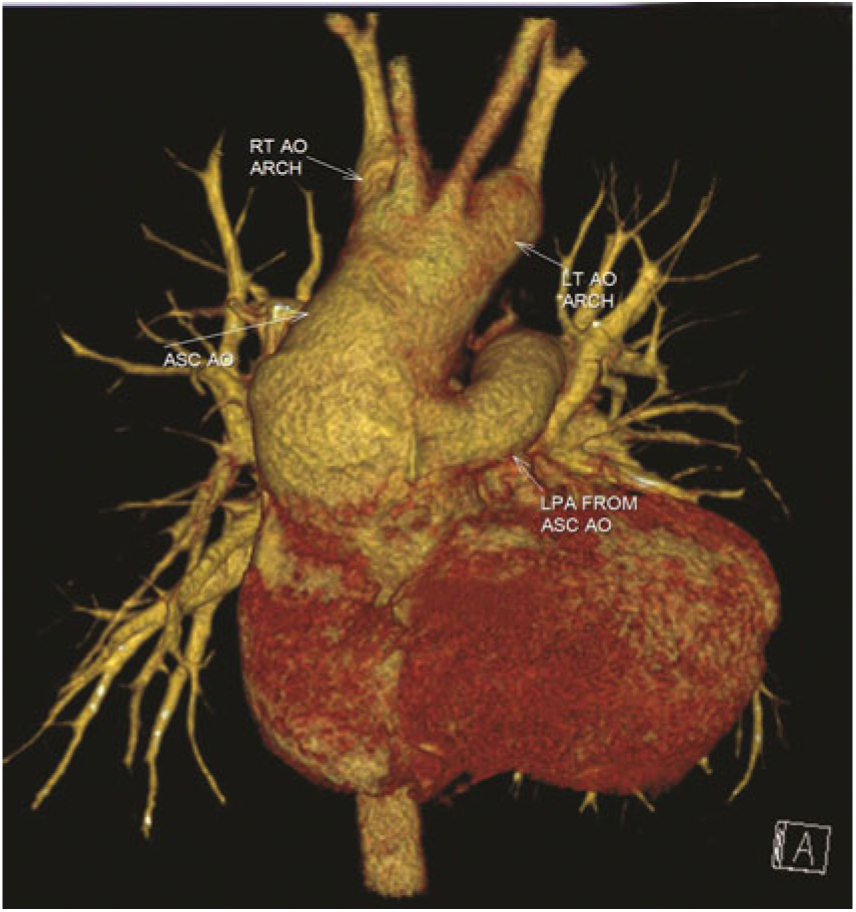
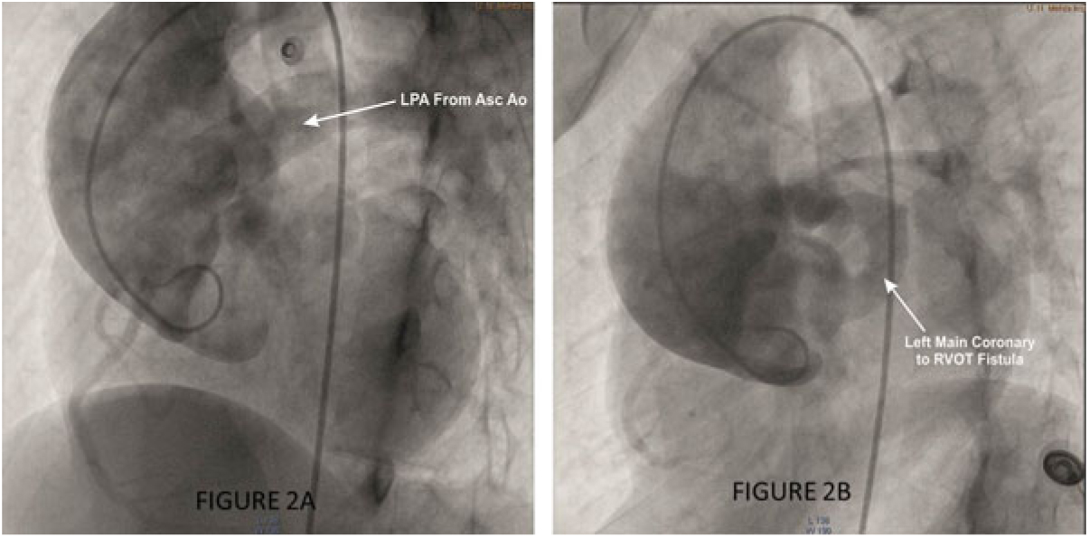
A female patient (14 years, 31 kg) with a history of easy fatigability was referred to our institute for surgical opinion. On examination, the patient was not clinically cyanotic, and room air arterial oxygen saturation was 96%. During auscultation, a grade 3/6, short-early-peaking ejection systolic murmur was best heard in the pulmonary area with a single second sound. The arterial pulses were normal, and heart rate was 110 bpm. The electrocardiogram showed sinus rhythm with signs of biventricular hypertrophy. In the chest X-ray, an obvious cardiomegaly with normal lung field was seen. The two-dimensional transthoracic echocardiography showed the features of TOF with atretic pulmonary valve, absence of left PA (LPA), and presence of major aortopulmonary collaterals. CT pulmonary angiogram confirmed the TOF with atretic pulmonary valve and hypoplastic main pulmonary trunk, along with anomalous origin of the LPA (AOLPA) from the ascending aorta, double aortic arch forming a complete vascular ring (both arch codominant), causing compression over the oesophagus, but there was no obstruction of trachea (Fig 1). There was also a dilated left main coronary artery with suspicion of coronary cameral fistula. A further evaluation by cardiac catheterisation revealed that the systolic pressure in both ventricles was 95 mmHg, and PA pressure in AOLPA was 97 mmHg. PA pressure in RPA was not measured as the pulmonary valve was atretic, and the main PA (MPA) was hypoplastic. It also confirmed the presence of fistulous tract from dilated left main to right ventricular outflow tract (RVOT) at subvalvular level (Fig 2). Qp/Qs was 2.4:1 and pulmonary vascular resistance index was 7.9 with significant reversibility after 100% oxygen. Since the MPA was not catheterised due to pulmonary valvular atresia and only the LPA was catheterised via the aorta and measurements taken, the values above represented only the left lung, and the right lung was considered not hypertensive due to limiting flow because of pulmonary atresia. Based on clinical diagnosis and investigations, a decision to perform total surgical correction of the anomaly was undertaken. The considerable risk involved in the surgery was discussed with the relatives.
3D reconstructed CT angiogram showing codominant double aortic arch with AOLPA from the ascending aorta.

Aortography showing ( a ) LPA arising from the ascending aorta, ( b ) dilated left main coronary artery due to RVOT fistula.

Operative procedure
Following midline sternotomy, the anatomy was confirmed. Careful dissection of the ascending aorta, PA, and LPA was undertaken with extensive mobilisation of left arch till descending thoracic aorta. Under hypothermic bypass and LPA snugged, left arch was divided distal to left subclavian artery (SCA). Following this, ascending aorta was cross-clamped and antegrade cold blood cardioplegia was given. Right ventriculotomy was performed. There was a fistulous opening in RVOT just beneath the pulmonary valve from where cardioplegia was trickling, and it was closed primarily by approximation with two interrupted pledgeted sutures. RV infundibular resection was carried out through right ventriculotomy. Subaortic ventricular septal defect (VSD) was closed by gortex patch. An additional 4 mm mid-muscular VSD was closed by direct approximation with the help of two interrupted pledgeted sutures. The LPA was detached from the aorta and the aortic defect closed with the help of gortex patch. The LPA and MPA were in close approximation with adjoining media of the two vessels. RVOT incision was extended across the pulmonary valve into the hypoplastic MPA and LPA. The LPA was used to reconstruct the wall of MPA, with augmentation of the anterior wall with autologous non-fixed pericardial patch. The pericardial patch extended across the RVOT, leaving the valve ring adequate for the 16-mm Hegar dilator (expected calibre for the patient’s body surface area). Nearly 5 mm surgically created defect in the interatrial septum was left open. The procedure was completed by atrial closure. Coming off bypass was uneventful, and the RV/aorta pressure ratio was 0.5. Following haemostasis and closure, the patient was shifted to the ICU in haemodynamically stable condition on minimal inotropes.
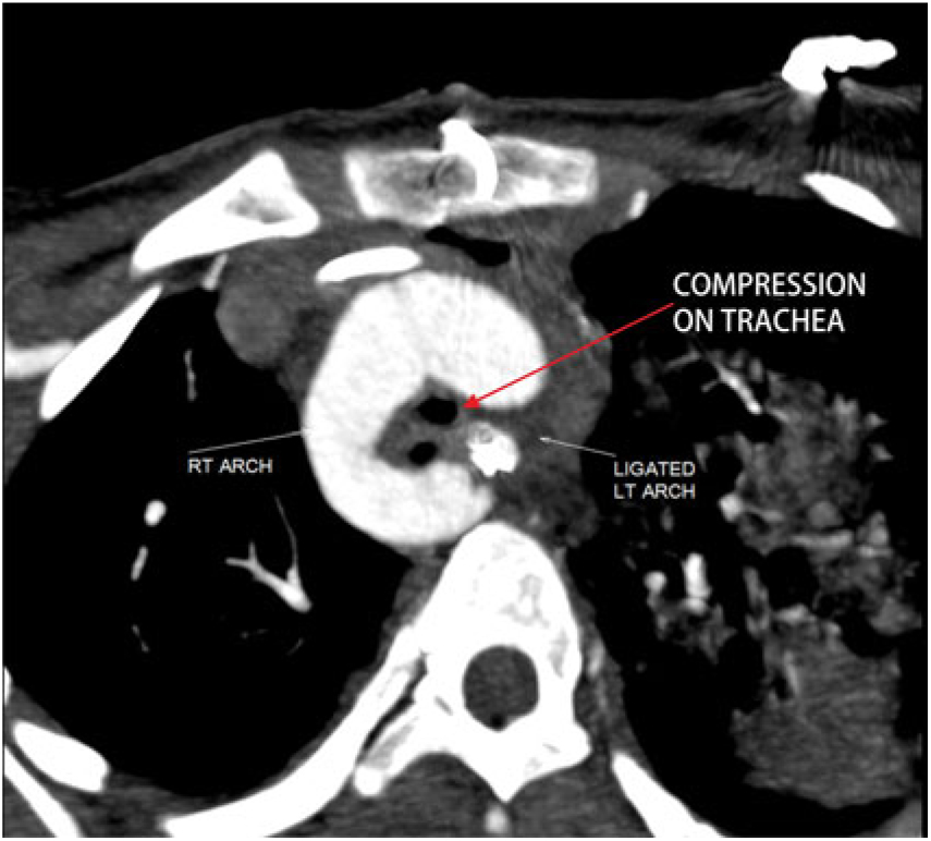
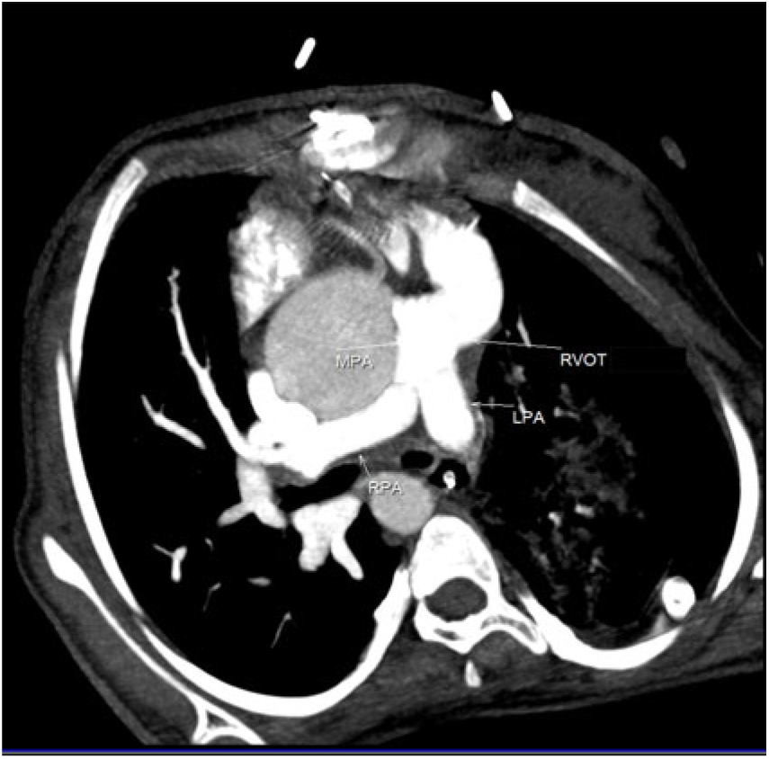
The immediate postoperative period was cautious, with the patient extubated after slowly weaning off the ventilator on the fourth postoperative day. Transthoracic echo revealed good biventricular function and no residual shunting. However, the patient developed right lower lobe consolidation and respiratory distress, needing reintubation on the fifth postoperative day. The patient failed two more trials of extubation in successive days. Flexible bronchoscopy was done, which suggested dynamic obstruction with significant reduction in tracheal lumen at the mid-tracheal level. Also mucous plug obstructing the right lower lobe was removed and bronchoalveolar lavage given, which on culture was positive for Klebsiella and Acinobacter. To evaluate tracheal obstruction, CT pulmonary and aortic angiography was done. It showed a mild compression over the trachea at D4 level (diameter 8 × 7 mm) between the right arch and left SCA origin (Fig 3). The visualised neo-constructed MPA, RPA, and LPA were of good size with no evidence of stenosis (Fig 4). No residual shunts were detected. There were diffuse consolidative patches over the entire left lung and right lower lobe. Since there was a mild compression and no evidence of tracheomalacia, no surgical intervention was mandated. Tracheostomy was performed, but the condition of the patient gradually deteriorated with signs of septicaemia and multi-organ dysfunction. The patient eventually died on the 19th postoperative day.
Postoperative CT scan showing a mild compression of the trachea.

Postoperative CT scan showing a well-reconstructed MPA and LPA.

Discussion
AOPA branch from the aorta is a rare congenital heart disease, in which one of the pulmonary branches arises from the ascending aorta. The condition is included in the group of aortic arch abnormalities.Reference Cucci, Doyle and Lewis 3 The absence of the RPA differs from AOLPA as the former occurs in normal hearts and the latter is accompanied by additional cardiovascular abnormalities, often of the Fallot type.Reference Aru, English, Gaymes and Heath 4
AOPA without RVOT obstruction will result in early congestive cardiac failure and the development of pulmonary arterial hypertension. In patients with RVOT obstruction, reduced pulmonary blood flow from the RV appears to be a protective factor in delaying the onset of pulmonary oedema. Although these patients may be better at first presentation, poorer functional outcome after surgery has been noted due to associated comorbidities.Reference Peng, Shanmugam, Macarthur and Pollock5 It is important to emphasise that an early diagnosis of this anomaly is critical for the long-term follow-up of these patients because of the possible development of pulmonary hypertension. Clinical suspicion of AOLPA is difficult, despite some reports mentioning a differential vascularity of lung in the chest X-ray.Reference Ananthakrishna, Moorthy, Rao and Nanjappa 6 The diagnosis is usually suspected on the echocardiogram, with the first clue being an apparent absence of the LPA. CT angiography is a non-invasive investigation of choice for the diagnosis of AOPA.Reference Liu, Juan and Chen 7 Cardiac catheterisation reaffirms the diagnosis by right ventriculography showing an isolated connection between the RV and the RPA without opacification of the LPA, which can be visualised through aortography.
Different surgical techniques have been performed to implant the AOPA to the MPA on the basis of the anatomy and origin site of the AOPA. While previously these techniques involved use of a conduit, recent trend is for direct implantation with or without augmentation by autologous pericardial patch, aortic flap, and simultaneous aortic and pulmonary flaps, depending on the length of the PA required.Reference Peng, Shanmugam, Macarthur and Pollock 5 , Reference Prifti, Crucean and Bonacchi 8 – Reference Garg, Talwar and Kothari 10 Prosthetic conduits will need replacement as the patient outgrows the conduit. Peng et alReference Peng, Shanmugam, Macarthur and Pollock 5 have shown relatively poorer long-term outcomes related to the use of a conduit for the repair of RVOT. Because the AOPA is close to the MPA, patients with AOLPA can be surgically treated with direct anastomosis. While performing direct anastomosis, Peng et alReference Peng, Shanmugam, Macarthur and Pollock 5 encountered stenosis at the anastomotic site requiring re-interventions, while Garg et alReference Garg, Talwar and Kothari 10 did not encounter the same. However, we preferred to augment the native PA with dilated AOLPA wall as well as with autologous pericardial patch, as in our case the MPA was small in calibre.
Conclusion
AOPA from the aorta usually is detected at an early age and treated as soon as presented. In developing countries, we sometimes come across such late presentations of anomaly. Definitive management of such cases is not well documented in literature. Based on clinical diagnosis and investigations of our patient, which was favourable for surgical correction, the decision for total correction was taken. The patient did well postoperatively with good anatomical correction, but finally succumbed due to sepsis and multi-organ dysfunction. The suitability of operability in such cases needs to be further discussed and studied.