Introduction
Obesity is increasingly prevalent in bipolar disorder (BD), in that over 60% of patients with BD may be overweight or obese (Goldstein et al. Reference Goldstein, Liu, Zivkovic, Schaffer, Chien and Blanco2011). Obesity has been associated with poor clinical outcomes in BD, including shorter time to recurrence during the maintenance phase of the illness (Fagiolini et al. Reference Fagiolini, Kupfer, Houck, Novick and Frank2003), more frequent depressive episodes and suicide attempts compared with the general population (Fagiolini et al. Reference Fagiolini, Kupfer, Houck, Novick and Frank2003, Reference Fagiolini, Kupfer, Rucci, Scott, Novick and Frank2004). Furthermore, patients with BD who are overweight and obese often have poor medical outcomes including greater risk of suffering from metabolic disturbances such as diabetes and cardiovascular disease (Magalhães et al. Reference Magalhães, Kapczinski, Nierenberg, Deckersbach, Weisinger, Dodds and Berk2012) and these are the leading causes of premature and excess mortality in BD (Osby et al. Reference Osby, Brandt, Correia, Correia, Ekbom and Sparén2001).
In the general population, previous structural magnetic resonance imaging (MRI) studies have found either no change in white-matter volume (Pannacciulli et al. Reference Pannacciulli, Del Parigi, Chen, Le, Reiman and Tataranni2006) or decreased total brain volume, grey-matter volume in overweight/obese individuals (Taki et al. Reference Taki, Kinomura, Sato, Inoue, Goto, Okada, Uchida, Kawashima and Fukuda2008; Soreca et al. Reference Soreca, Rosano, Jennings, Sheu, Kuller, Matthews, Aizenstein and Gianaros2009) as well as greater loss of brain volume over time compared with individuals with normal weight (Enzinger et al. Reference Enzinger, Fazekas, Matthews, Ropele, Schmidt, Smith and Schmidt2005; Bobb et al. Reference Bobb, Schwartz, Davatzikos and Caffo2012). Recent studies using diffusion tensor imaging (DTI) in otherwise healthy adults found that obesity is associated with disruptions in white-matter integrity as indicated by reductions of fractional anisotropy (FA) within the corpus callosum (Mueller et al. Reference Mueller, Anwander, Möller, Horstmann, Lepsien, Busse, Mohammadi, Schroeter, Stumvoll, Villringer and Pleger2011) and involving midbrain and brainstem white-matter tracts (Verstynen et al. Reference Verstynen, Weinstein, Schneider, Jakicic, Rofey and Erickson2012). Furthermore, individuals with high body mass index (BMI) exhibited deficits in attention, speed of processing, and executive functioning on neuropsychological testing compared with those with normal BMI (Gunstad et al. Reference Gunstad, Paul, Cohen, Tate and Gordon2006, Reference Gunstad, Paul, Cohen, Tate, Spitznagel and Gordon2007), indicating that elevated BMI may have observable adverse impacts on neural substrates.
To the best of our knowledge, there is a paucity of neuroimaging studies that have specifically evaluated the underlying brain structural changes that may mediate the impact of BMI in BD. The only study was a recent structural MRI report by Bond et al. (Reference Bond, Lang, Noronha, Kunz, Torres, Su, Honer, Lam and Yatham2011) that documented decreased brain white-matter volume and temporal lobe volume in adults newly diagnosed with BD and with elevated BMI. There are to date no data on the effect of BMI on brain white-matter integrity in BD patients. However, in the context of pharmacotherapy, previous studies investigating the impact of psychotropic medication use on brain structures have found associations of grey-matter volume increase with use of mood stabilizers (Atmaca et al. 2008; Kempton et al. Reference Kempton, Geddes, Ettinger, Williams and Grasby2008) and increase of glial cell density and non-reduction of white-matter volume with antipsychotic administration (Selemon et al. Reference Selemon, Lidow and Goldman-Rakic1999; Bartzokis et al. Reference Bartzokis, Lu, Amar, Raven, Detore, Altshuler, Mintz, Ventura, Casaus, Luo, Subotnik and Nuechterlein2011). Thus, based on these sparse extant data, we sought to examine whether excess weight is associated with changes of brain white-matter integrity in adults with BD who have achieved symptomatic remission from a first episode of mania. The emphasis on newly diagnosed BD patients decreases the likelihood of confound by effects of illness, chronicity and treatment. In view of the findings of Bond et al. (Reference Bond, Lang, Noronha, Kunz, Torres, Su, Honer, Lam and Yatham2011) involving white-matter volume and temporal lobe volume in patients with first-episode mania (FEM) and elevated BMI, we hypothesized that increased BMI in remitted patients with FEM is associated with reduced FA in the frontal, temporal, parietal and occipital brain regions.
Method
Participants
The study sample comprised 54 participants (31 men and 23 women) who gave written informed consent to participate in the study after a detailed explanation of the study procedures between June 2008 and June 2010. A total of 26 patients with remitted FEM were recruited from the Institute of Mental Health, Singapore. All diagnoses were made by a psychiatrist (K.S.) using information obtained from the existing medical record, clinical history, mental status examination, interviews with the patients and their significant others as well as the administration of the Structured Clinical Interview for DSM-IV disorders (SCID) – Patient Version (First et al. Reference First, Spitzer, Gibbon and Williams1994) and Young Mania Rating Scale (YMRS; Young et al. Reference Young, Biggs, Ziegler and Meyer1978). All patients were in remission from mania (i.e. YMRS score <10) at the time of recruitment and on the day of neuroimaging (Tohen et al. Reference Tohen, Frank, Bowden, Colom, Ghaemi, Yatham, Malhi, Calabrese, Nolen, Vieta, Kapczinski, Goodwin, Suppes, Sachs, Chengappa, Grunze, Mitchell, Kanba and Berk2009; Chan et al. Reference Chan, Yang, Chia, Woon, Lee, Keefe, Sitoh, Nowinski and Sim2010), were maintained on a stable dose of psychotropic medication for at least 2 weeks prior to the recruitment and did not have their medication withdrawn for the purpose of the study. In addition, 28 age-, gender-, handedness- and education-matched healthy controls (HC) were administered the SCID – Non-Patient Version (First et al. Reference First, Spitzer, Gibbon and Williams2002). None of the participants had a history of significant and/or unstable/untreated medical illnesses such as seizure disorder, head trauma or cerebrovascular accidents. Moreover, no subjects had a current or past history of substance use or alcohol use disorder, metabolic disorder such as diabetes, dyslipidaemia or previous diagnosis and/or treatment for another psychiatric disorder. This study was approved by the Institutional Review Boards of the Institute of Mental Health, Singapore, as well as the National Neuroscience Institute, Singapore.
BMI and weight evaluation
Each participant had their height and weight recorded. The BMI was calculated using the formula BMI = weight (kg)/height (m)2. For the purpose of analyses, participants were categorized into normal weight (BMI of 18.5 to <23.0 kg/m2; n = 25), overweight (BMI 23.0 to <25.0 kg/m2; n = 12) and obese (BMI > 25.0 kg/m2; n = 17) according to the International Association for the Study of Obesity (World Health Organization, International Association for the Study of Obesity, International Obesity Task Force, 2000) and the World Health Organization (WHO) proposed BMI cut-off points for obesity in adult Asians (WHO Expert Consultation, 2004).
Brain imaging acquisition and data processing
Brain imaging was performed using a 3-Tesla whole body scanner (Philips Achieva; Philips Medical Systems, The Netherlands) with a SENSE head coil at the National Neuroscience Institute, Singapore. High-resolution T1-weighted magnetization prepared rapid gradient recalled echo (MP-RAGE) images were required [repetition time (TR) = 7.2 ms; echo time (TE) = 3.3 ms; flip angle = 8°]. Each T1-weighted volume consisted of 180 axial slices of 0.9 mm thickness with no gap (field of view, 230 mm × 230 mm; acquisition matrix, 256 × 256 pixels). For DTI, single-shot echo-planar diffusion tensor images were obtained (TR = 3725 ms; TE = 56 ms; flip angle = 90°, b = 800 s/mm2) with 15 different non-parallel directions (b = 800 s/mm2) and the baseline image without diffusion weighting (b = 0 s/mm2). The acquisition matrix was 112 × 109 pixels with a field of view of 230 mm × 230 mm, which was zero-filled to 256 × 256 pixels. A total of 42 axial slices of 3.0 mm thickness were acquired parallel to the anterior–posterior commissure line. The T1-weighted and DTI data were sequentially acquired in a single session scan time without position change. Stability of a high signal to noise ratio was assured through a regular automated quality-control procedure.
The structural MRI images were processed using the Free Surfer software package to delineate the different brain regions (Athinoula A. Martinos Center for Biomedical Imaging, Massachusetts General Hospital, Harvard University; http://surfer.nmr.mgh.harvard.edu/). The Free Surfer software reformatted each brain volume image into a 1 cubic mm isovoxel volume image (Fischl et al. Reference Fischl, Salat, Busa, Albert, Dieterich, Haselgrove, van der Kouwe, Killiany, Kennedy, Klaveness, Montillo, Makris, Rosen and Dale2002). Within each subject, the diffusion-weighted images (DWIs) of each subject were corrected for motion and eddy current distortions using affine transformation to the baseline image without diffusion weighting (b = 0). FA maps were acquired from the DTI images using DTI Studio (Jiang et al. Reference Jiang, van Zijl, Kim, Pearlson and Mori2006) and were then co-registered automatically to the structural MP-RAGE images. Mean FA values in the frontal, parietal, temporal and occipital lobes were then computed and used in the following statistical analyses.
Statistical analyses
As the number of subjects who were categorized as overweight (BMI 18.5 to <23.0 kg/m2) or obese (BMI⩾23.0 kg/m2) was relatively low, we combined the two groups and compared those with remitted FEM who were normal weight with those who were overweight/obese. Both groups were also compared with a HC group. Sociodemographic variables between FEM and HC were compared using the two-sample Student's t test and χ 2 test for continuous and categorical variables; respectively. The diagnosis effect, BMI effect and BMI × diagnosis interactions on brain white-matter integrity (FA) were examined using two-way analysis of variance. The BMI effect, diagnosis effect and diagnosis × BMI interactions were further analysed using two-way analysis of covariance to control for covariates such as age, gender, years of education and medications in terms of antipsychotic use (mean daily chlorpromazine equivalents) and prescription of mood stabilizer. When a significant diagnosis × BMI interaction was found, differences between the two groups of FEM and HC were further evaluated separately. All statistical tests were performed using PASW for Windows, version 18.0 (SPSS Inc., USA). The significance level for statistical tests was set at two-tailed p < 0.05 as our analyses were exploratory on the different brain regions at this stage.
Results
Sociodemographic and clinical characteristics
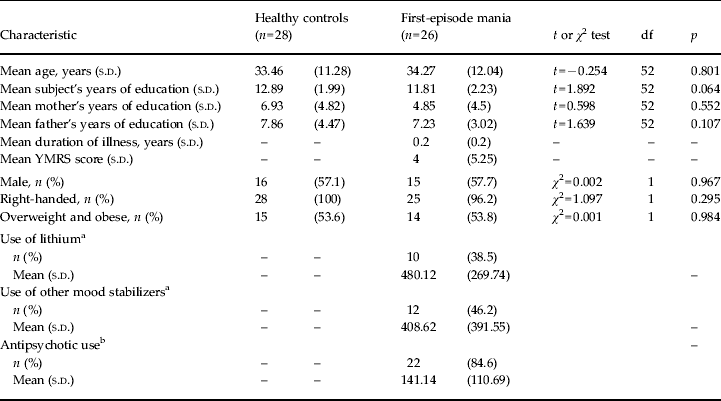
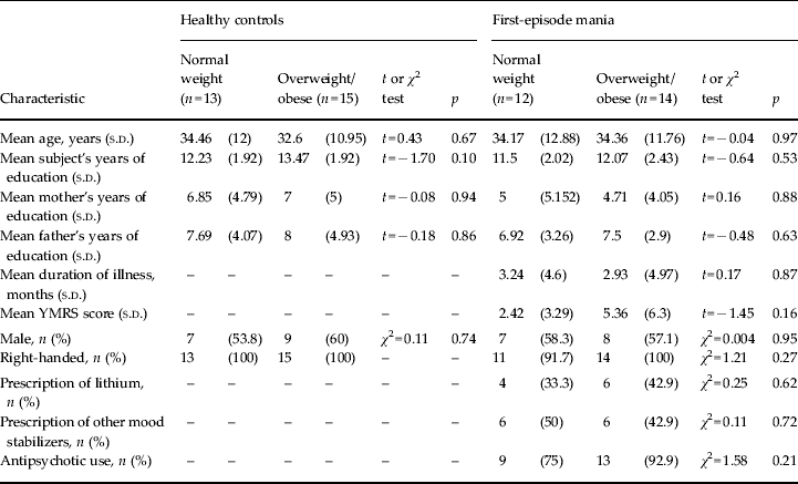
There was no significant difference in age, gender, handedness, subject and parental years of education between patients with FEM and HC. Of the subjects, 46% (n = 25, 12 patients with FEM and 13 controls) of the subjects had normal weight while the rest of the subjects were either overweight or obese (n = 29; 14 patients with FEM and 15 controls). The majority of the patients received medications such as mood stabilizers, namely valproate (n = 12) and lithium (n = 10), as well as antipsychotics (n = 22; two typicals, 20 atypicals), while the remaining four patients did not receive any medication at the time of recruitment. The sociodemographic and clinical features are shown in Table 1. Table 2 shows the details comparing between normal-weight and overweight/obese groups within both patients and HC. Essentially, there was no significant difference in terms of age, gender, years of education, parental years of education and handedness between normal-weight and overweight/obese groups within the patients and HC. Within the patient group, there was no significant difference between normal-weight and overweight/obese groups in terms of duration of illness, YMRS scores, lithium, mood stabilizers and antipsychotic medications prescribed.
Demographic and clinical characteristics of the participants

df, Degrees of freedom; s.d., standard deviation; YMRS, Young Mania Rating Scale.
a Frequency and mean dose of lithium or other mood stabilizers prescribed.
b Frequency and mean dose of antipsychotic prescribed in mean daily chlorpromazine equivalents.
Demographic and clinical characteristics of the participants by weight group

s.d., Standard deviation; YMRS, Young Mania Rating Scale.
Effect of BMI on brain white-matter integrity
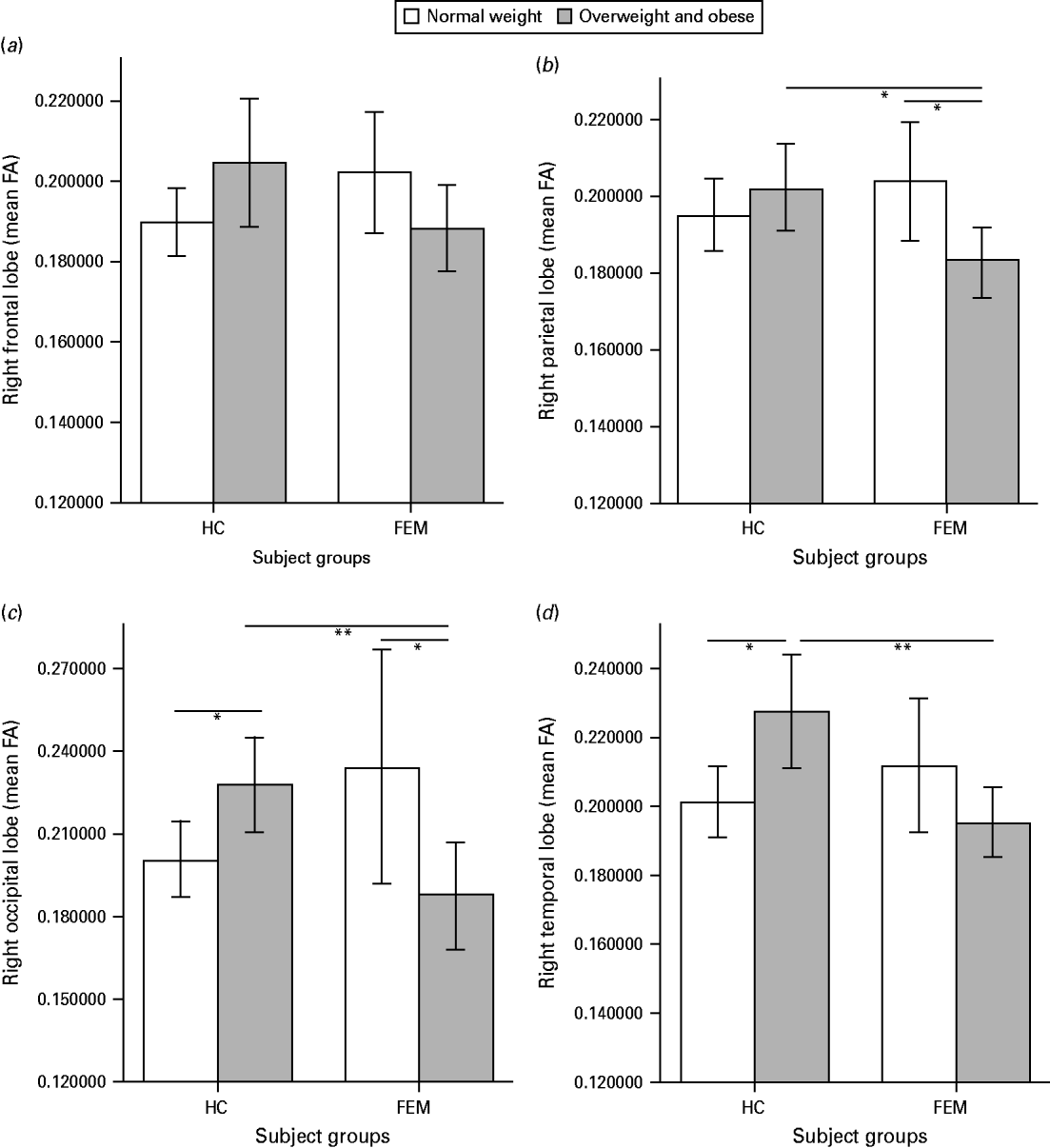
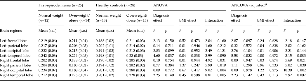
There were significant effects of diagnosis × BMI interactions observed in the right frontal lobe (F 1,50 = 4.92, p = 0.031), right parietal lobe (F 1,50 = 5.90, p = 0.019), right occipital lobe (F 1,50 = 9.02, p = 0.004) and temporal lobe (F 1,50 = 8.81, p = 0.005). These interactions remained significant after controlling for covariates (right frontal lobe: adjusted, F 1,48 = 5.69, p = 0.021; right parietal lobe: adjusted, F 1,48 = 5.02, p = 0.030; right occipital lobe: adjusted, F 1,48 = 10.30, p = 0.002; right temporal lobe: adjusted, F 1,48 = 7.92, p = 0.007). As the diagnosis × BMI interactions were found to be significant for the right frontal, parietal, temporal and occipital lobes (Table 3), we analysed the effects of BMI on these four brain regions between patients with FEM and controls as well as separately within patients with FEM and controls (Fig. 1). Specifically, decreased FA was found in the right parietal (F 1,23 = 5.864, p = 0.023) and occipital (F 1,23 = 4.397, p = 0.047) lobes with standardized effect sizes of −1.1 and −1.2, respectively, within overweight/obese patients compared with normal-weight patients with FEM. Compared with overweight/obese controls, decreased FA was observed in right parietal (F 1,25 = 6.708, p = 0.015), temporal (F 1,25 = 10.751, p = 0.003) and occipital (F 1,25 = 9.531, p = 0.005) regions with standardized effect sizes of −1.0, −1.65 and −2.3, respectively, in overweight/obese patients with FEM.
Fractional anisotropy (FA) in the (a) right frontal, (b) parietal, (c) occipital and (d) temporal lobes in healthy subjects (HC) and first-episode mania patients (FEM). Values are means, with standard errors represented by vertical bars. Significant mean FA differences: * p < 0.05, ** p < 0.01.

Effect of BMI on brain white-matter integrity (mean fractional anisotropy)

BMI, Body mass index; ANOVA, analysis of variance; ANCOVA, analysis of covariance; s.d., standard deviation.
a Adjusted for age, gender and years of education.
We performed additional analyses on the possible effect of medication on these brain regions including antipsychotic use (in mean daily chlorpromazine equivalents), use of lithium and valproate. Apart from the right frontal region (F 1,47 = 3.068, p = 0.086), the diagnosis × BMI interactions remained largely unchanged in the other brain regions after including medication as covariates (right parietal: F 1,47 = 4.668, p = 0.036; right occipital: F 1,47 = 7.224, p = 0.003; right temporal: F 1,47 = 8.017, p = 0.007). Backward regression was performed to further analyse the effect of medication in the right frontal region, in which we found that only mean daily chlorpromazine equivalents significantly predicted mean FA compared with lithium and valproate (β = − 0.460, t = − 2.523, p = 0.020).
Discussion
To our knowledge, this is the first study to specifically examine the impact of weight status on brain white-matter integrity in patients with FEM. There were several observations in the sample examined herein. First, reductions of FA in the right parietal and occipital regions were found in FEM patients who were overweight/obese compared with those who had normal weight. Second, reductions of FA were seen in the right parietal, occipital and temporal regions within overweight/obese FEM patients compared with controls, suggesting that increased BMI early in the course of BD is associated with disruptions of white-matter integrity involving temporal–parietal–occipital brain circuitry.
The observed reductions in FA within FEM patients who were overweight/obese compared with normal weight occur in brain regions previously found to be implicated in the patho-aetiology of BD. Earlier structural MRI studies of patients with BD have reported increased rates of white-matter hyperintensities in BD, with a recent meta-analysis noting up to 2.5 times increased odds of having deep white-matter hyperintensities in patients with BD (Kempton et al. Reference Kempton, Geddes, Ettinger, Williams and Grasby2008). Structural MRI studies of FEM patients found reductions of brain white-matter volume but not grey-matter volume (Vita et al. Reference Vita, De Peri and Sacchetti2009). DTI studies that specifically examine brain white-matter integrity have implicated disruptions in FA within parietal and occipital regions in patients with BD (Chan et al. Reference Chan, Yang, Chia, Woon, Lee, Keefe, Sitoh, Nowinski and Sim2010; Heng et al. Reference Heng, Song and Sim2010). Our findings of FA reductions specifically within parietal and occipital brain regions suggest that similar brain regions are affected early in the course of the illness in FEM patients who are overweight/obese.
Compared with the overweight/obese healthy adults, we also found FA reductions in right parietal, temporal and occipital regions in FEM patients who were overweight/obese, providing evidence that FEM patients with elevated BMI may be more susceptible to brain white-matter changes. Of note, the overweight/obese patients with FEM had lower FA in the right occipital and temporal lobes compared with normal-weight patients with FEM, but overweight/obese controls had higher FA in the similar brain regions compared with normal-weight controls. The observed higher FAs within the temporal and occipital lobes in overweight/obese controls compared with normal-weight controls need replication, although these results may be consistent with findings of an earlier MRI study which reported increased white-matter volume in temporal and occipital brain regions of obese subjects (Haltia et al. Reference Haltia, Viljanen, Parkkola, Kemppainen, Rinne, Nuutila and Kaasinen2007). However, other studies have found no difference in brain white-matter volume (Pannacciulli et al. Reference Pannacciulli, Del Parigi, Chen, Le, Reiman and Tataranni2006), no difference in FA between overweight and normal-weight controls (Stanek et al. Reference Stanek, Grieve, Brickman, Korgaonkar, Paul, Cohen and Gunstad2011), reductions in FA within the corpus callosum in obese female controls (Mueller et al. Reference Mueller, Anwander, Möller, Horstmann, Lepsien, Busse, Mohammadi, Schroeter, Stumvoll, Villringer and Pleger2011) or especially found within midbrain and brainstem white-matter tracts (Verstynen et al. Reference Verstynen, Weinstein, Schneider, Jakicic, Rofey and Erickson2012). In this regard, elevated BMI may have differential aetiological mechanisms amongst individuals with FEM and controls and may be a marker of greater illness severity in BD, hence accounting for the FA differences in controls and FEM with elevated BMI compared with those individuals with normal weight. Future studies are needed to better determine the relationship between elevated BMI and brain white-matter structure as it may affect different brain structures differently in controls and within the context of BD.
The neurobiological pathways linking obesity, mania and brain white-matter changes are still unclear and await further elucidation. Earlier studies have found reduced white-matter integrity in patients with obesity-associated co-morbidity such as diabetes, hypertension, metabolic syndrome and stroke (Hoth et al. Reference Hoth, Tate, Poppas, Forman, Gunstad, Moser, Paul, Jefferson, Haley and Cohen2007; Kodl et al. Reference Kodl, Franc, Rao, Anderson, Thomas, Mueller, Lim and Seaquist2008). Postulated biological mechanisms subserving the relationship between obesity and brain white-matter changes include endothelial dysfunction, insulin resistance and inflammatory processes (Hoth et al. Reference Hoth, Tate, Poppas, Forman, Gunstad, Moser, Paul, Jefferson, Haley and Cohen2007; Rosenberg, Reference Rosenberg2009; Katsumata et al. Reference Katsumata, Otori, Nishiyama, Okubo, Nishiyama, Nagayama, Ueda, Utsumi, Yamazaki, Komaba, Katsura and Katayama2010). Genetic imaging studies have highlighted the relationship between risk alleles at rs1421085 and rs17817449 of the fat mass and obesity-associated (FTO) gene and volume reductions of 8% in the bilateral frontal and 12% in the bilateral occipital lobes, respectively (Ho et al. Reference Ho, Stein, Hua, Lee, Hibar, Leow, Dinov, Toga, Saykin, Shen, Foroud, Pankratz, Huentelman, Craig, Gerber, Allen, Corneveaux, Stephan, DeCarli, DeChairo, Potkin, Jack, Weiner, Raji, Lopez, Becker, Carmichael and Thompson2010), indicating that genetic factors relevant to obesity may contribute to susceptibility towards brain structural changes including brain white matter. In addition, there are some data to support the roles of the pleiotropic peptides insulin, insulin-like growth factor 1 (IGF-1) and incretins in neuronal and glial cell function including neurogenesis, myelination and modulation of synaptic plasticity (McIntyre et al. Reference McIntyre, Vagic, Swartz, Soczynska, Woldeyohannes, Voruganti and Konarski2008). Alterations in insulin/IGF-1 signalling can result in downstream effects of decreased cellular integrity and survival in brain regions affecting neurocognition and other brain functions (McIntyre et al. Reference McIntyre, Vagic, Swartz, Soczynska, Woldeyohannes, Voruganti and Konarski2008). BD is associated separately with disruptions in white-matter integrity in the putative regions (Heng et al. Reference Heng, Song and Sim2010), which may be further exacerbated in the context of elevated BMI. In addition, leptin from adipose tissue (Farooqi et al. Reference Farooqi, Bullmore, Keogh, Gillard, O'Rahilly and Fletcher2007) and gut hormones such as ghrelin, peptide YY (peptide tyrosine-tyrosine) and glucagon-like peptide (Gibson et al. Reference Gibson, Carnell, Ochner and Geliebter2010) have been associated with activation of different cortical brain areas related to visual presentation of food, suggesting the complex inter-relationships between neurobiological factors within the gut–brain axis in affecting brain white matter.
In addition, the diagnosis × BMI interactions and reductions of FA occur mainly within brain regions in the right hemisphere, suggesting laterality effect. This laterality effect is consistent with the findings of a meta-analysis of 10 whole-brain DTI studies (Vederine et al. Reference Vederine, Wessa, Leboyer and Houenou2011), which reported significant clusters of decreased FA in the right hemisphere although not taking into account the weight status. One possible explanation is that brain white-matter composition may be dynamic (May, Reference May2011) and altered white-matter fibres in the corpus callosum of patients with BD may have contributed to asymmetrical impact on white-matter integrity within the different brain regions. Reduced white-matter integrity in the corpus callosum has been observed in obese healthy adults (Stanek et al. Reference Stanek, Grieve, Brickman, Korgaonkar, Paul, Cohen and Gunstad2011), and obesity may have additionally contributed to the laterality effect of affected brain white-matter regions.
There are several limitations in this study. First, the study sample is small and the findings need to be replicated in other populations and in a larger sample. Second, the findings may be confounded by the effects of medications such as mood stabilizers and antipsychotics. However, the patients with FEM were recruited at an early phase of their illness with no previous treatment with medications and had been receiving their treatment for a minimal period of time. In addition, available data suggest that psychotropic medications used such as lithium and valproate are associated with grey-matter volume increases and are less likely to contribute to changes in white matter (Moore et al. Reference Moore, Bebchuk, Wilds, Chen and Manji2000; Atmaca et al. 2008; Kempton et al. Reference Kempton, Geddes, Ettinger, Williams and Grasby2008), as is also consistent with our findings which remained largely unchanged when we included medications as covariates in our analyses. Exposure to antipsychotics (typical and atypical) in animal models has been associated with an increase in glial cell density as well as promotion of oligodendrocyte differentiation and myelin repair (Selemon et al. Reference Selemon, Lidow and Goldman-Rakic1999). Adherence to antipsychotic treatment with remission of illness is also thought to result in better trajectory of myelination and non-reduction of brain white-matter volume (Bartzokis et al. Reference Bartzokis, Lu, Amar, Raven, Detore, Altshuler, Mintz, Ventura, Casaus, Luo, Subotnik and Nuechterlein2011). Third, this is a cross-sectional study and the longitudinal follow-up of these subjects would proffer further insights into the complex interaction between weight status, illness factors, treatment and changes in brain white-matter integrity. Fourth, the functional and behavioural implications of our findings may be relevant to neurocognitive function. Unfortunately, we did not include measures of neurocognitive function in our sample. Extant data indicate that individuals with BD exhibit clinically significant and broad-based neurocognitive deficits that often persist during periods of euthymia. Neurocognitive deficits in BD are known to cause and maintain functional impairment. A separate body of evidence has reported that obesity and diabetes are associated with cognitive deficits (McIntyre et al. Reference McIntyre, Kenna, Nguyen, Law, Sultan, Woldeyohannes, Adams, Cheng, Lourenco, Kennedy and Rasgon2010). Moreover, preliminary evidence indicates that the metabolic syndrome may be associated with neurocognitive deficits in bipolar populations (Yim et al. Reference Yim, Soczynska, Kennedy, Woldeyohannes, Brietzke and McIntyre2012).
In conclusion, we found that elevated BMI is associated with disruptions of brain white-matter integrity affecting right parietal, temporal and occipital regions in FEM patients who were overweight/obese. These findings add to a growing confluence of study results indicating that somatic health status is associated with alterations in central nervous system (CNS) structure and function. In addition to replication and extension of our results, elucidating effector systems that mediate the association between BD, obesity, and white-matter integrity would be a future research vista. Practitioners providing care for individuals with BD need to consider the CNS effects associated with overweight/obesity in BD and recommendations for screening, prevention, and treatment of excess weight and BMI are supported by these data.
Acknowledgements
We thank all of the patients, their families and our hospital staff for their support of this study. This study was supported by the Singapore Bioimaging Consortium (RP C-009/2006) and National Healthcare Group Small Innovative Grant (11003; 05186) research grants awarded to K.S.
Declaration of Interest
R.S.M. has received research funding from AstraZeneca, GlaxoSmithKline, Merck, Servier and Wyeth and has served as a consultant to and on the speaker's boards for AstraZeneca, Eli Lilly, GlaxoSmithKline, Lundbeck, Organon, Ortho-McNeil-Janssen, Oryx, Pfizer, Prestwick and Wyeth.