Introduction
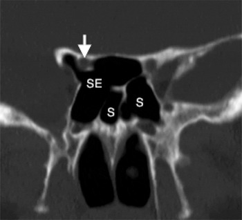
A sphenoethmoidal cell, first described by Adolf Onodi in 1903, is a posterior ethmoid cell that pneumatises superiorly and/or laterally to the sphenoid sinus.Reference Lund, Stammberger, Fokkens, Beale, Bernal-Sprekelsen and Eloy1, Reference Onodi2 This normal variant is of relevance to the ENT surgeon because the optic nerve may lie within the sphenoethmoidal cell or be exposed along the superolateral wall (Figure 1). This close relationship can lead to visual symptoms in the presence of sinus disease.
Coronal computed tomography scan showing the right optic nerve (arrow) running through a sphenoethmoidal cell. SE = sphenoethmoidal cell; S = sphenoid sinus

We report four cases of chronic rhinosinusitis involving a sphenoethmoidal cell, two with visual loss and two without. We discuss the management of this problem and review the current literature.
Case reports
Case one
A 20-year-old man with a longstanding history of chronic rhinosinusitis with nasal polyps had undergone bilateral nasal polypectomy 1 and 2 years previously, with only short-term improvement in his symptoms of nasal blockage and anosmia. There had been no visual symptoms at any point.
A computed tomography (CT) scan of his sinuses showed complete opacification and an expanded left sphenoethmoidal cell (consistent with a mucocele), with bony erosion over the adjacent skull base, optic nerve canal and orbit.
Medical treatment was commenced, and functional endoscopic sinus surgery (FESS) was undertaken two weeks later in view of the potential risk of both intracranial and orbital complications. This involved the clearance of nasal polyps and opening of all sinuses, including the large left sphenoethmoidal cell. The latter contained thick, inspissated mucus suggestive of eosinophilic fungal sinusitis.
A follow-up CT scan performed four months later showed progressive expansion of the left sphenoethmoidal cell. Further surgery was undertaken via a combined midfacial degloving and endoscopic approach. This involved complete clearance of the inspissated secretions and wider opening of the sphenoethmoidal cell which extended around the optic nerve and into the anterior clinoid process. Mucosa was revealed to be intact over both the dura and the optic nerve. A midfacial approach was used as the internal carotid was also exposed and it was felt prudent to adopt a wider exposure in case significant haemorrhage was encountered.
Despite ongoing medical treatment with a gradually reduced dose of prednisolone, the patient's sinonasal symptoms worsened after an upper respiratory tract infection (URTI). Therefore, empirical treatment with oral itraconazole was given for three months with good effect. This was followed by a three-month course of low-dose macrolide antibiotics. Further acute exacerbations secondary to URTIs were all managed with short-term increases in prednisolone. Prednisolone was finally stopped completely after four years.
The patient remains well after 12 years; his symptoms are well controlled with topical nasal steroid drops. He has had no further surgery and no visual symptoms at any point.
Case two
A 42-year-old woman presented to another hospital with rapid loss of vision in the left eye over a 2-hour period following a 3-week history of an URTI. There were no signs of orbital infection. She was known to have chronic rhinosinusitis with nasal polyps and aspirin-sensitive asthma, also known as Samter's triad.Reference Samter and Beers3 She had undergone a nasal polypectomy 15 years earlier and FESS 5 years earlier.
The patient was commenced on intravenous antibiotics and steroids, and underwent an urgent CT scan. This showed widespread opacification, with an expanded left sphenoethmoidal cell. In addition, there was loss of the lateral bony wall over the optic nerve canal and orbital apex. She underwent urgent endoscopic drainage of this left sphenoethmoidal mucopyocele, but unfortunately her vision did not improve and she remains blind in the left eye.
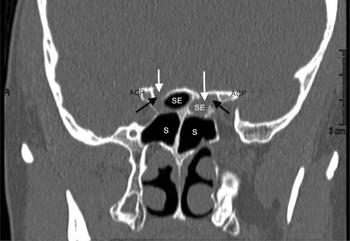
The patient was referred to our department eight months later regarding further management, as she was understandably concerned about the possibility of a similar complication occurring on the contralateral side. A CT scan performed at that time confirmed a contralateral (right) sphenoethmoidal cell with partial dehiscence of the optic nerve canal, but with no opacification or evidence of mucocele formation (Figure 2).
Coronal computed tomography scan showing an opacified left sphenoethmoidal cell with superolateral bony erosion (white arrow) adjacent to the optic nerve (black arrows), and an aerated right sphenoethmoidal cell with bony dehiscence of the optic nerve canal (white arrow). R = right; ACP = anterior clinoid process; SE = sphenoethmoidal cell; S = sphenoid sinus

The options of medical treatment versus ‘prophylactic’ surgical treatment were discussed, along with the potential risk of injury to the right optic nerve during any FESS. She elected to continue with medical treatment in the form of nasal douches and topical intranasal steroid drops, and underwent topical lysine-aspirin desensitisation, with good results.
The patient's sinonasal symptoms remain well controlled after five years. She is monitored with annual magnetic resonance imaging (MRI) of the sinuses to check for any signs of expansion of the right sphenoethmoidal cell, which have not occurred to date.
Case three
A 54-year-old man presented with longstanding sinonasal symptoms. He was subsequently diagnosed with chronic rhinosinusitis with nasal polyps on a background of asthma and marked allergic rhinitis. He was commenced on maximum medical treatment with douching, oral and topical steroids, and clarithromycin.
A CT scan showed generalised opacification. Bilateral sphenoethmoidal cells were also noted, with dehiscence of the medial wall of the right optic nerve canal within the right sphenoethmoidal cell; the cell itself was aerated.
His nasal symptoms and endoscopic appearances improved significantly with medical treatment. He had never had any visual symptoms.
After discussing the options, he chose to be followed up with annual MRI to ensure no mucocele development within the sphenoethmoidal cells. He has had no problems to date (at 18 months following presentation).
Case four
A 40-year-old man, known to have chronic rhinosinusitis with nasal polyps with aspirin-sensitive asthma, had undergone 3 previous nasal polypectomies elsewhere, the last of which was performed 6 years earlier. A CT scan performed seven years previously had identified a right sphenoethmoidal mucocele with loss of bone superiorly. Surgical treatment had been advised but not undertaken.
Five months prior to being seen in our department, he developed acutely reduced vision in the right eye, which improved with parenteral steroids. The same occurred four months later and again resolved with parenteral steroids.
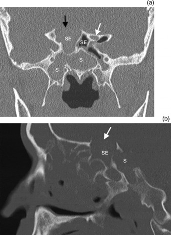
Magnetic resonance imaging and CT scans conducted two months prior to being seen in our department showed marked expansion of the ethmoid sinuses and dehiscence of the lamina papyracea, especially in relation to the left posterior ethmoid mucocele, with further enlargement of the right sphenoethmoidal mucocele (Figure 3). The generalised sinus opacification was heterogeneous, suggestive of possible eosinophilic fungal sinusitis.
(a) Coronal computed tomography (CT) scan showing bony erosion (black arrow) in the roof of an opacified right sphenoethmoidal cell; the left sphenoethmoidal cell is aerated with the optic nerve visible (white arrow). (b) Sagittal CT scan showing bony erosion (arrow) in the roof of a right sphenoethmoidal cell. SE = sphenoethmoidal cell; S = sphenoid sinus

In view of the episodes of visual loss, urgent surgery was advised. Functional endoscopic sinus surgery with image guidance was performed. This involved complete clearance of bilateral grade three nasal polyps and opening of the maxillary, sphenoid, ethmoid and frontal sinuses bilaterally. Pus was released from the left posterior ethmoid. The right sphenoethmoidal cell was opened, but contained only sterile mucus. The right optic nerve was clearly seen traversing the lateral wall of the sphenoethmoidal cell beneath intact mucosa.
Fourteen months post-surgery, the patient has had no further visual problems.
Discussion
Sphenoethmoidal cells are found in approximately 5–17 per cent of Caucasians, but are significantly more common in the Chinese population where they occur in 20–30 per cent of individuals.Reference Badia, Lund, Wei and Ho4 These prevalence figures are based on CT findings, and are generally accepted within the literature; however, a recent paper reported a much higher prevalence of 65.3 per cent based on 170 CT scans.Reference Tomovic, Esmaeili, Chan, Choudhry, Shukla and Liu5 Interestingly, cadaveric studies have also shown a higher prevalence, of up to 60 per cent, in Asian cadavers.Reference Thanaviratananich, Chaisiwamongkol, Kraitrakul and Tangsawad6, Reference Yeoh and Tan7 The discrepancy may reflect a lack of resolution on imaging. No significant difference has been found in terms of the presence of sphenoethmoidal cells in patients with chronic rhinosinusitis compared to normal controls.Reference Jones, Strobl and Holland8
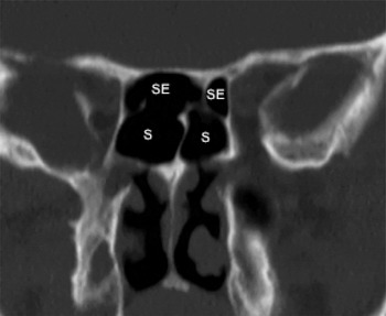
The optic nerve is not exposed in all sphenoethmoidal cells. In a study of 999 CT scans, sphenoethmoidal cells were identified in 16 per cent of cases and were bilateral in two-thirds of cases, as indicated by a characteristic cruciform appearance on coronal CT images (Figure 4).Reference Ozturan, Yenigun, Degirmenci, Aksoy and Veyseller9 The optic nerve protruded into 80 per cent of sphenoethmoidal cells compared to only 18 per cent of normal sphenoid sinuses (p < 0.01). The nerve was dehiscent in 36 per cent of cases with sphenoethmoidal cells compared to 0 per cent without (p < 0.01). In another review, of 170 CT scans, the rate of optic nerve dehiscence within a normal sphenoid sinus was reported as 2 per cent.Reference Tomovic, Esmaeili, Chan, Shukla, Choudhry and Liu10 Hence, there appears to be a higher risk of optic nerve dehiscence in the presence of a sphenoethmoidal cell. Interestingly, the internal carotid artery has been reported as dehiscent in 19 per cent of cases with sphenoethmoidal cells, compared to 6 per cent of cases without,Reference Ozturan, Yenigun, Degirmenci, Aksoy and Veyseller9 as evidenced by our first case.
Coronal computed tomography scan showing bilateral sphenoethmoidal cells with a more inferiorly placed sphenoid sinus, giving a characteristic cruciform appearance. SE = sphenoethmoidal cell; S = sphenoid sinus

There have been several case reports of pathology within a sphenoethmoidal cell causing visual loss. Mucoceles are the most commonly reported cause of optic neuropathy.Reference Ogata, Okinaka and Takahashi11–Reference Victores, Foroozan and Takashima18 This can present with sudden, or occasionally gradual, unilateral loss of vision, often with associated pain. In one reported case, the visual loss was bilateral because the large sphenoethmoidal cell extended laterally around both optic nerves.Reference Fukuda, Chikamatsu, Ninomiya, Yasuoka, Miyashita and Furuya14 The optic neuropathy is thought to be caused by: direct mechanical compression of the optic nerve, vascular thrombosis due to compression, or optic neuritis secondary to the inflammatory reaction.Reference Fukuda, Chikamatsu, Ninomiya, Yasuoka, Miyashita and Furuya14, Reference Nickerson, Lane, Subramanian and Izbudak17 In the majority of cases reported, surgical decompression of the mucocele was undertaken promptly, with subsequent improvement in vision. In one case, vision returned even though the surgery took place five weeks after the onset of symptoms.Reference Wu, Sun, Cannon, Jianbo and Selva16 Infection involving a sphenoethmoidal cell without mucocele formation has also been implicated as a cause of optic neuropathy and orbital apex syndrome.Reference Chee and Looi19, Reference Taflan, Gungor, Cengal Kurnaz and Gungor20 Visual loss from sphenoid sinus disease in the absence of a sphenoethmoidal cell is much less commonly reported.Reference Bizzoni, Bolzoni Villaret, Lombardi, Tomenzoli, Danzi and Semeraro21
• A sphenoethmoidal cell is a posterior ethmoid cell that pneumatises superiorly and/or laterally to the sphenoid sinus
• The optic nerve may lie within it or be exposed along the superolateral wall, leading to visual loss in the presence of sinus disease
• Optic neuropathy may be mechanical, thrombotic or inflammatory
• Prompt surgical decompression of the cell may restore vision
In our series of four patients, one had sudden and irreversible complete visual loss secondary to an acute mucopyocele within an expanded sphenoethmoidal cell. This occurred despite urgent surgical decompression and parenteral steroid treatment. The fourth patient experienced two episodes of acute reduction in vision; this improved in both instances with steroid treatment administered prior to surgical drainage. Two patients had no visual symptoms at any point, despite extensive sinonasal disease and the presence of sphenoethmoidal cells with bony dehiscence over the adjacent optic nerve. One of these patients is effectively cured, and the other is monitored with MRI to detect any involvement or expansion of the sphenoethmoidal cells which may lead to visual problems. If such a cell is found incidentally in patients with marked sinonasal disease, the options of elective surgical treatment versus monitoring should be discussed carefully with the patient.
Conclusion
Pathology within a sphenoethmoidal cell must be considered in cases of optic neuropathy. The presence of these cells may be relevant even in cases of seemingly uncomplicated chronic rhinosinusitis, as there is a higher rate of optic nerve protrusion and dehiscence within a sphenoethmoidal cell compared to a normal sphenoid sinus. Ophthalmologists and otolaryngologists alike should be aware of this anatomical variant. Treatment in such cases is prompt surgical decompression of the cell, which in many cases may be anticipated to restore vision.