Case report
A previously well 18-year-old man presented with hyponasal speech, nasal regurgitation of fluids and peripheral sensory symptoms. These symptoms appeared one week after eating a chicken sandwich and were attributed to an episode of Campylobacter jejuni enteritis, proven on stool culture. The patient first noticed hyponasal speech over a period of two hours, followed eight hours later by tingling in the finger tips. These symptoms did not progress over the following 24 hours. Four days later, however, the patient complained of nasal regurgitation of fluids, diplopia at the limit of horizontal gaze and mild imbalance.
On presentation at our department, the patient appeared generally well and had a normal gait. Temperature and other vital signs were normal. Examination of the ears and nose was normal. The oropharynx revealed mild fasciculations in the tongue and complete paralysis of the soft palate. Flexible fibre-optic nasendoscopy revealed a normal pharynx and larynx, with normal vocal fold mobility. A general head and neck examination was normal.
A detailed neurological examination was undertaken. Findings included a right abducens nerve palsy. All other eye movements were normal. Dysfunction of the motor branch of the trigeminal nerve was indicated by bilateral palatal paralysis. Facial and vestibulocochlear nerves were functioning normally. Bilateral glossopharyngeal impairment was indicated by reduced sensation of the posterior tongue and pharynx. The absent gag reflex was noted and attributed initially to the loss of glossopharyngeal sensation bilaterally. However, the concomitant complete palatal paralysis suggested the potential for bilateral vagal nerve involvement as well. Peripheral examination identified bilaterally reduced biceps and supinator reflexes; the ankle jerk reflexes were absent bilaterally. Glove distribution paraesthesia was noted up to the mid-forearms and to the ankles, bilaterally.
A diagnosis of Miller Fisher syndrome was made on the basis of a descending, symmetrical polyneuropathy secondary to Campylobacter jejuni gastroenteritis.
Initial management included intravenous fluid therapy and nasogastric feeding. The intensive care team were informed of the patient's condition. Intravenous mixed immunoglobulin (Ig) was administered over three days at a dose of 0.4 g per kg per day. Daily serial vital capacity measurements and routine blood results were within normal parameters. Cerebrospinal fluid (CSF) protein sampling and nerve conduction studies were not performed.
Over the following 48 hours, an improvement in the patient's diplopia, speech, peripheral dysaesthesia and gag reflex was reported. Lower back pain was noted and was attributed to difficulty adjusting to a hard hospital mattress, rather than to the neuropathy. After 72 hours, a neurology review identified persistently absent ankle reflexes. The absence of ataxia was noted. Within five days, all symptoms were improving and the patient was discharged as oral intake was adequate. Full recovery was achieved by the 10th day.
Discussion
To our knowledge, Miller Fisher syndrome has not previously been reported in the ENT literature. It is an important condition to be aware of, as it may lead to respiratory failure and other organ system dysfunction. Its presentation may lead to ENT referral, as this report demonstrates.
Background
Charles Miller Fisher first described this acute, idiopathic, inflammatory, demyelinating polyneuropathy, a rare variant of Guillain–Barré syndrome.Reference Fisher1 Comprising a classic triad of ophthalmoplegia, areflexia and ataxia, the condition is characterised by a descending paralysis in which the cranial nerves are primarily affected.Reference Overell and Willison2 Miller Fisher syndrome is synonymous with ophthalmoplegia-ataxia-areflexia syndrome, polyradiculoneuritis and acute, disseminated, encephalo-myeloradiculopathy.
Epidemiology
Patients with Miller Fisher syndrome make up approximately 5 per cent of cases of Guillain–Barré syndrome. The male to female ratio is three to two. The median age at onset is 42 years, with peaks at 30–39 and 50–59 years.Reference Ito, Odaka, Koga, Hirata and Yuki3
Aetiology
The condition commonly follows viral infection. However, bacteria, such as Campylobacter jejuni, are recognised causative agents.Reference Overell and Willison2
Pathogenesis
The link between infection and Miller Fisher syndrome can be explained by the presence of monoclonal antibodies that react with Campylobacter jejuni lipo-oligosaccharides and nerve axon or Schwann cell gangliosides.Reference Chiba, Kusunoki, Obata, Machinami and Kanazawa4 Pathogenesis involves pre- and post-synaptic neuromuscular blockade,Reference Chiba, Kusunoki, Obata, Machinami and Kanazawa4 with auto-antibodies found in highest concentration within the oculomotor, trochlear and abducens nerves. Antibodies react with complex carbohydrate components of myelin (GQ1b or GT1b).Reference Chiba, Kusunoki, Obata, Machinami and Kanazawa4
Miller Fisher subtype is particularly associated with GQ1b, which is an antigen reported in Campylobacter jejuni strains, as well with GT1a and GD3.Reference Yuki, Taki, Takahashi, Saito, Yoshino and Tai5, Reference Koga, Gilbert, Li, Koike, Takahashi and Furukawa6Haemophilus influenzae antigens resembling GM1 and GQ1b explain its own association with polyneuronitis.Reference Koga, Gilbert, Li, Koike, Takahashi and Furukawa6 The anatomical distribution of ganglioside types does correlate with clinical features, such as the presence of GQ1b in the ocular motor nerves. However, the density, configuration and accessibility of gangliosides is important to remember. The optic nerve contains equivalent amounts of GQ1b; however, the blood–brain barrier offers some protection, which explains the functional preservation of the brain.Reference Chiba, Kusunoki, Obata, Machinami and Kanazawa4
Anti-GQ1b antibodies have been shown to elicit complement-mediated damage to motor nerve terminals.Reference Goodyear, O'Hanlon, Plomp, Wagner, Morrison and Veitch7–Reference O'Hanlon, Plomp, Chakrabarti, Morrison, Wagner and Goodyear9 A significant release of acetylcholine caused a conduction block in a mouse phrenic nerve-diaphragm preparation.Reference Goodyear, O'Hanlon, Plomp, Wagner, Morrison and Veitch7 Due to the lack of post-mortem data, affected parts of the nervous system cannot be biopsied, and the overall pathology of Miller Fisher syndrome therefore remains unclear.Reference Hughes and Cornblath10
Clinical features
The clinical picture is a classic triad of ophthalmoplegia, areflexia and ataxia. Ophthalmoplegia is the most common feature. Symptoms progress over five to 10 days. Around half of the patients seen have additional symptoms, including generalised muscle weakness, respiratory failure, oropharyngeal weakness and/or unintelligible speech, facial sagging, tingling or numbness, and dizziness. Dysautonomia, bladder dysfunction and ileus are occasionally seen.Reference Meythaler11 Wide fluctuations of pulse rate and blood pressure occur in severe cases, signifying autonomic failure.
Coexistent bulbar, facial and generalised symptoms may be considered to represent an overlap between autoimmune demyelinating polyneuropathy and Miller Fisher syndrome.
Investigations
Definitive diagnosis can be demonstrated with electromyelography and nerve conduction studies. The primary electrophysiological finding is of sensory conduction abnormality secondary to sensory peripheral nerve demyelination.Reference Guiloff12, Reference Fross and Daube13 Demonstration of elevated protein levels in the CSF, in the absence of a leucocytosis, is seen in up to 60 per cent of patients.Reference Overell and Willison2 Serological evidence of recent campylobacter or haemophilus infection may also be sought. Measurement of serum IgG antibodies to GQ1b ganglioside has also been described and has been considered informative.Reference Overell and Willison2 An increased magnetic resonance imaging signal intensity from the brainstem, cerebellum and cranial nerves has also been reported.Reference Battaglia14
Differential diagnosis
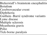
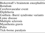
The differential diagnoses for an acute bulbar palsy is listed in Table I.
Causes of acute bulbar palsy
Management
Clinical observation for any deterioration is vital. Serial vital capacity measurement is undertaken to detect any respiratory deterioration, which can occur in up to 25 per cent of patients. Respiratory failure is more likely in rapidly progressing cases with bulbar palsy, upper limb involvement and dysautonomia. The early involvement of intensive care anaesthetists, to assess the requirement for prophylactic intubation, is recommended. Cardiac arrhythmia and deep vein thrombosis should be considered in severe cases.
Targetted treatment of the condition has been shown to reduce the duration of the illness. Plasma exchange of about five plasma volumes over two weeks has been reported to be beneficial during the first four weeks after presentation, particularly when administered early in severe cases.15, Reference McKhann, Griffin, Cornblath, Mellits, Fisher and Quaskey16 In patients with moderate disease, there was no difference in outcome between those who received four 1.5 plasma volume exchanges and those who received six exchanges.17 The costs associated with plasma exchange are offset by avoiding intensive care treatment and shortening the patient's hospital stay.Reference Espérou, Jars Guincestre, Bolgert, Raphael and Durand-Zaleski18
Intravenous mixed IgG has replaced plasma exchange as the standard treatment. In a Cochrane review of intravenous Ig,Reference Hughes, Raphael, Swan and van Doom19 the two treatments were comparable in terms of improvement in disability after four weeks, duration of mechanical ventilation, death and residual disability. Intravenous Ig is favoured because of its convenience. Using intravenous Ig after either plasma exchange or immunoabsorption has not been shown to provide extra benefit.Reference Hughes, Raphael, Swan and van Doom19, Reference Hughes, Wijdicks and Barohn20 Intravenous Ig is likely to have a multifactorial mechanism of action, possibly involving blockade of Fragment constant receptors, provision of anti-idiotypic antibodies, interference with complement activation and T-cell regulation.Reference Dalakas21
• Charles Miller Fisher was the first to describe an acute, idiopathic, inflammatory, demyelinating polyneuropathy, a rare variant of Guillain–Barré syndrome
• The condition commonly follows viral infection. However bacteria, such as Campylobacter jejuni, are recognised causative agents
• This paper describes a case of Miller Fisher syndrome presenting in an ENT setting. Referral was made on the basis of worsening nasal regurgitation following Campylobacter jejuni enteritis
• Early diagnosis is crucial and confers a favourable prognosis
Severely affected children are usually treated with intravenous Ig because of its greater convenience, and the small amount of evidence available supports this practice.Reference Korinthenberg, Schessl, Kirschner and Montning22 Trials of intravenous Ig have included patients within two weeks of disease onset, but the effect of intravenous Ig beyond two weeks remains untested. The question of how patients without signs of recovery by two weeks should be treated is debatable. One series has reported intravenous Ig to be effective.Reference Farcas, Avnun, Herishanu and Wirguin23 A newer technique involves antibody absorption upon a filter column with specific affinity for the individual ganglioside.Reference Willison, Townson, Veitch, Boffrey, Issacs and Andersen24
Steroids, commonly used in treating the demyelinated disease, have less encouraging results.Reference Hughes and van der Meche25 A possible explanation is that they adversely affect macrophage clearance of myelin debris, inhibiting remyelination, or that they aggravate the damage of denervated muscle fibres.Reference Rich, Pinter, Craner and Barchi26 A study using beta interferon-1a failed to show a significant effect, possibly due to the small sample size.Reference Pritchard, Gray, Idrissova, Lecky, Sutton and Swan27 Protection of the axons by sodium channel blockade was a successful strategy in experimental autoimmune neuritis, and this should be considered in cases of Guillain–Barré syndrome.Reference Bechtold, Yue, Evans, Davies, Gregson and Smith28
Supportive care from a multidisciplinary team is vital; intravenous fluids and nasogastric feeding are often required because of dysphagia.Reference Merkies, Schmitz, Samijn, van der Meché and van Doom29, Reference Bernsen, De Jager, Schmitz and van der Meché30 Organisations such as the Guillain Barré Syndrome Foundation International in the USA and the Guillain–Barré Syndrome Support Group provide ongoing patient support in UK.
Recovery
A full recovery is seen in the majority of patients, yet up to 20 per cent of patients are left with some degree of disability, irrespective of treatment. In most cases, recovery begins within four weeks and is almost complete after an average period of 10 weeks. Some patients are left with residual deficits. Less than 3 per cent suffer relapses, with these being classed as chronic demyelinating cases.
Conclusion
Miller Fisher syndrome is an uncommon condition with serious consequences. It may present to an ENT department, particularly if nasal regurgitation or dysphagia are prominent symptoms. It is important to undertake a thorough neurological examination and to involve the medical team early, as deterioration can be rapid. We draw the attention of the ENT community to this neurological disorder, which on presentation may easily be mistaken for an ENT disorder.
Acknowledgements
We would like to thank Dr I Ferguson (Consultant Neurologist, Southmead Hospital) and Dr A Turner (Consultant Neurologist, Southampton General Hospital) for their advice.