Introduction
In the 1980s, Barker observed that in the 20th century, the highest rate of infant mortality and coronary heart disease-caused mortality was found in the same regions of England. The main cause of infant death at that time was low birthweight; therefore, the risk of developing coronary heart disease appeared to be higher in adults who had a low birthweight.Reference Barker and Osmond 1 – Reference Barker, Winter, Osmond, Margetts and Simmonds 3 These among other observations discussed below led to the ‘Barker hypothesis’. Testing this hypothesis established that nutritional restriction of the mother results in a low-energy intrauterine environment that hinders fetal organ development and increases the offspring’s risk of developing non-communicable diseases (NCDs) later in life.Reference Ellison 4 , Reference de Boo and Harding 5
NCDs are non-genetic non-infectious medical conditions which progress slowly and are present throughout life such as atherosclerosis or diabetes.Reference Roura and Arulkumaran 6 They constitute a major problem because they are the current leading cause of death around the world, according to the World Health Organization. Approximately, 36 million people die every year due to NCDs and around 9 million of these are premature deaths.Reference Roura and Arulkumaran 6 Moreover, NCDs pose a huge economic threat because they require expensive treatment and reduce the working population, either through incapacitation or premature death. For example, in 2005, India and Brazil lost $9 and $3 billion, respectively, due to diabetes, heart conditions and strokes.Reference Roura and Arulkumaran 6 As a consequence, identification of the causes and prevention of NCDs are of high importance.
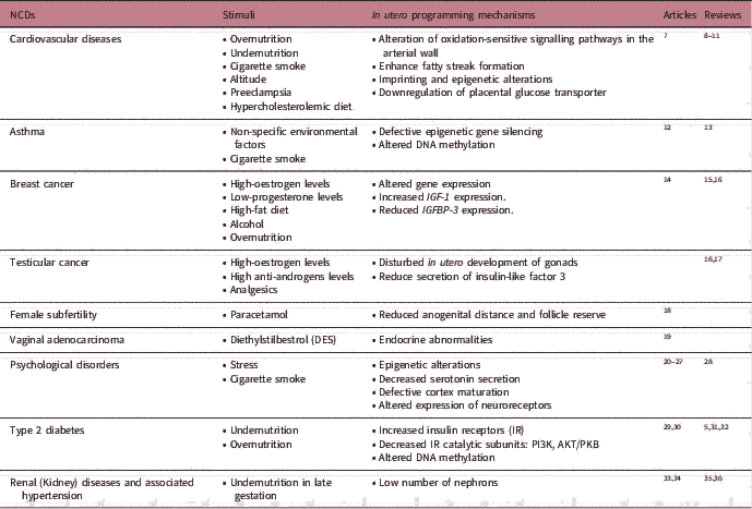
An increasing amount of evidence supports the hypothesis that defective in utero programming is the origin of a broad range of NCDs (Table 1). In fact, multiple studies have corroborated the Barker hypothesis. For instance, people conceived during the Dutch famine of 1944 and 1955 were more likely to suffer excessive weight gain, hypertension and glucose intolerance later in life.Reference Roseboom, van der Meulen and Osmond 37 – Reference de Rooij, Painter and Phillips 39 In addition, nutritional and non-nutritional stimuli such as a caloric-restricted diet and cigarette smoke could also affect offspring at multiple stages of development and increase NCDs risk in adulthood. As a result, the ‘Barker hypothesis’ has developed into the ‘Developmental Origins of Health and Disease’ (DOHaD). This theory proposes that stimuli at different life stages can augment predisposition to NCDs later in life and includes the concept of in utero programming by which programming of the embryo would promote offspring’s risk to NCDs later in life.Reference Roura and Arulkumaran 6 , Reference Ramirez-Velez 40
Examples of non-communicable diseases (NCDs) caused by defective in utero programming

Our aim is to provide a comprehensive review of the scientific literature on in utero programming and NCDs focusing on potential strategies utilizing this programming to reduce the incidence of NCDs in the offspring. First, the concept and mechanisms involved in in utero programming are discussed. Second, the negative role of in utero programming on the increased predisposition of NCDs in the offspring is briefly assessed. This introduces the first line of defence against NCDs: a passive medical approach consisting of the avoidance or reduction in preventable common negative stimuli. Finally, active medical approaches to protect against these diseases later in life are examined; the majority of these strategies involve the novel induction of positive in utero programming.
DOHaD and in utero programming
DOHaD stipulates that environmental stimuli at critical periods of development augment the risk of NCDs in adulthood due to subtle changes in biological functions.Reference Roura and Arulkumaran 6 These critical periods happen at different periods of the life of the individual, including in utero, neonatal and postnatal. This review focuses on the effect of in utero programming on the predisposition to NCDs. ‘Programming’ is the process by which a stimulus at a critical period of development results in a lifelong effect. This critical period during in utero programming corresponds to a window in which the biological system has a high degree of plasticity and is sensitive to the uterine environment provided by the mother. Once this window has passed, the plasticity disappears and the functional capacity is stabilizedReference de Boo and Harding 5 , Reference Roura and Arulkumaran 6 , Reference Barker 31 – plasticity might reappear at later critical periods.
Embryonic development, which starts with the fertilization of an egg and ends with a fully formed organism, is an extremely complex process. This process involves modifications of DNA and histone proteins to control gene expression and facilitate precise cell fate specifications in a spatio-temporal manner. Therefore, in utero programming might result in a broad range of lasting effects that can predispose fetuses to NCDs in adulthood. The basis of fetal programming remains largely unknown, and further studies are needed. Nevertheless, current research has elucidated three potential main mechanisms by which stimuli can have an effect; these can be classified as anatomical disruptions, epigenetic modifications and microbiota alterations.
Anatomical disruptions
Embryonic development involves the proliferation and differentiation of cells to form all tissues and organs of the organism. Any stimulus during this process might affect the anatomy of the biological systems currently developing and for the ones developing later; it might lead to suboptimal organs and tissues of disproportionate size. The disruption of the normal structure might have a subtle effect on the physiology because it alters the number of cells, which can result in an inability to fulfil the biological needs reducing the net performance. Consequently, the physiological function of the affected organ or tissue might be hindered increasing the risk of NCDs later in life.Reference Barker 31
As an illustration, prolonged undernutrition of the embryo in late gestation diminishes the replication of nephrons in the kidney, which cannot proliferate after birth. Brenner proposed that this reduction in renal cells is associated with an increased pressure in the glomerular capillaries leading to glomerular sclerosis, a condition that causes the further death of nephrons. This anatomical disruption could result in hypertension and extensive glomerular damage in later life.Reference Mackenzie and Brenner 35 Consistent with this, low birthweight in humans is associated with a low number of nephrons, which increases glomerular volume and hypertension.Reference Hughson, Farris, Douglas-Denton, Hoy and Bertram 33 , Reference Hoy, Douglas-Denton and Hughson 34 , Reference Hoy, Hughson, Bertram, Douglas-Denton and Amann 36 Loss of kidney mass due to accidents and resections is also associated with hypertension and further kidney damage.Reference Hoy, Hughson, Bertram, Douglas-Denton and Amann 36 , Reference Mei-Zahav, Korzets and Cohen 41 Similarly, the transplantation of small kidneys to large humans is linked to hypertension.Reference Hoy, Hughson, Bertram, Douglas-Denton and Amann 36 , Reference Brenner and Milford 42
Epigenetic modifications
Epigenetics is the process that regulates gene expression without modifying the DNA sequence. This regulation is mainly achieved through four epigenetic modifications: non-coding RNAs, histone modifications, chromatin remodellers and DNA methylation. DNA methylation has been the most widely researched epigenetic modifications in in utero programmingReference Babenko, Kovalchuk and Metz 28 , Reference Maccani and Knopik 43 (for more detailed information on the other epigenetic modifications, see Vliet et al., Kaikkonen et al. and Schmitz et al. Reference van Vliet, Oates and Whitelaw 44 – Reference Schmitz, Grote and Herrmann 46 ). In mammals, methylation of DNA takes place mainly on cytosines of CpG dinucleotides and represses the expression of the sequence. This repressive role is important during embryogenesis and is involved in a variety of events such as X-chromosome inactivation and genomic imprinting.
The epigenetic landscape changes considerably through embryonic development. There are two reprogramming events in which DNA is demethylated and progressively re-methylated. The first event, which occurs when the primordial cells migrate to the gonads, is the sex-specific marking of the imprinting genes that indicates if the DNA of the gonads belongs to a female or a male. If the gametes contribute to a future pregnancy, these methylations will mark the maternal or paternal origin of the DNA. These imprinting genes control fetal and placental growth during in utero development of the offspring. The second reprogramming event takes place between the zygote and the blastocyst stage. DNA is actively demethylated at fertilization to restore totipotency to all cells. This is followed by passive demethylation until blastocyst stage when DNA de novo methylation patterns are restored to facilitate cell fate specifications. Imprinted genes escape this second event.Reference Santos and Dean 47
Evidence suggests that epigenetic modifications are sensitive to environmental stimuli. Ollikainen and colleagues demonstrated that the intrauterine environment alters the epigenetic profile by analysing the DNA methylation patterns of dizygotic (DZ) and monozygotic (MZ) twins.Reference Ollikainen, Smith and Joo 48 They observed that DZ and MZ twins exhibited 82% and 54% of methylation discordances at birth, respectively; MZ twins share the same genome and were expected to have the same epigenome, therefore, the epigenetic differences observed suggested in utero environment can modify the epigenome of the offspring.Reference Ollikainen, Smith and Joo 48 In addition, undernutrition changes the epigenetic profile of the offspring. For example, individuals conceived during the Dutch famine exhibited 5.6% less methylation of insulin-like growth factor II (IGF2) differentially methylated region (DMR) before exon 3 than their siblings conceived before or after the famine.Reference Heijmans, Tobi and Stein 49 Hypomethylation of this DMR leads to increased expression of IGF2. IGF2 is a growth factor that promotes embryonic and placental growth during in utero development; it is maternally imprinted and only the paternal allele is expressed. IGF2 overexpression leads to overgrowth, a phenotype observed in individuals conceived during the Dutch famine.Reference Heijmans, Tobi and Stein 49
Due to the changing epigenetic landscape during embryonic development, any epigenetic alteration could have a broad range of effects on gene expression influencing the risk of NCDs in adulthood. IGF2 DMR hypomethylation increases the risk of colorectal cancer and NCDs associated with perinatal overgrowth such as prostate and breast cancer.Reference de Boo and Harding 5 , Reference Heijmans, Tobi and Stein 49 , Reference Cui, Onyango and Brandenburg 50 Exposure to maternal tobacco in utero also induces epigenetic changes as reviewed in Suter et al. Reference Suter, Anders and Aagaard 51 It is currently under study whether or not these modifications are associated with an increase in embryo growth restriction and associated NCDs.
Microbiota alterations
The human microbiota refers to the set of symbiotic micro-organisms that cohabitate in the human body. The gut microbiota is the most studied subset. Microorganisms in the gut are essential for development since they are involved in the programming of metabolism and immune response in the offspring. Studies have demonstrated that prenatal disruption of the gut microbiota in animals is associated with NCDs later in life, including colorectal cancer and asthma.Reference Couturier-Maillard, Secher and Rehman 52 , Reference Russell, Gold and Willing 53 In humans, a reduced allergy risk was observed in the offspring of mothers exposed to stables or barns during pregnancyReference Ege, Bieli and Frei 54 – this environment is rich in bacteria and most likely can affect the maternal microbiome composition. The role of microbiota alterations on in utero programming in humans remains largely unknown; studies have been mainly undertaken on postnatal programming (for the effect of the microbiome on NCDs development, see Vuillermin et al., Mulligan and Friedman and JenmalmReference Vuillermin, Macia and Nanan 55 – Reference Jenmalm 57 ).
Maternal microbes are transmitted to the offspring during childbirth. There is also some evidence that gut colonization might start in utero due to the microbiota in the female reproductive tract; however, this has not been conclusively demonstrated.Reference Collado, Rautava, Aakko, Isolauri and Salminen 58 , Reference Chen, Song and Wei 59 Intrauterine microbiota could arise from the gut microbiota or via the vaginal microbiota. Translocation of maternal gut bacteria to other body areas like the mammary gland in humans and mice has already been proven.Reference Perez, Dore and Leclerc 60 Perturbations of maternal gut microbiota could disrupt offspring microbiota and hinder metabolic and immune programming. For example, in fly larvae, maternal G418 antibiotic (stimuli) eliminates Acetobacter species in the maternal gut leading to delayed development in the offspring. External administration of Acetobacter species restores normal offspring development.Reference Fridmann-Sirkis, Stern and Elgart 61 It is possible that modifications of the maternal microbiota could protect the offspring against diverse NCDs.
Passive strategy against NCDs: negative in utero programming
As previously introduced, negative stimuli in the maternal uterine environment can lead to detrimental in utero programming resulting in NCDs in adulthood. The first line of defence against these NCDs is to avoid or reduce the adverse in utero programming induced by common maternal lifestyle choices. The International Federation of Gynaecology and Obstetrics (FIGO) recognizes the importance of supporting pregnant women to avoid defective in utero programming and reduce the incidence of NCDs. Consequently, FIGO with other agencies aims to improve prenatal care programmes ensuring early detection and treatment of maternal diseases as well as promoting the proper education of mothers about the risks of environmental and nutritional exposures.Reference Roura and Arulkumaran 6 In this review, we focus on basic scientific studies that gave rise to these FIGO approaches. We introduce the concept of negative in utero programming through two important maternal stimuli: diet and environmental chemicals.
Diet
Undernutrition is related to an increased risk of hypertension, type 2 diabetes, obesity and immune and coronary heart diseases in the offspring. This association has been strongly demonstrated through a series of epidemiological studies summarized in the study of Barker, 1998, and Ojha et al., 2013.Reference Barker 31 , Reference Ojha, Robinson, Symonds and Budge 62 When the nutritional demand of the fetus is higher than the placental supply, the embryo initially starts catabolism. If the nutritional restriction is prolonged, the embryo first decreases its metabolic rate and substrate usage, thereby slowing fetal growth. Second, the embryo blood flow is redistributed by organ-specific vasodilatation to protect the most important organs like the brain, and finally, the fetal secretion of hormones and interaction with the placenta and mother are altered.Reference Barker 31 , Reference Yoshimura, Masuzaki, Miura, Gotoh and Ishimaru 63 The placenta is also modified to ease survival.Reference Sandovici, Hoelle, Angiolini and Constancia 8 Altered organ growth and maturation seem to affect the physiology of several biological components such as liver, pancreas, muscle, hypothalamic–pituitary–anterior axis, kidney, vasculature and heart. This leads to hyperlipidaemia, hypertension and diabetes, which increase the risk of cardiovascular diseases as reviewed in the study of Boo and Harding.Reference de Boo and Harding 5
Slow fetal growth and blood redistribution in late gestation cause disruption of kidney anatomy leading to hypertension (Section ‘Anatomical disruptions’).Reference Barker 31 Hypertension may also be caused by altered vascular function and renin–angiotensin system homeostasis. Fleming’s group demonstrated that maternal low-protein diet during pregnancy or before implantation resulted in reduced vasodilatation in male mice. This diet also led to increased angiotensin-converting enzyme activity in the serum of female mice and lungs of male mice.Reference Watkins, Lucas and Torrens 64 Angiotensin-converting enzyme produces angiotensin II, a potent vasoconstrictor associated with hypertension.
The thrifty phenotype hypothesis proposed by Hales and BarkerReference Hales and Barker 32 stipulates that the embryo reduces insulin secretion and increases insulin resistance during undernutrition to conserve more glucose, which is directed to the brain and heart instead of insulin-dependent tissues. It has been shown that nutritional deficiency in utero in sheep is associated with an increase in insulin receptors (IRs) but a decrease in downstream catalytic subunits in the skeletal muscle.Reference Thorn, Regnault and Brown 29 The expression of the IR’s downstream proteins phosphatidylinositol 3-kinase (PI3K) and protein kinase-B (AKT/PKB) was reduced by 40%. It has been proposed that later in life, the abundance of nutrients combined with this insulin intolerance could lead to diabetes.Reference Thorn, Regnault and Brown 29 This study provided evidence of in utero alterations in the insulin metabolic pathway that, if conserved in adulthood, could lead to diabetes. Caution must be used when interpreting the results because the outcomes of animal studies might not be relevant for humans.Reference Barry and Anthony 65 Animal studies should be complemented with human epidemiological studies and in vitro studies using human tissues to more accurately determine the effect of stimuli on in utero programming and NCDs development in Homo sapiens, although in vitro studies have their own limitations. Furthermore, they have used a placental insufficiency intrauterine growth restriction (PI-IUGR) sheep model created by exposure to high temperature. The diet of the mothers was optimal for the development of the fetus. Thus, the study was not focused on the exclusive effects of undernutrition. Although placental insufficiency (PI) leads to reduced transport of nutrients including branched-chain aminoacids and this would mimic an undernutrition status, PI also provokes hypoxia that can also have negative effects on the fetus. To corroborate this study, the expression of IR, PI3K and AKT/PKB should be determined in ewes exposed exclusively to diet restriction. The expression levels of these proteins could also be analysed in humans known to have been exposed to undernutrition in utero. It would be interesting to follow up the sheep in this study and observe if the offspring develop diabetes in adulthood.
Undernutrition can alter the epigenome of the progeny. Male offspring (F1) of female mice subjected to caloric restriction exhibited hypomethylation of 17 regions in the sperm.Reference Radford, Ito and Shi 30 This suggests that the re-acquisition of methylation in progenitor cells may have been hindered in utero. Interestingly, the affected regions were close to genes whose expression patterns were also altered in F1 including Sstr3, Tbc1d30, Sur1 and Kcnj11; these genes are known to be involved in metabolism and glucose tolerance. For instance, Kcnj11 encodes two subunits of a β-cell pancreatic potassium receptor required for the regulation of insulin secretion; polymorphisms on this gene are linked to type 2 diabetes.Reference Radford, Ito and Shi 30 This study demonstrates a transgenerational effect; in utero programming-induced modifications may extend to and affect several generations, suggesting predisposition to NCDs might be transmitted through the family. It is thought that this effect could be caused by alteration of the paternal metabolic environment that would affect F2 or by epigenetic inheritance. In this study, F1 offspring depicted low birthweight, impaired muscle stem cell population as well as pancreatic function, and glucose intolerance. F2 generation through the male line also exhibited the same alterations including glucose intolerance and low birthweight without having being exposed to the adverse stimulus. Consequently, the authors analysed the methylome of the F2 generation focusing on the DMRs found in F1 to identify the transgenerational mechanism involved.Reference Radford, Ito and Shi 30 This might be seen counter-intuitive since the second reprogramming event in the zygote is thought to demethylate the whole genome excepting the imprinted regions. However, recent studies suggest that other domains of the genome also resist demethylation at this reprogramming event.Reference Borgel, Guibert and Li 66 – Reference Smith, Chan and Mikkelsen 68 Almost half of the DMRs analysed in this study were resistant. The methylome study of F2 offspring did not exhibit the previous hypomethylation suggesting that they might affect very early developmental processes before re-methylation with yet unknown outcome later in life.Reference Radford, Ito and Shi 30 Future work should further investigate the observed transgenerational effect; the involvement of the hypomethylated genes on early embryonic development could be studied with transgenic animals. Future work should also analyse whether the DMRs identified in F1 are secondary to other alterations in the DNA. This study does not demonstrate that undernutrition is directly inducing the demethylation; it might be affecting other chromatin elements such as transcription factors and histones.
Overnutrition is strongly associated with obesity, a NCD whose prevalence has skyrocketed in recent years.Reference Ojha, Robinson, Symonds and Budge 62 In utero overnutrition increases the risk of obesity, diabetes, hypertension, cardiovascular diseases and early adult death.Reference de Boo and Harding 5 Children of obese women who lost weight show a different risk of being overweight in adulthood; children born when their mothers were at optimal weight have half the risk of becoming obese than their older siblings born when the mothers were obese.Reference Smith, Cianflone and Biron 69 The developmental overnutrition hypothesis suggests that fetal overnutrition leads to different adaptations affecting control of the appetite, development of adipocytes, metabolism and endocrine axis. For example, obese ewes overfed during pregnancy resulted in offspring with increased expression of fatty acid transporters and increased rate of fatty acid synthesis in adipose tissue – these ewes were fed 150% of the national recommendations (USA).Reference Long, Rule, Tuersunjiang, Nathanielsz and Ford 70 Bitter taste receptors have recently been proved to be in the heart and agonists of these receptors cause relaxation of heart muscles, reduced ventricular pressure and increased pressure of the aorta.Reference Foster, Blank and See Hoe 71 Interestingly, maternal obesity due to high-fat diet during pregnancy and lactation results in a decrease in bitter taste receptors in hearts of rats.Reference Raipuria, Hardy, Bahari and Morris 72 The decrease in these receptors might increase offspring risk of cardiovascular diseases in adulthood. In addition, maternal overnutrition induces an epigenetic modification of the hypothalamic anorexigenic neuropeptide proopiomelanocortin (POMC) gene.Reference Marco, Kisliouk, Tabachnik, Weller and Meiri 73 POMC encodes α-melanocyte-stimulating hormone that suppress the appetite. The offspring of rats fed with high-fat diets exhibited increased levels of 5-methylcytosine (5mC) in Pomc promoter, which was associated with high bodyweightReference Marco, Kisliouk, Tabachnik, Weller and Meiri 73 . On the contrary, offspring from control rats presented high levels of 5-hydroxymethylcytosine but low 5mC in this promoter. Increased 5mC allows binding of methyl-binding domain 1 (MBD1) to Pomc promoter. MBD1 then binds to SETDB1 methyltransferase that bi-methylates histones (H3K9me2); this histone modification decreases Pomc expression. Therefore, maternal obesity modifies the methylation pattern of Pomc promoter in the offspring leading to histone modifications that decrease the expression of this gene. The resulted Pomc downregulation could hinder the suppression of the appetite contributing to the obese phenotype in the offspring.Reference Marco, Kisliouk, Tabachnik, Weller and Meiri 73
Environmental chemicals
People are exposed to environmental chemicals from common sources such as cosmetic products and pharmaceutical treatments. These chemicals might influence in utero programming, which increases the risk of NCDs in the offspring. A good example is bisphenol A (BPA) that increases offspring risk of infertility.Reference Savabieasfahani, Kannan, Astapova, Evans and Padmanabhan 74 – Reference Veiga-Lopez, Beckett, Abi Salloum, Ye and Padmanabhan 76 Certain analgesics like paracetamol, aspirin and indomethacin also augment offspring risk of NCDs (Table 1).
BPA is an endocrine disrupting chemical found in plastic and has been found in air, water, dust and in some biological samples such as hair, breast milk and placenta.Reference Liao and Kannan 77 In sheep, BPA exposure in utero, at doses relevant to humans, causes shortening of the time between the rise in oestradiol and the Luteinizing hormone (LH) surge and changes in ovarian follicular dynamics; this results in variable occurrence of follicular waves and alteration of follicular counts.Reference Veiga-Lopez, Beckett, Abi Salloum, Ye and Padmanabhan 76 Shortening of the oestradiol–LH surge interval suggests that the positive feedback of oestradiol to generate the LH surge and promote ovulation is altered. This seems to be caused by an increased sensitivity of the hypothalamic–pituitary axis to oestrogen and an earlier upregulation of gonadotropin-releasing hormone receptor in the pituitary. BPA increases the expression of oestrogen receptor 1 and reduces the expression of oestrogen receptor 2 in the hypothalamus, which promotes and inhibits LH surge, respectively.Reference Veiga-Lopez, Beckett, Abi Salloum, Ye and Padmanabhan 76 It would be interesting to expose human pituitary cells to BPA in vitro at concentrations relevant to humans. This experiment could be done using a human pituitary adenoma cell line and would determine whether the expressions of oestrogen receptors 1 and 2 are altered by BPA.
The presence of BPA in multiple human reproductive fluids and tissues such as ovarian follicular fluid, fetal plasma and placental tissue may result in a transgenerational effect of infertility. BPA exposure to pregnant mice modified the reproductive capacity of offspring multiple generations later.Reference Ziv-Gal, Wang, Zhou and Flaws 78 Pregnant mice (F0) were exposed to BPA concentrations relevant to environmental doses. Their female offspring (F1) generated the F2 generation and F2 females gave birth to the F3 generation. F3 depicted delayed pubertal onset and first oestrus and reduced fertility. Moreover, F1, F2 and F3 exhibited a decreased ability to maintain pregnancy with increasing age.Reference Ziv-Gal, Wang, Zhou and Flaws 78 This transgenerational effect could be studied in humans; an epidemiological study could investigate the associations between subfertile women and previous BPA exposure in the family. Moreover, in utero BPA exposure had a dimorphic effect in mice livers, increasing oestrogen receptors and 17α-hydrolase in females but reducing them in males. On the contrary, cyclooxygenase 1 was decreased in the liver of female mice but increased in male mice.Reference Ilagan, Mamillapalli, Goetz, Kayani and Taylor 79
The underlying mechanisms of BPA’s negative effect on in utero programming and NCDs development are controversial. One study showed that BPA exposure in utero in mice did not affect DNA methylation of the liver in offspring.Reference van Esterik, Vitins and Hodemaekers 80 On the contrary, BPA exposure in utero has also been shown to reduce global DNA methylation and to increase DNA methylation at Gck promoter in liver and sperm of rats.Reference Ma, Xia and Wang 81 , Reference Li, Chang and Xia 82 The differences between these studies’ outcomes might be due to the windows of exposure, the species studied and the doses of BPA administered. It is important to use doses relevant to human exposure in studies about environmental chemicals since different doses might have notably different effects on in utero programming. However, the relevant doses might be difficult to determine whether the substance has a rapid clearance ratio or if its exposure concentration is highly variable between populations.
Active strategies against NCDs: positive in utero programming
In utero programming could potentially be used to prevent NCDs in later life. The increasing knowledge about NCDs’ mechanisms and defective in utero programming origin allows to actively target against the causes of these diseases. These active approaches will not only decrease the rise in NCDs, like the passive approach but could potentially reduce notably and faster their incidence. Importantly, the characteristic transgenerational effect of in utero programming suggests that these active approaches could induce NCDs protection through multiple generations. These active approaches are new and the majority of the information available is based upon animal studies – although there are some nutritional modification studies undertaken in humans. As mentioned earlier, animal studies cannot be extrapolated to humans and future in vitro studies with human tissue need to be performed to determine whether these approaches could protect against NCDs. If they show a protective effect, extensive clinical trials should be undertaken to corroborate their effect in human beings and to discard any potential side effects. This section provides a comprehensive summary of the current literature on positive in utero programming; it does not suggest these treatments should be recommended to pregnant women because of the above reasons. The aim is to provide a starting point for the research of active in utero programming in the fight against NCDs. These active approaches can be divided into direct in utero programming modifications and personalized lifestyle pregnancy programs (PLPPs).
Direct in utero programming modifications
The defective in utero programming origin of NCDs allows the development of treatments that preclude this negative programming. In addition, understanding NCD’s mechanisms, it is possible to positively manipulate in utero programming to protect against them in the future.
Preclusion of negative in utero programming
This active strategy hinders in utero programming by adverse stimuli to which the mother could be exposed. For instance, it is well known that maternal hypercholesterolemia predisposes offspring to atherosclerosis in later life due to increased lipid peroxidation and fetal fatty streak generation; in animal models, maternal treatment with cholesterol-lowering agents such as cholestyramine as well as with antioxidants such as vitamin E reduces offspring’s risk of developing this disease.Reference Palinski and Napoli 9
Recently, maternal prebiotic treatment was shown to protect against the increased risk of offspring obesity induced by maternal overnutrition.Reference Paul, Bomhof, Vogel and Reimer 83 Treatment of rats with oligofructose altered the composition of the maternal gut flora decreasing gestational weight gain and adiposity as well as reducing maternal energy intake. Bacterial fermentation of oligofructose induces the secretion of satiety hormones in the intestine inhibiting appetite.Reference Paul, Bomhof, Vogel and Reimer 83 In addition, resveratrol, an anti-inflammatory phytochemical, protects against lipopolysaccharide (LPS) caused adverse in utero programming in the developing brain of rats.Reference Rose, Parmar and Cavanaugh 84 LPS is an endotoxin found on gram-negative bacteria that activate astrocytes and microglia in the developing brain generating an inflammatory response. This leads to a deficit of dopamine, 3,4-dihydroxyphenylacetic acid, homovanillic acid and tyrosine hydroxylase; feeding the pregnant rats with a resveratrol-supplemented diet maintained normal expression levels of these proteins after LPS injection. This is important because dopamine deficit and chronic inflammation are markers of neurological NCDs like Parkinson’s disease.Reference Rose, Parmar and Cavanaugh 84 Maternal supplementation with germinated brown rice also protected the offspring against insulin resistance due to maternal high-fat diet in rats.Reference Adamu, Imam and Ooi 85
Continuous maternal exposure to light during pregnancy induces hypertension in male offspring of rats.Reference Tain, Lin, Chan, Lee and Hsu 86 Recently, maternal melatonin or agomelatine supplementation during pregnancy and lactation was observed to prevent offspring programmed hypertension. Interestingly, melatonin and agomelatine stopped upregulation of sodium transporters and the renin–angiotensin system genes, such as Slc9a3, Slc12a3, Agtr2 and Mas1. They also downregulated core clock genes like Bam1 and Clock. All these genes are important in programmed hypertension.Reference Tain, Lin, Chan, Lee and Hsu 86 Since the dams were supplemented with melatonin or agomelatine during pregnancy and lactation, it is unknown whether the protective effect is due to in utero programming, postnatal programming or both. Future studies could further investigate the role of these programmings.
Positive direct manipulation of in utero programming
Positive in utero programming treatments can potentially prevent the development of NCDs risk factors through life like high blood LDL levels.
In 2006, maternal immunization against oxidized LDL (oxLDL) was shown to prevent atherosclerosis in the offspring of hypercholesterolemic and normocholesterolemic rabbits and mice.Reference Yamashita, Freigang and Eberle 87 Increased numbers of IgM against oxLDL epitopes and IgM-LDL immune complexes were found in the offspring of the immunized mothers. Moreover, atherosclerosis was reduced by 56% in the offspring of oxLDL-immunized females compared to control. Even a single immune challenge with oxLDL stimulated a significant rise in IgM and IgM-LDL complexes in naive rabbits never exposed to hypercholesterolemic diets. The results suggested that oxLDL maternal immunization prevents atherosclerosis by three mechanisms: reducing the exposure of the fetus to maternal hypercholesterolemic, reducing the amount of oxLDL in maternal circulation and programming the immune response in the offspring. OxLDL maternal immunization positively changed postnatal humoral response to reduce this NCD.Reference Yamashita, Freigang and Eberle 87
The concept of in utero immune programming might be quite controversial since, up until now, the only known mechanisms to transmit immunological protection to the offspring consists of the transfer of maternal IgG antibodies through the placenta and the transmission of enterocytes coated with antibodies during lactation; the latter does not exist in humans. Thus, it is completely unknown whether this therapy would be applicable to humans. Nevertheless, if in utero immune programming is possible, the development of multiple immune-related disorders could be prevented. A recent study showed that immunization of hypercholesterolemic mice with oxLDL induces antibody-mediated immune response against a broad range of oxidation-specific epitopes in the offspring, which decreases risk to atherosclerosis and also insulin resistance and type 2 diabetes.Reference Eberle, Merki and Yamashita 88 Therefore, it would reduce the offspring’s risk of cardiovascular diseases later in life.
Low doses of formaldehyde during pregnancy in rats were observed to protect offspring against asthma by interfering with inflammation of the lung.Reference Maiellaro, Correa-Costa and Vitoretti 89 Maternal inhalation of formaldehyde in rats, at doses deemed harmless for human health, resulted in decreased recruitment of immune cells in the offspring’s lungs, blood and bone marrow after sensitization with ovalbumin. The synthesis of anaphylactic antibodies was also reduced as well as tracheal hyperresponsiveness.Reference Maiellaro, Correa-Costa and Vitoretti 89 Despite the protective effect of formaldehyde against asthma, intake of this substance during pregnancy is not recommended. Formaldehyde is a dangerous chemical used to fix tissues and cells. It is associated with multiple side effects in adults such as bronchitis, dermatitis and chest pain. Furthermore, in the previous study, the offspring of formaldehyde-treated rats depicted low birthweight. However, a new synthetic chemical could be engineered based upon formaldehyde structure to maintain the beneficial effects but eliminate the adverse ones.
Interestingly, manipulation of the maternal microbiota during pregnancy can protect against the development of autoimmune diabetes in the offspring.Reference Hu, Jin and Peng 90 Type-1 diabetes is a genetic disease in which the pancreatic islets are destroyed by T cells. Maternal treatment with neomycin modified the microorganism population of the mother, which increases the number of Bacteroidetes and Cyanobacteria in the intestine of non-obese diabetic mice. These changes in the maternal gut microbiota resulted in variations in the offspring microbiota that could influence the immune programming. The antigen-presenting cells of the neomycin-treated mice’s offspring exhibited weaker stimulation of CD8+ and CD4+ T cells. In addition, neomycin perinatal exposure reduced the incidence of autoimmune diabetes in the offspring including the second generation of mice.Reference Hu, Jin and Peng 90 This study suggests that positive in utero programming could be used to treat genetic diseases of offspring, which, with soaring levels of diabetes and treatment costs escalating, is well worth exploring further.
PLPPs
Healthy life habits of the mother can also induce positive in utero programming. PLPP programs could be developed for pregnant women to protect their offspring against common NCDs and high-risk ones, based upon family history.
Maternal diet
Certain diets contain bioactive compounds that reduce offspring’s risk of developing certain NCDs such as cancer or cardiovascular diseases. They might affect epigenetics processes and the maternal/fetus microbiota. Identification of these biological products and consumption of them at the developmental stages when in utero programming is more susceptible to their action might become an efficient preventive measure against NCDs.Reference Li, Saldanha and Tollefsbol 91 An important strength of this approach is that specific diet recommendations are easily applicable.
Illustrations of bioactive compounds are genistein, sulphoraphane and resveratrol.Reference Li, Saldanha and Tollefsbol 91 Genistein is a phytoestrogen found in soya bean products that seems to modify the hypermethylated phenotype of the agouti mice decreasing obesity and protecting against mammary gland cancer later in life. Sulphoraphane is an isothiocyanate found in certain vegetables, such as broccoli and cauliflower, which can reduce the risk of some cancers. A maternal sulphoraphane-enriched diet prevents the offspring of a mouse model of spontaneous mammary gland cancer to develop this disease.Reference Li, Saldanha and Tollefsbol 91 Resveratrol is a polyphenol found in plants such as cranberries and blueberries. As previously mentioned (Section ‘Preclusion of negative in utero programming’), it can be used to directly suppress the adverse in utero programming of LPS in the brain. In addition, resveratrol regulates histone methylation and acetylation modifying the epigenome.Reference Vahid, Zand, Nosrat-Mirshekarlou, Najafi and Hekmatdoost 92 For example, resveratrol activates sirtuin 1 (SIRT1), a histone deacetylase involved in healthy aging that promotes renewal of cells in the retina and protects retinal neurons. Abnormal localization of SIRT1 increases the apoptosis of retinal photoreceptors, as reviewed in Pennington and DeAngelis.Reference Pennington and DeAngelis 93 Consequently, maternal supplementation with resveratrol could also promote acute vision in the offspring. The regulatory role of dietary bioactive substances is extensively reviewed in the study of Vahid et al. Reference Vahid, Zand, Nosrat-Mirshekarlou, Najafi and Hekmatdoost 92
Maternal diets rich in fruits and vegetables during pregnancy are associated with reduced risk of certain cancers in the offspring including childhood brain tumours, lymphoblastic leukaemia and germ cell tumours.Reference Jensen, Block and Buffler 94 – Reference Pogoda, Preston-Martin and Howe 96 An epidemiological study showed a maternal diet rich in fruits but low in fried foods and cured meats decreased offspring’s risk of unilateral retinoblastoma. The latter disease was also prevented by maternal high intake of vegetables and fruits but low consumption of fried foods, sweets and red and cured meats. The main weaknesses of this epidemiological study were that food questionnaires were performed 13 years after pregnancy and that the responses could be biased depending on the mothers’ perception of a healthy diet.Reference Lombardi, Ganguly and Bunin 97
Consumption of fats might also be beneficial to the offspring. Although, different types of fats appear to have different effects. Oils containing high amounts of n-6 fatty acids appear to increase the risk of mammary tumorigenesis in the offspringReference Hilakivi-Clarke, Clarke and Onojafe 98 , Reference de Assis, Khan and Hilakivi-Clarke 99 while oils with high levels of n-3 fatty acids seems to reduce it.Reference Stark, Kossoy, Zusman, Yarden and Madar 100 – Reference Ion, Akinsete and Hardman 102 A lard-based high-fat diet (37% saturated, 38% monounsaturated, 24% polyunsaturated fatty acids) during pregnancy or pregnancy and lactation decreased the risk of mammary cancer in rats. The consumption of this diet during pregnancy in rats resulted in reduced incidence and multiplicity of mammary cancer, as well as longer tumour latency. The lard-based high-fat diet also induced an increase in H3K9me3 methylation and expression of p21 as well as p27, which are involved in G1-checkpoint of mitosis.Reference de Oliveira Andrade, Fontelles and Rosim 103 Interestingly, maternal consumption of conjugated linoleic acid (CLA) – an-6 fatty acid – during pregnancy and lactation prevented increased adiposity in offspring from high-fat diet dams.Reference Segovia, Vickers, Gray, Zhang and Reynolds 104 The optimal combinations of fatty acids in the pregnant maternal diet to prevent the development of NCDs in the offspring should be elucidated as this has far-reaching effects for women’s health.
Taurine supplementation during pregnancy and lactation protects against type 2 diabetes in fetal undernutrition.Reference Arany, Strutt and Romanus 105 – Reference Tang, Marchand and Lam 108 Taurine is a sulphur-containing non-essential amino acid that works as an antioxidant and a neurotransmitter; its concentration in fetal blood is 1.5-fold higher than in the mother. In rats, maternal supplementation with taurine promotes proliferation and decreases apoptosis of β-cell in pancreatic islet. It also increases glucose metabolism and insulin sensitivity. Interestingly, taurine consumption during pregnancy leads to certain gender-specific differences. It improves β-cell function more in males than in females and improves insulin sensitivity in peripheral tissues more in females than in males.Reference Tang, Marchand and Lam 108 Type 2 diabetes and fetal low-protein nutrition are also associated with altered mitochondrial DNA (mtDNA). mtDNA is reduced in type 2 diabetes and insulin resistance.Reference Lee, Lee and Lee 107 Moreover, gestational protein restriction in mice was shown to induce significant changes in gene expression in liver and skeletal muscle; the most abundant changes were in mitochondrial genes especially those involved in oxidative phosphorylation.Reference Mortensen, Olsen and Frandsen 106 In fact, mitochondria with abnormal shapes and reduced number of secretory granules were observed in β-cell of in utero malnourish rats. These mitochondria also exhibited decreased cytochrome c oxidase subunit I (COX I) expression. COX I is a protein encoded in the mitochondrial DNA that belongs to complex II of the oxidative phosphorylation. Maternal taurine supplementation restored all the above mitochondrial alterations.Reference Lee, Lee and Lee 107 In rats, maternal taurine supplementation exclusively in late pregnancy increases insulin resistance and obesity in the offspring.Reference Hultman, Alexanderson and Manneras 109 Therefore, these data indicate the importance of taurine during pregnancy. It would be advantageous to determine whether supplementation in early pregnancy has negative or positive effects to develop accurate taurine supplementation guidelines during pregnancy. Modifications of mtDNA through positive in utero programming present a great potential since mtDNA is transmitted maternally and beneficial modifications are likely to be transmitted to several generations.
It is worth mentioning that diabetes often leads to diabetic retinopathy that causes loss of vision. This loss of vision is due to the destruction of retinal vasculature, neo-vascularization and rupture of the blood–retinal barrier. Low-salt maternal diets protect against these effects reducing the risk of diabetic retinopathy and related diseases like retinopathy of prematurity.Reference Deliyanti, Armani and Casely 110 It would be interesting to assess the beneficial effect of maternal low-salt diets in human offspring.
As previously mentioned, prebiotics prevent against the adverse in utero programming induced by maternal obesity (Section ‘Preclusion of negative in utero programming’). Prebiotic diets can also reduce the risk of allergies later in life.Reference Lyons, O’Mahony and O’Brien 111 – Reference Bouchaud, Castan and Chesne 115 In wheat-immunized mice, a maternal diet containing galacto-oligosaccharide and inulin prevented wheat allergies in the offspring.Reference Bouchaud, Castan and Chesne 115 Similarly, maternal intake of short-chain galacto- and long-chain fructo-oligosaccharides protected against egg allergies in ova-immunized mice.Reference Hogenkamp, Knippels, Garssen and van Esch 112 , Reference Hogenkamp, Thijssen, van Vlies and Garssen 113 Prebiotic diet during pregnancy also prevented asthma in house dust mite-immunized mice offspring.Reference Verheijden, Willemsen and Braber 114 These beneficial effects of prebiotics are due to reduced induction of immunoglobulins E and histamine, damped Th2 immune response and increased number of T regulatory cells. This immune regulatory effect of prebiotics could be used to protect against immune-related NCDs in the offspring. It would be very interesting to test whether maternal prebiotic consumption can protect against certain immunological reproductive diseases. Maternal immune tolerance is essential for a successful pregnancy and its failure leads to rejection of the embryo preventing implantation or damaging the embryo through gestation. Prebiotic diets might increase maternal and offspring’s immune tolerance, which might reduce the risk of infertility due to recurrent implantation failure or miscarriage. A detailed study of three bacterial strains showed that, in mice, perinatal consumption of Bifidobacterium longum significantly increased the numbers of Foxp3+ T regulatory cells involved in immune regulation.Reference Lyons, O’Mahony and O’Brien 111 Direct administration of these bacteria during pregnancy might protect offspring against immune-related NCDs.
Importantly, clinical studies showed a potential benefit of maternal nutritional supplementation to prevent NCDs in humans. A reduction in low birthweight was observed after maternal supplementation with a cocktail of multiple micronutrientsReference Vaidya, Saville and Shrestha 116 and supplementation with iron and folate.Reference Pena-Rosas, De-Regil, Dowswell and Viteri 117 , Reference Pena-Rosas, De-Regil, Dowswell and Viteri 118 A balanced protein energy supplementation during pregnancy is also associated with increased birthweight in the offspring of undernourished women, which reduces the risk of low birthweight.Reference Imdad and Bhutta 119 Given the importance of birthweight on NCDs development in the offspring, maternal supplementation could be a good approach to prevent them. In addition, folate supplementation is associated with reduced levels of microalbuminuria in the offspringReference Stewart, Christian and Schulze 120 – this is considered to protect against neuronal damage.
Maternal exercise
Maternal exercise is widely recommended for a healthy pregnancy. The American College of Obstetricians and Gynaecologists recommends daily half-hour of moderate exercise during pregnancy. Water exercises are desirable to help regulate body temperature and blood flow. In animals, maternal physical activity has long-term benefits in the offspring. Increased spatial memory and the number of hippocampus cells were observed in the offspring of pregnant rats that swam. Maternal swimming also enhanced neurogenesis and short memory and augmented antioxidants activity and mitochondria biogenesis in the brain of offspring rats; these mechanisms may underlie the neuroprotection.Reference Marcelino, Longoni and Kudo 121 Therefore, maternal exercise during pregnancy might protect against neuronal damage later in life, this would reduce the incidence of neurodegenerative NCDs such as Alzheimer’s disease and Glaucoma. Until now, the positive in utero programming of exercise has only been studied in animals. Human epidemiological studies could assess the association between maternal sports habits before and during pregnancy with the incidence of neurodegenerative diseases later in life.
The mechanisms underlying the positive in utero programming of exercise also remain to be elucidated. Interestingly, regular physical exercise in adult rats is associated with increased brain expression of neurotrophic and growth factors as well as epigenetic modifications.Reference Solvsten, de Paoli, Christensen and Nielsen 122 For instance, the expression of vascular endothelial growth factor A (VegfA) gene is increased in the hippocampus of male rats after 2 weeks of voluntary running; this is associated with CpG hypomethylation in VegfA promoters and exon 1.Reference Solvsten, de Paoli, Christensen and Nielsen 122 Therefore, the neurogenesis in offspring enhanced by maternal exercise during pregnancy in rats might be the results of epigenetic modifications during in utero programming.
Conclusion
In utero programming origin of NCDs allows the development of several medical strategies to reduce their incidence. This programming is based upon the great plasticity of embryonic development in which stimuli can lead to anatomical disruptions, epigenetic modifications and microbiota alterations.
Adverse stimuli lead to negative in utero programming causing alterations with subtle detrimental effects on tissue physiology that increase offspring risk of NCDs later in life. Unhealthy diet and environmental chemicals increase the risk of cardiovascular diseases, atherosclerosis and infertility in the offspring. They represent maternal lifestyle choices that could be avoided or reduced as a passive defence against NCDs. Consequently, FIGO plans to improve maternal education to prevent pregnant women from getting in contact with those risk stimuli for NCDs. Nevertheless, many unknowns remain to be elucidated to optimize this passive approach. Future studies should determine whether preventable stimuli other than those mentioned herein might negatively affect in utero programming. Most importantly, future research should investigate whether in utero programming has more, the same or less relevance than postnatal programming for the development of NCDs
The increase in knowledge about in utero programming and NCDs development gives rise to the opportunity to actively target NCDs, which has the potential to significantly and rapidly reduce NCD incidence. Furthermore, active strategies might provide transgenerational protection. These strategies can be divided into direct in utero programming modifications and PLPP. The former might involve the preclusion of negative in utero programming by adverse stimuli or the induction of positive in utero programming that prevents the development of NCDs risk factors later in life. PLPP programs include beneficial maternal diet and exercise that protect offspring against these diseases. The active medical approach is promising since it could be used to prevent common NCDs and high-risk ones, according to family history. However, research on the active use of in utero programming to protect against NCDs is at a very early stage and future studies need to determine whether this approach is effective in humans and any potential side effects before being considered for clinical practice.
Acknowledgements
None.
Financial Support
This research received no specific grant from any funding agency, commercial or not-for-profit sectors.
Conflicts of Interest
None.