Introduction
Due to their short reproductive cycle and reduced maintenance cost, mice are widely used as an experimental model in reproductive biology research (Rochester, Reference Rochester2013; Gioiosa et al., Reference Gioiosa, Palanza, Parmigiani and vom Saal2015). Japanese fancy mouse, mini mouse or pet mouse are common names used to refer to domesticated strains of mice that present with different colour varieties and coat types (Davies, Reference Davies1912). The Japanese fancy mouse has many coloured spots or patches, irregularly placed on a white background and was established from the original Japanese wild mouse, Mus musculus molossinus (Davies, Reference Davies1912; Minezawa et al., Reference Minezawa, Moriwaki and Kondo1979; Koide et al., Reference Koide, Moriwaki, Uchida, Mita, Sagai, Yonekawa, Katoh, Miyashita, Tsuchiya, Nielsen and Shiroishi1998). Some studies have suggested that this strain is a hybrid between the ancestral colonies of M. m. musculus and M. m. castaneus, rather than an independent subspecies (Yonekawa et al., Reference Yonekawa, Moriwaki, Gotoh, Watanabe, Hayashi, Miyashita, Petras and Tagashira1988).
Although testis anatomy and organization are similar in mammals, each species may present specific morphofunctional features, such as those related to phylogenetic aspects and reproductive strategies (Kerr et al., Reference Kerr, Loveland, O'Bryan, Kretser and Neill2006; Setchell & Breed, Reference Setchell, Breed and Neill2006; Costa et al., Reference Costa, Leal, Silva, Ferreira, Guimarães and França2010a). Comprehensive studies on testis morphology and function have been conducted on very few living mammalian species (Leal & França, Reference Leal and França2009; Cordeiro-Júnior et al., Reference Cordeiro-Júnior, Costa, Talamoni and França2010; Costa et al., Reference Costa, Leal, Ferreira, Guimarães and França2010b). Knowledge of male reproduction is crucial for comparative studies (Comizzoli et al., Reference Comizzoli, Mermillod and Mauget2000; Pukazhenthi et al., Reference Pukazhenthi, Comizzoli, Travis and Wildt2006).
Extensive asynapsis of chromosomes in meiosis has been demonstrated for male and female hybrids between two closely related mouse subspecies (leading to pachytene arrest) (Bhattacharyya et al., Reference Bhattacharyya, Gregorova, Mihola, Anger, Sebestova, Denny, Simecek and Forejt2013). Based on the difficulty in generating large cohorts of offspring by natural mating, recently it was reported that the Japanese fancy mouse (hybrid) has poor reproductive performance (Hasegawa et al., Reference Hasegawa, Mochida, Matoba, Yonezawa, Ohta, Watanabe, Taya and Ogura2012). Although many genetic studies involving spotting phenotype on the coat have been performed on fancy mouse (Steingrímsson et al., Reference Steingrímsson, Copeland and Jenkins2006; Yoshiki & Moriwaki, Reference Yoshiki and Moriwaki2006; Moriwaki et al., Reference Moriwaki, Miyashita, Mita, Gotoh, Tsuchiya, Kato, Mekada, Noro, Oota, Yoshiki, Obata, Yonekawa and Shiroishi2009; Reissmann & Ludwig, Reference Reissmann and Ludwig2013), to our knowledge there are no quantitative data available regarding its testis structure and spermatogenic efficiency. In this context, the main aims of this study were to conduct a detailed testis biometric, histological and stereological analysis as well as to estimate the duration of the spermatogenic cycle, and the Sertoli cell and spermatogenic efficiencies in the Japanese fancy mouse.
Materials and Methods
Animals
Ten sexually mature Japanese fancy mice (JF1/MsJ, ~3.5 months of age) weighing ~15 g were used in this study. The animals were approximately 7–13 cm of length (from the tip of the nose to the tip of the tail) and were obtained from commercial breeders (Sete Lagoas City, Minas Gerais State, Brazil). The typical animal phenotype consisted of a black spotted white coat, and black eyes with a grey-ringed iris (Mouse Phenome Database, The Jackson Laboratory, USA). The animals were kept under controlled temperature (23°C) and lighting conditions (12L:12D photoperiod). Experiments were conducted following approved instructions for the ethical treatment of animals (CEUA/UFMG).
Thymidine injections and tissue processing
To estimate spermatogenesis duration, three animals received i.p. injections of tritiated thymidine (82.0 Ci/mmol, Amersham, Life Science, Buckinghamshire, UK), a tracer for proliferating cells. Injections of 100 µCi 3H-thymidine were performed using a hypodermic needle and the animals that received two thymidine injections were sacrificed at approximately 9 days and 1 h after injections.
Testes were perfused-fixed (through the left ventricle) by gravity with 0.9% saline containing 4% buffered glutaraldehyde, over 30 min (Sprando, Reference Sprando, Russell, Ettlin, Sinha-Hikim and Clegg1990). All animals received i.p. injection of heparin (125 IU kg/BW) and pentobarbital (50 mg kg/BW) before surgery. After fixation, testes were weighed, and cut with a razor blade into small fragments. Afterwards, the fragments were immersed, for 12 h, in 4% buffered glutaraldehyde. The tissue samples (~2 mm thickness) were prepared and embedded in plastic (glycol methacrylate) for histological and morphometric analyses.
For the autoradiographic analysis, unstained testis sections were immersed in a specific emulsion (Kodak NTB-2, Eastman Kodak Company Rochester, NY, USA) at 45°C. After drying for 1 h at 25°C, the testis sections were stored at 4°C over the course of 4 weeks. Afterwards, these sections were developed using Kodak D-19 solution (Eastman Kodak Company, Rochester, NY, USA) (Bundy, Reference Bundy, Stumpf and Solomon1995) and, afterwards, stained with toluidine blue. The subsequent evaluations of the sections were performed to identify the most differentiated germ cells type marked (when five grains were present over the nucleus) (França et al., Reference França, Ogawa, Avarbock, Brinster and Russell1998) after the radioisotope injection.
Testis morphometry
The volume densities of the different testis structures were calculated by light microscopy using a grid (441-intersection) placed in the microscopic ocular. Each animal scored 15 fields (6615 points) during counts at ×400 magnification. The seminiferous tubule diameter, as well as the epithelium height, were measured using a ruler placed in the microscopic ocular. Round tubule cross-sections (n = 30) were measured for each animal. The whole length of seminiferous tubules (metres) was achieved using the cylinder formula, considering the seminiferous tubule volume as well as the half of tubular diameter (radius) (Johnson & Neaves, Reference Johnson and Neaves1981).
Seminiferous epithelium cycle
Seminiferous epithelium cycle (SEC) stages were characterized using acrosome and spermatid nuclear morphology. Stage frequencies were calculated analysing 150 seminiferous tubule cross-sections per animal. The spermatogenic cycle duration was estimated taking into consideration the most differentiated germ cell type marked at different periods (times) after radioisotope injection. The duration of spermatogenesis was estimated taking into account that 4.5 cycles are necessary for the completion of the entire process (Amann, Reference Amann1983).
Cell counts and cell numbers
Germ and Sertoli cells
All Sertoli cell nucleoli and germ cell nuclei were counted per animal in 10 round seminiferous tubule cross-sections at stage VII of the cycle. These counts were normalized considering the section thickness (4 µm) and nucleus/nucleolus diameter (Abercrombie, Reference Abercrombie1946; Amann, Reference Amann1962). The Sertoli cell nucleolus diameter (10 nucleoli per animal) was measured because it makes the identification of this cell easier as the nuclear shape is irregular. In addition, the germ cells nuclei (n = 10) were measured per animal and the cell ratios were found from the normalized counts.
Considering that there are no significant apoptosis during spermiogenesis (Russell & Clermont, Reference Russell and Clermont1977), the round spermatids number obtained in this species was considered to be the final population of spermatozoa. Sertoli cell number was calculated for each testis and per testis gram from the normalized counts of Sertoli cell nucleoli and the seminiferous tubules length (Hochereau-de-Reviers & Lincoln, Reference Hochereau-de-Reviers and Lincoln1978). The daily sperm production for each testis and per testis gram was calculated based on Sertoli cell number, efficiency and seminiferous stage frequencies using the formula described by França (Reference França and Dieleman1992).
Leydig cells
The proportion between nucleus and cytoplasm was calculated to estimate the individual Leydig cell volume. Its volume was calculated from the mean nuclear diameter as the nucleus is spherical. In this regard, 30 Leydig cell nuclei were measured per animal and the nuclear volume was achieved using the volume formula of a sphere, in which radius = nuclear diameter/2. The nuclear and cytoplasmic percentages were obtained placing a grid (1000 points) over the Leydig cells. The individual cell size and the entire volume occupied by these cells in the testis parenchyma allowed the achievement of the Leydig cell number per testis and per gram of testis.
Results
Biometric data and testis volume density
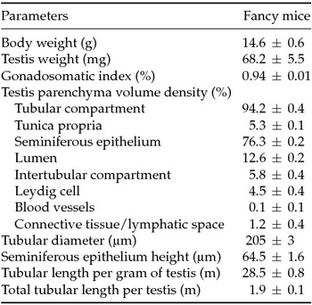
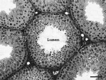
Mean body and testis weights were ~15 g and 68 mg respectively, giving almost 1% of gonadosomatic index (testis mass in relation to body weight) (Table 1). As shown in Table 1, the tunica albuginea percentage (data obtained after the removal of testis parenchyma) was approximately 4%, while the volume density of the tubular and intertubular compartments were ~94% and 6%, respectively. In these compartments, the seminiferous epithelium occupied ~76% while the Leydig cells volume occupancy was 4.5% (Fig. 1). The tubular diameter and height of seminiferous epithelium were 205 and 65 µm respectively (Table 1). The length of the seminiferous tubule per gram of testis was approximately 29 m, whereas about 2 m of tubules were found per testis.
Stereological biometric and testis data in fancy mice [mean ± standard error of the mean (SEM)]

Cross-section showing tubular and interstitial compartments of the Japanese fancy mouse testis. The tubular compartment is comprised by tunica propria (TP), seminiferous epithelium (SE) and lumen. In the intertubular compartment, the Leydig cells (LC) are closely associated with the blood vessels (BV) and surrounded by a lymph-filled space (LS). Figure magnification = ×200; scale bar = 50 µm.

Stages of seminiferous epithelium cycle and their relative frequencies
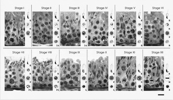
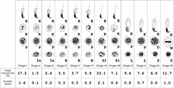
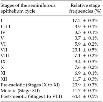
Twelve stages of SEC were classified based on acrosome morphology in spermatids (Fig. 2). The germ cell composition in the 12 stages and their relative frequencies are shown in Fig. 3 and Table 2. The frequencies of stages II and III were presented together because of their very low occurrence and stage VII (~23%) showed the highest frequency. The frequencies of pre-meiotic (stages IX–XI), meiotic (stage XII) and post-meiotic (I–VIII) phases were approximately 24%, 12% and 64% respectively.
Stages I to XII of the seminiferous epithelium cycle, based on the development of the acrosome, in spermatids and overall germ cell association are shown in Japanese fancy mouse. The individual germ cell nuclei shown in the right column represent the germ cells found in each particular stage. Type A spermatogonia (A); intermediate spermatogonia (In); Type B spermatogonia (B); pre-leptotene (Pl); leptotene (L); zygotene (Z); pachytene (P); diplotene (D) spermatocytes; meiotic figure (M); secondary spermatocyte (II); round spermatids (R); elongating/elongated spermatids (E); and Sertoli cell (SC). Figure magnification = ×1000, scale bar = 10 µm.

Diagram showing the germ cell composition for each of the 12 stages (roman numerals at the bottom) of the cycle, characterized based on the development of the acrosome in the spermatids nuclei. Letters within each column indicate the different germ cell types present at each stage of the cycle. Intermediate (In) and Type B (B) spermatogonia; preleptotene (Pl), leptotene (L), zygotene (Z), pachytene (P) and diplotene spermatocytes (D); meiotic figures (M); round (R) and elongating/elongated spermatids (E). The stage frequencies and duration (in days) of each stage are also shown at the bottom in this figure.

Stages of the seminiferous epithelium cycle and relative stage frequencies in Japanese fancy mice (mean ± SEM)

Seminiferous epithelium cycle duration
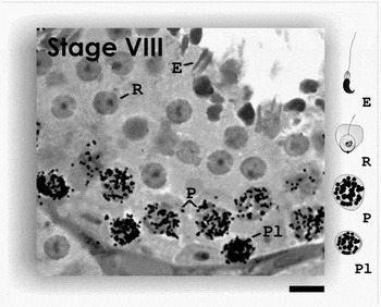
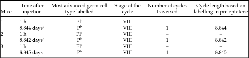
The most differentiated germ cell types (3H-thymidine positive) observed at the different time periods evaluated post radioisotope administrations, are shown in Fig. 4 and Table 3. Approximately 1 h after injection, the labelled cells were preleptotene spermatocytes that were located in the basal compartment at stage VIII (Fig. 4). Nine days after injection, labelled pachytene spermatocytes were found in the same stage (Fig. 4). Considering the most advanced labelled germ cell, the SEC duration was calculated as 8.8 ± 0.01 days. In our study, the interval between the germ cells labelling (preleptotene to pachytene spermatocytes) corresponded to approximately one cycle of the seminiferous epithelium (Fig. 4). Taking into consideration that approximately 4.5 cycles are required for the entire spermatogenesis, the total length of this process was estimated to be 39.8 ± 0.01 days.
Most advanced labelled germ cells found at the two different time intervals after thymidine injections in Japanese fancy mouse. As can be observed the labelled cells were preleptotene (Pl) and pachytene (P) spermatocytes found at the stage VIII respectively at 1 h and 8.8 days. R = round spermatids; E = elongated spermatids. Figure magnification = ×1000, scale bar = 10 µm.

The length (days) of seminiferous epithelium cycle (mean ± SEM)

Mean duration of the cycle = 8.84 ± 0.01 days.
a Pl/L, Preleptotene primary spermatocytes.
b P, pachytene spermatocytes.
c Total time after thymidine injection minus 1 h.
Testis stereology
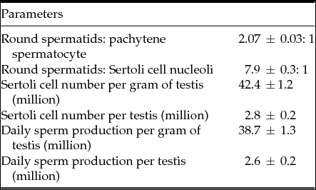
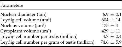
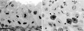
The data related to the testis stereology are displayed in Tables 4 and 5. The number of round spermatids that originated from each pachytene primary spermatocyte (meiotic index), was approximately 2.1. This result demonstrated that almost 50% of germ cell loss occurred in the course of the two meiotic divisions (Fig. 5). Almost eight round spermatids are supported by each Sertoli cell (Sertoli cell efficiency). The Sertoli cell number per testis gram was 42 × 106, whereas this figure per testis was approximately 3 × 10⁶. The daily sperm production per testis gram and per testis were about 39 × 106 and 3 × 10⁶ respectively. The Leydig cell nuclear volume was 175 µm3 while its individual size was 604 µm3. The data obtained for Leydig size and volume occupancy allowed us to estimate that the number of this steroidogenic cell per gram of testis (~75 millions) and per testis (~5 millions).
Cell counts, cell ratios and sperm production in Japanese fancy mouse (mean ± SEM)

Leydig cell morphometry in Japanese fancy mice (mean ± SEM)

Germ cell apoptosis during meiotic divisions in the Japanese fancy mouse. Seminiferous tubule cross-sections displaying morphologically normal meiotic divisions (A; white arrowheads) and commonly observed apoptotic meiotic figures (B; black arrowheadheads). Figure magnification = ×1000, scale bar = 10 µm.

Discussion
This study is the first to perform a detailed stereological/morphometric evaluation of the testis and to estimate testis key parameters such as the duration of spermatogenesis, Sertoli cell and spermatogenic efficiencies in the Japanese fancy mice. We demonstrated a high index of apoptotic spermatocytes during the meiotic divisions and, as a consequence of this germ cell loss, the values found for Sertoli cell and spermatogenic efficiencies were lower when compared with other laboratory mice strains investigated (Clermont & Trott, Reference Clermont and Trott1969; Avelar et al., Reference Avelar, Leal, França, Levy, Taskén and Hansson2004).
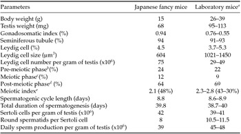
Table 6 is a summary of key parameters comparing the data herein obtained with those related to laboratory mice strains (Clermont & Trott, Reference Clermont and Trott1969; Avelar et al., Reference Avelar, Leal, França, Levy, Taskén and Hansson2004; Hess & França, Reference Hess, França and Cheng2007). However, the spermatogenic efficiency in the Japanese fancy mice is still relatively high in comparison with other evaluated mammalian species (França & Russell, Reference França, Russell, Martýnez and Regadera1998; Johnson et al., Reference Johnson, Varner, Robert, Smith, Keillor and Scrutchfield2000; França et al., Reference França, Avelar and Almeida2005; Hess & França, Reference Hess, França and Cheng2007).
Comparative parameters related to the testis stereology and spermatogenic events

a Clermont & Trott, Reference Clermont and Trott1969; Avelar et al., Reference Avelar, Leal, França, Levy, Taskén and Hansson2004; Hess & França, Reference Hess, França and Cheng2007.
b After spermiation and prior to meiosis.
c Meiosis I through meiosis II.
d After completion of meiosis until spermiation.
e Measured as the number of round spermatids produced per pachytene primary spermatocyte (presumptive germ cell loss in parenthesis).
Sertoli cell number (Sharpe et al., Reference Sharpe, Fraser, Brougham, McKinnell, Morris, Kelnar, Wallace and Walker2003; França et al., Reference França, Avelar and Almeida2005; Holsberger & Cooke, Reference Holsberger and Cooke2005) determines the magnitude of sperm production per testis (spermatogenic efficiency) (Johnson et al., Reference Johnson, Varner, Robert, Smith, Keillor and Scrutchfield2000; Hess & França, Reference Hess, França and Cheng2007). Therefore, this cell number was used to quantify and analyse daily sperm production, as Sertoli cell efficiency is relatively constant for each species (França & Russell, Reference França, Russell, Martýnez and Regadera1998; Johnson et al., Reference Johnson, Varner, Robert, Smith, Keillor and Scrutchfield2000; França & Hess, Reference França, Hess, Skinner and Griswold2005; França et al., Reference França, Avelar and Almeida2005; Gerber et al., Reference Gerber, Heinrich and Brehm2016). Although Sertoli cell number per gram of testis found in Japanese fancy mouse is very similar to that obtained for laboratory mice, probably due to high incidence of apoptosis in the second phase of the spermatogenesis the values obtained for Sertoli cell efficiency in this hybrid are lower. The data obtained here corroborate with the data of Bhattacharyya et al. (Reference Bhattacharyya, Gregorova, Mihola, Anger, Sebestova, Denny, Simecek and Forejt2013) in which meiotic asynapsis of heterospecific homologous chromosomes was found for hybrids of mice (Mus m. musculus × Mus m. domesticus) leading to pachytene arrest and lower reproductive rates. This situation probably occurs because meiosis is carefully monitored through checkpoints, avoiding therefore the creation of defective gametes with an aberrant number of chromosomes (Subramanian & Hochwagen, Reference Subramanian and Hochwagen2014).
Regarding the stage frequencies, our study corroborates with several studies suggesting that phylogenetically close related species present a similar stage frequencies distribution when these frequencies are grouped in pre-meiotic and post-meiotic phases (Hess & França, Reference Hess, França and Cheng2007; Costa et al., Reference Costa, Leal, Silva, Ferreira, Guimarães and França2010a, Reference Costa, Leal, Ferreira, Guimarães and Françab). For instance, as shown in Table 6 the frequencies of these phases are very close when different mice strains are compared. Although the length of spermatogenesis is controlled by the germ cell genome (França et al., Reference França, Ogawa, Avarbock, Brinster and Russell1998) and not phylogenetically determined (Clermont, Reference Clermont1972; Amann & Schanbacher, Reference Amann and Schanbacher1983), the values observed for the SEC duration are very similar among fancy and laboratory mice strains (Table 6).
Regarding testis interstitium, the fancy mouse followed the pattern of organization already described for rodents, i.e. presented extensive peritubular lymphatic sinusoids and groups of Leydig cells clustered with blood vessels (Kerr et al., Reference Kerr, Loveland, O'Bryan, Kretser and Neill2006). Although the Leydig cell morphology and volumetric density are very similar between fancy and laboratory mouse, the individual Leydig cell volume in fancy mouse represents approximately half of the cell volume observed for laboratory mice. This characteristic leads therefore to approximately two-fold higher number of these steroidogenic cells per gram of testis in the fancy mouse (Clermont & Trott, Reference Clermont and Trott1969; Avelar et al., Reference Avelar, Leal, França, Levy, Taskén and Hansson2004). To our knowledge, an explanation about what determines the Leydig cell volume density and its size is not available in the literature.
In conclusion, most of the testis parameters investigated in this study for Japanese fancy mice were similar to those obtained for laboratory mice. Although there is a high incidence of germ cell loss during the meiotic divisions, the combination of high tubular compartment volume, fast duration of spermatogenesis and high number of Sertoli cells allows high sperm production in the Japanese fancy mouse, though not as high as the laboratory mice.
Author contributions
GMJC, MCL and LRF performed the experiments, analysed the data and wrote the paper.
Conflict of interest statement
The author(s) declare(s) that there is no conflict of interest regarding the publication of this paper.
Acknowledgements
Technical help from Mara L. Santos is highly appreciated.
Financial support
This work was supported by the Brazilian National Council for Research (CNPq); Minas Gerais State Foundation (FAPEMIG); and Coordination for the Improvement of Higher Level Personnel (CAPES) and PRPq.