Introduction
Human lymphatic filariasis is a highly debilitating disease caused by Wuchereria bancrofti and Brugia malayi. It affects 1.2 billion people and bancroftian cases alone account for 90% of the infections. The World Health Organization (WHO) initiated the Global Program to Eliminate Lymphatic Filariasis (GPELF) – by the year 2020 – through mass drug administration (MDA) and to alleviate disability and the disease (Ottensen, Reference Ottensen2000). There is much emphasis on developing rapid diagnostic assays to measure the prevalence of the disease in endemic and non-endemic areas. Antibody- and antigen-based assays are very useful in the early identification of filarial infections. They would be useful in assessing endemicity, exposure in children and travellers, and monitoring of MDA and the surveillance programme. The recombinant Wuchereria bancrofti SXP-1 (rWbSXP-1) antigen-based immunoassay developed earlier is capable of detecting circulating antibodies to both bancroftian and brugian infections (Rao et al., Reference Rao, Eswaran, Ravi, Gnanasekhar, Narayanan, Kaliraj, Jayaraman, Marson, Raghavan and Scott2000; Lalitha et al., Reference Lalitha, Eswaran, Gnanasekar, Rao, Narayanan, Scott, Nutman and Kaliraj2002). The immunoassay was later converted to a rapid, field applicable format for the diagnosis and surveillance of the filarial elimination programme (Baskar et al., Reference Baskar, Srikanth, Suba, Mody, Desai and Kaliraj2004; Lammie et al., Reference Lammie, Weil, Noordin, Kaliraj, Steel, Goodman, Lakshmikanthan and Ottesen2004). However, to identify active filarial infection in endemic populations, it is important to detect the circulating filarial antigens in infected human blood.
Currently, there are two diagnostic tests available: Og4C3 enzyme-linked immunosorbent assay (ELISA) (More & Copeman, Reference More and Copeman1990; Weil et al., Reference Weil, Lammie and Weiss1997) and an immunochromatographic card test (ICT) (Weil et al., Reference Weil, Lammie and Weiss1997) that detect circulating antigens of W. bancrofti in serum or whole-blood samples. However, the commercial assays have certain issues such as stability, cost and specificity in field applications. The ICT format shows 25% of microfilaria-negative individuals as being positive for circulating filarial antigens. Furthermore, differences in batch production have affected the effectiveness of the kits (Melrose et al., Reference Melrose, Durrheim and Burgess2004). Moreover, the sensitivity of the Og4C3 test is lower than that of the ICT test (Pani et al., Reference Pani, Hoti, Vanamail and Das2004). The Og4C3 ELISA is time consuming, needs skilled manpower and equipment, and may not suit field conditions.
Thus it is imperative to develop an additional method to detect brugian and bancroftian antigens. In the study presented in this paper, monoclonal antibodies against recombinant WbSXP-1 were developed and characterized for detection of the circulating filarial antigens.
Materials and methods
Microorganism and plasmid
The recombinant filarial antigen WbSXP-1 (Gene accession number AFO98861 cloned in pRSETB) was expressed as a His-tag fusion protein in salt-inducible Escherichia coli GJ1158 (Bhandari & Gowrishankar, Reference Bhandari and Gowrishankar1997), provided courtesy of Dr J. Gowrishankar, CDFD, Hyderabad, India. The protocol for large-scale production of recombinant E. coli GJ1158 was carried out as optimized and described by Janardhan et al. (Reference Janardhan, Pandiaraja, Thirugnanam, Balamurali, Kennedy, Mody, Desai, Meenakshisundaram and Kaliraj2007).
Protein purification
Purification of rWbSXP-1 protein was carried out by immobilized metal affinity chromatography (IMAC) on chelating Sepharose fast flow (Ni2+) matrix using an AKTA FPLC system (Amersham Pharmacia Biosciences, Sweden), under native conditions. The base buffer comprised 50 mm sodium phosphate, 25 mm Tris, 500 mm NaCl, pH 8.0. The protein was eluted in 250 mm imidazole, in the base buffer, desalted and buffer exchanged, freeze dried and purified further using gel filtration chromatography (Superdex-100) in AKTA FPLC using 10 mm sodium phosphate, 10 mm Tris buffer, pH 7.5.
Bancroftian and brugian serum samples
Serum samples from filarial patients were collected from distinct geographical locations for bancroftian and brugian filariasis, such as Wardha, Maharastra, through the Mahatma Gandhi Institute of Medical Sciences, India, and endemic regions of Chennai, India, such as Tambram, Chengalpet and Vellore, through the Department of Public Health and Preventive Medicine, Tamil Nadu, India. Sera were collected from various clinical groups: endemic normals (ENs), microfilaraemics (MFs) and those with chronic pathology (CPs).
To obtain microfilarial (mf) antigens, 5 ml samples of heparinized venous night blood of MFs were collected and filtered using 3 μm polycarbonate filters (Nucleopore filters, Whatman, UK) as described previously (Rao et al. Reference Rao, Eswaran, Ravi, Gnanasekhar, Narayanan, Kaliraj, Jayaraman, Marson, Raghavan and Scott2000; Baskar et al., Reference Baskar, Srikanth, Suba, Mody, Desai and Kaliraj2004). The mf antigen was prepared by resuspending the filtered mf in 500 μl phosphate-buffered saline (PBS), pH 7.2, and sonicating, using a small probe (Sartorius AG, Goettingen, Germany), for 10 min at 10 cycles/min.
Immunoreactivity of expressed rWbSXP-1 protein
Western blotting was performed as described by Towbin et al. (Reference Towbin, Stabelin and Gorden1979). The recombinant WbSXP-1 protein or mf antigens (~2 μg) were electrophoresed on polyacrylamide gels and transferred to a nitrocellulose membrane. To confirm rWbSXP-1 reactivity, the sera of MFs (either Bm or Wb), CPs, ENs, non-endemic normals (NENs) (1:200) and normal mice (1:200) were used as primary antibody. Pooled sera comprised ten human sera samples exhibiting reactivity with the antigens in ELISA. Rabbit antibodies to either human-IgG or mouse-IgG conjugated to alkaline phosphatase (1:1000) were used as the secondary antibodies. All the dilutions were made using PBS, pH 7.2. The colour was developed using nitro-blue tetrazolium chloride/5-bromo-4-chloro-3′-indolylphosphate p-toluidine salt (NBT/BCIP; Sigma-Aldrich Co., St. Louis, Missouri, USA) as substrate.
Monoclonal (MAb) and polyclonal antibodies to rWbSXP-1
Establishment and selection of hybridoma
The generation of hybridoma was carried out as described by Gangatirkar et al. (Reference Gangatirkar, Gangadharan, Narendranath, Nagpal, Salunke and Karande2002). Briefly, 6- to 8-week-old female BALB/c mice were immunized subcutaneously (s.c.) with 50 μg of purified rWbSXP-1 protein emulsified in Freund's complete adjuvant, followed by administration of 25 μg of the antigen in Freund's incomplete adjuvant. After resting for a month, 4 days prior to fusion, the mice were injected with 250 μg of rWbSXP-1, intraperitoneally in saline.
Fusion
Spleen was excised from the mice, splenocytes were prepared as a single-cell suspension and fused with myeloma line SP2/0-Ag14 at a ratio of 5:1 using PEG4000 (Merck, Rahway, New Jersey, USA). Cells were then aliquoted in 96-well plates in Iscove's Modified Dulbecco's Medium (IMDM) supplemented with 20% fetal bovine serum, 50 μm β-mercaptoethanol (βME) and HAT (10 mm hypoxathine, 4 × 10− 5 m aminopterin, 1.6 mm thymidine). When the hybridoma cells reached >50% confluency, the supernatant from each well was tested by ELISA for the presence of rWbSXP-1 antigen-specific antibodies.
The antigen-specific hybridoma clones were expanded and subsequently subcloned to monoclonality by the method of limiting dilution. Monoclonality was confirmed by subclass isotyping using MAb isotyping kit II (ImmunoPure, Pierce Biotechnology, Rockford, Illinois, USA).
Polyclonal antibodies to rWbSXP-1
Laboratory-bred rabbits were immunized with purified rWbSXP-1 as described by Harlow & David (Reference Harlow and David1998).
ELISA
Initially, the concentration and the dilution of the antigen and antibody were optimized by checkerboard ELISA as follows. ELISA plates (Nunc™, Thermo Fisher Scientific, New York, USA) were coated with rWbSXP-1 (10 μg/ml) suspended in 50 mm carbonate buffer (pH 9.6) and incubated for 2 h at 37°C. Blocking of the unoccupied sites was done with 0.5% gelatin in PBS for 2 h, followed by incubation with monoclonal or polyclonal antibodies for 1 h at 37°C. Plates were washed three times with PBS-T (PBS containing 0.05% Tween-20), then incubated with the respective secondary antibody conjugated to horseradish peroxidase (HRP) (Dako, Glostrup, Denmark) for 1 h. The plates were washed extensively and the bound peroxidase activity was detected using the substrate diaminobenzidine (DAB) or with tetramethyl benzidine (TMB) at a concentration of 60 μg/ml in citrate phosphate buffer, pH 5.5, containing 0.03% H2O2. The reaction was stopped with 1 m H2SO4, and absorbance was read at 450 nm using a multiscan reader (MTX, Lab Systems, Inc., Virginia, USA).
Sandwich ELISA
Briefly, purified polyclonal rabbit anti-rWbSXP-1 was coated on microtitre plate wells (Nunc Maxisorp™) and incubated with standard antigen/protein. Detection was achieved with monoclonal mouse anti-rWbSXP-1 antibodies, followed by rabbit antibodies to mouse Ig-conjugated to HRP (Dako). The peroxidase activity was determined as described above.
Purification of MAbs and polyclonal antibodies
The hybridoma culture supernatant (containing the monoclonal antibody of IgG1 isotype) was equilibrated with 50 mm glycine–Tris buffer (pH 8.5), containing 3 m NaCl and loaded on a protein A–Sepharose column (Amersham). The unbound proteins were washed with the equilibration buffer. The bound antibody was eluted with 0.1 m glycine–HCl, pH 3.0, and neutralized with 1 m Tris–HCl, pH 8.0. Antibodies were biotinylated as described by Harlow & David (Reference Harlow and David1998).
Results
Antigens
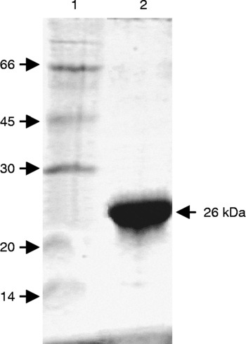
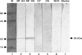
The filarial antigen, recombinant WbSXP-1, was expressed in E. coli GJ1158. The identity of purified rWbSXP-1 was established by SDS-PAGE (fig. 1). That the recombinant protein was of parasite origin was confirmed by Western blot analysis using infected human sera as the source of the corresponding antibodies. As can be seen from fig. 2, rWbSXP-1 reacted with the sera from infected individuals and not with that from normal or chronically infected individuals.
Purified rWbSXP-1 protein. Lanes: 1, protein molecular weight marker (kDa); 2, rWbSXP-1 (5 μg).

Western blot analysis of rWbSXP-1 protein. WbSXP-1 protein was probed with 1:200 diluted pooled human sera (ten samples/pool) and developed. Lanes: M, molecular weight marker lane; MF Bm, B. malayi microfilaraemics; MF Wb, W. bancrofti microfilaraemics; CP, chronic pathology; EN, endemic normal; NEN, non-endemic normal; and Murine, mouse anti-serum.

Hybridoma and monoclonal antibodies and selection of monoclones
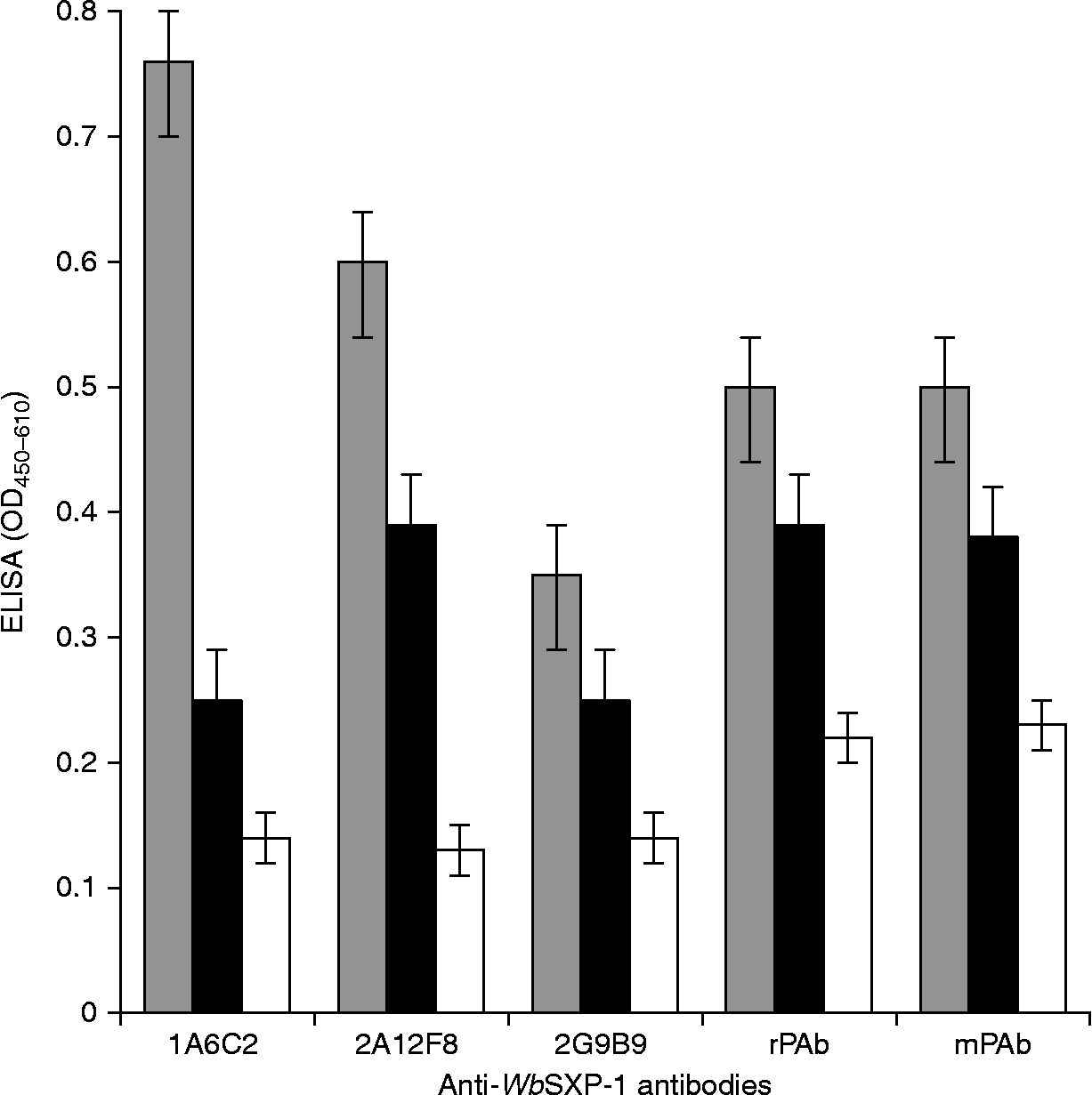
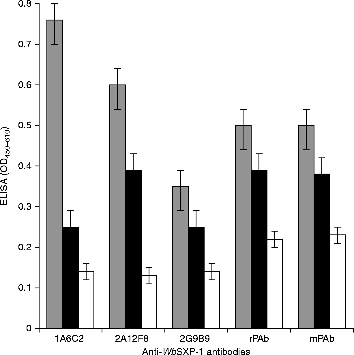
Mice were immunized with the purified protein for hybridoma development. Several hybridomas were established and antibody-secreting clones were first identified by ELISA for specific binding to rWbSXP-1 and expanded. The selected hybridoma clones were cultured over a few days, during which they were tested for binding to the recombinant protein. The stable clones were then cloned to monoclonality and tested for reactivity to W. bancrofti and B. malayi native mf antigens. Two clones, 1A6C2 and 2A12F8, which bound to both rWbSXP-1 and circulating mf proteins, giving high values in sandwich ELISA, were selected. As can be seen from fig. 3, the MAbs 1A6C2 and 2A12F8 showed better reactivity with the mf proteins than the polyclonal antisera raised in rabbit and mice. The 2A12F8 antibody, however, exhibited lower binding than 1A6C2 antibody.
Reactivity of MAbs with filtered mf antigens from infected sera. Sonicated samples (3 μg) from human serum membrane-retentates were coated (ELISA) and incubated with 1A6C2, 2A12F8, 2G9B9 culture supernatants. SP2/0 cell supernatant was used as the negative control. Polyclonal antibodies to WbXSP-1: rPAb, rabbit; mPAb, murine. n = 5 (mean of triplicates ± 3SD). Keys in order: grey bars, microfilaraemics; black bars, chronic pathology; white bars, endemic normals.
Characterization of monoclones
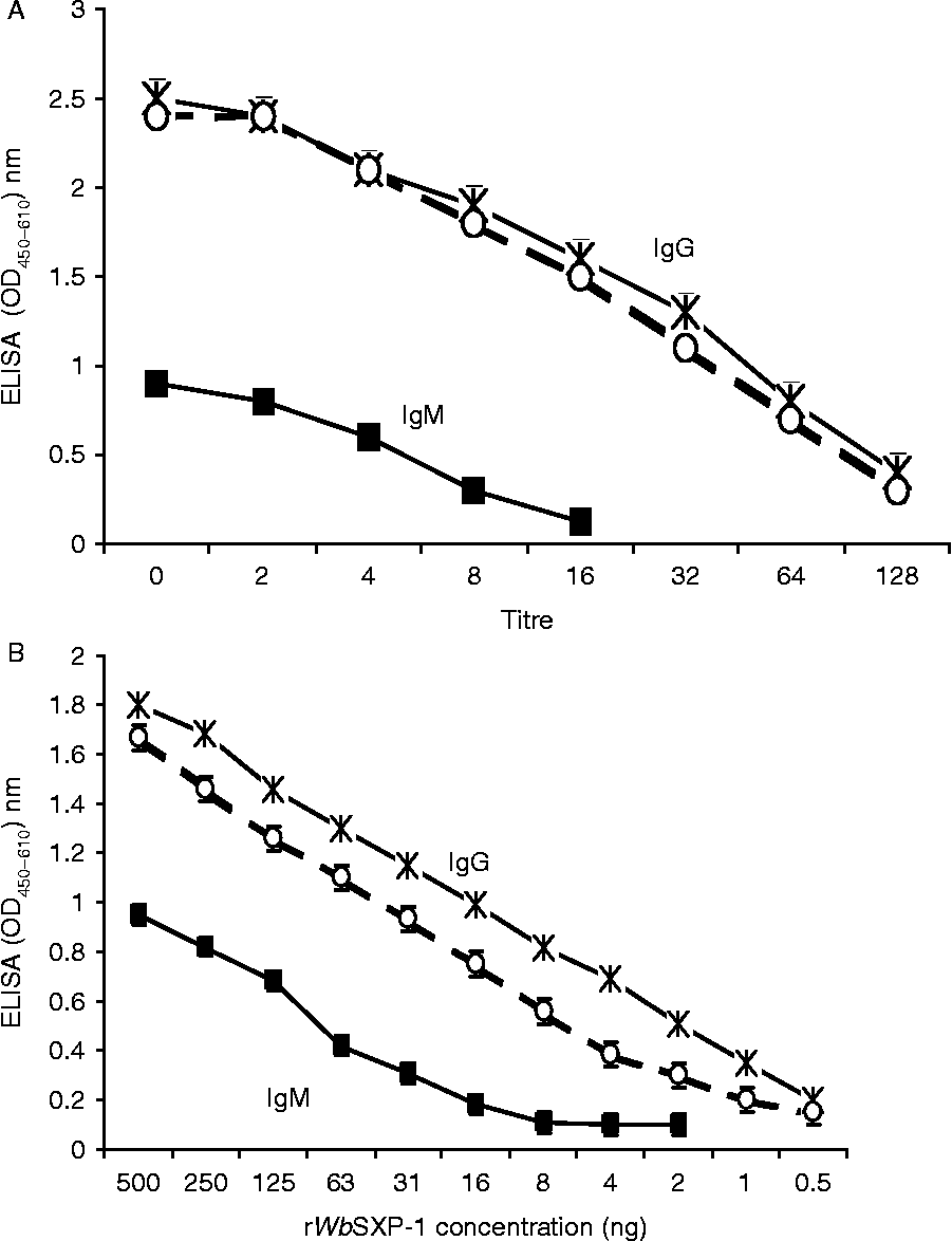
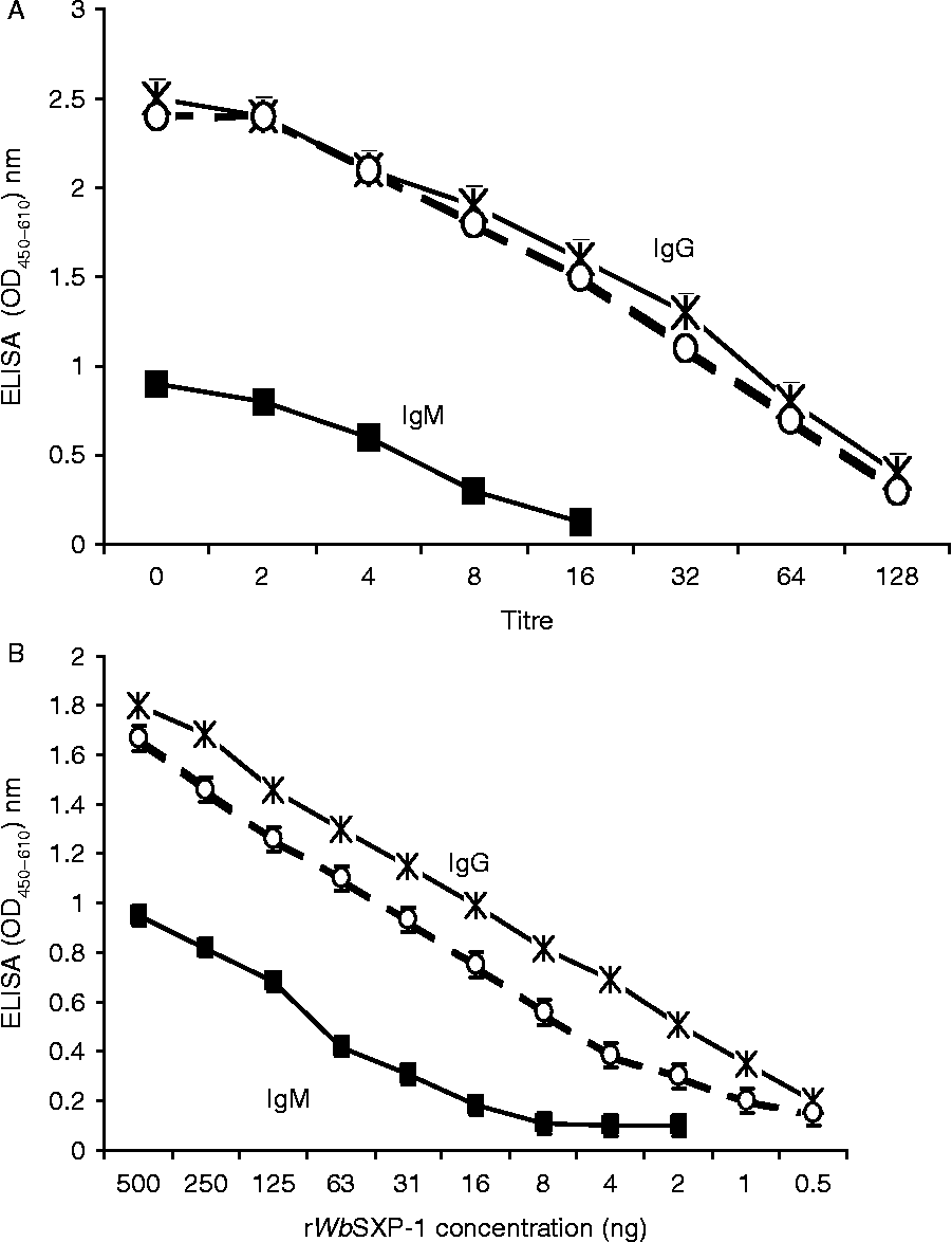
The dilution curves of the three MAbs, 1A6C2, 2G9B9 and 2A12F8, are shown in fig. 4A. Although the titres of the two MAbs 1A6C2 and B9C9 were comparable, as mentioned earlier, only MAb 1A6C2 bound to the native mf protein of the parasite. The MAb 2A12F8 exhibited a low titre. The sensitivity of MAbs to rWbSXP-1 was determined by the antigen dilution curve of rWbSXP-1 antigen ELISA (fig. 4B). The results showed that MAb 1A6C2 had a higher affinity to rWbSXP-1 when compared to the other clones. The lowest concentration of the antigen that could be detected with confidence was 4 ng for MAb 1A6C2. Based on the affinity and reactivity with the circulating mf antigens, 1A6C2 MAb was considered for further expansion and characterization. The clones 1A6C2 and B9C9 were identified to be of IgG1 subclass with kappa light chain.
Reactivity of monoclonal antibodies against rWbSXP-1. (A) rWbSXP-1 protein (1 μg) was coated (ELISA) and incubated with the hybridoma culture supernatants 1A6C2, 2A12F8 and 2G9B9 (serially double diluted). (B) Wells were coated with varying concentrations (500 to 1 ng) of rWbSXP-1 protein and incubated with undiluted hybridoma culture supernatants. The assay data plotted are mean values of triplicates ± SD. IgG:  , 1A6C2 and ○, 2G9B9; IgM: ■, 2A12F8.
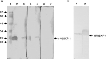
The reactivity of MAb 1A6C2 was then studied by Western blot analysis to detect rWbSXP-1 and SXP in Wb-mf and Bm-mf isolated by ultrafiltration from the infected serum (fig. 5). As is clear from the figure, the MAb recognized a protein band corresponding to the native antigen from infected serum. Mouse polyclonal antibodies were the positive control and SP2/0 culture supernatant was the negative control.
(A) Western blot analysis of 1A6C2 MAb with Wb-mf. Antigens were incubated with the primary antibodies and developed. Lanes: 1, molecular weight marker (kDa); 2, 4 and 6, purified rWbSXP-1; 3, 5 and 7, Wb-mf-SXP crude antigen. Lanes 2 and 3 were probed with 1A6C2 MAb, lanes 4 and 5 with anti-WbSXP-1 mouse polyclonal antiserum and lanes 6 and 7 with SP2/0 supernatant (negative control). (B) Western analysis of Bm-mf with 1A6C2 MAb. Lanes: 1, Bm-mf-SXP crude antigen; 2, rWbSXP-1 pure protein.

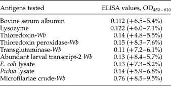
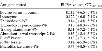
The specificity of the 1A6C2 monoclonal antibody was further established by testing its reactivity to several different recombinant antigens of the filarial parasite and other proteins, using ELISA (table 1). It was observed that the MAb 1A6C2 was highly specific for Wb antigens from microfilariae.
Establishing specificity of MAb to filarial antigens. Various purified bancroftian antigens, proteins and cell lysates (2 μg) were immobilized (ELISA) followed by incubation with the MAb 1A6C2 and developed. The assay data plotted are mean values of triplicates±deviations.
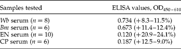
Further, the MAb 1A6C2 was evaluated for recognition of SXP-1 antigens in the serum from infected individuals. The selectivity and specificity of both brugian and bancroftian microfilaria-positive cases was studied using sandwich ELISA. Endemic normal and chronic pathology groups were used as controls. Although the level of antigen in serum samples and its bound state as immuno-complexes play a major role in detection, the MAb 1A6C2 could detect the circulating antigens only in positive cases of brugian and bancroftian infections (table 2). This confirmed that rWbSXP-1 is a prospective candidate for the development of antigen-based diagnostic assays for lymphatic filariasis.
Specific reactivity of monoclonal antibody 1A6C2 using sandwich ELISA with polyclonal rabbit anti-WbSXP-1 IgG as capture antibody. PcAbs were coated in the ELISA wells, followed by incubation with serum from infected and uninfected individuals. The antigens retained were probed with the biotinylated 1A6C2 followed by avidin–peroxidase and finally addition of the appropriate substrate. The experimental data (n=3) plotted are mean values of triplicates±deviations.
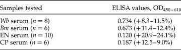
CP, chronic pathology; EN, endemic normal.
Discussion
Rapid diagnostic procedures for human lymphatic filariasis are essential and emphasized by the WHO for proper surveillance and eradication of the disease. It is estimated that out of 1.2 billion cases, 40% live in India. Wuchereria bancrofti accounts for more than 90% of the total filarial infections in India. Currently, Og4C3 ELISA (More & Copeman, Reference More and Copeman1990; Weil et al., Reference Weil, Lammie and Weiss1997) and an immunochromatographic card test (ICT) (Weil et al., Reference Weil, Lammie and Weiss1997) are available to detect circulating antigens of W. bancrofti in serum or whole blood. Recently, the sensitivity of the ICT test showed positive for highly infected MF individuals, and 25% of microfilaria-negative individuals were positive for circulating filarial antigens (Pani et al., Reference Pani, Hoti, Elango, Yuvaraj, Lall and Ramaiah2000). Also, the kit showed lower sensitivity (71.2%) when compared to the filtration technique (Pani et al., Reference Pani, Hoti, Elango, Yuvaraj, Lall and Ramaiah2000). Similarly, the sensitivity of the Og4C3 test was lower than that of the ICT test (Pani et al., Reference Pani, Hoti, Elango, Yuvaraj, Lall and Ramaiah2000). Nevertheless, the main drawback is that both are not effective in detection of B. malayi antigens. However, these kits were the only types that were commercially available and used in the GPELF. Thus, there is a need for better diagnostics for brugian and bancroftian infections (Pani et al., Reference Pani, Hoti, Vanamail and Das2004).
In this situation, recombinant W. bancrofti SXP-1 (rWbSXP-1) protein antigen, an orthologue of BmSXP-1 was identified and used to develop an immunoassay for diagnosis. The assay could detect circulating antibodies for both bancroftian and brugian infections (Rao et al., Reference Rao, Eswaran, Ravi, Gnanasekhar, Narayanan, Kaliraj, Jayaraman, Marson, Raghavan and Scott2000; Lalitha et al., Reference Lalitha, Eswaran, Gnanasekar, Rao, Narayanan, Scott, Nutman and Kaliraj2002). An improved rWbSXP-1 antigen-based immunoassay to detect circulating antibodies to filarial antigens, was found to be more specific and had rapid field-applicable formats for diagnosis, surveillance and elimination programmes (Baskar et al., Reference Baskar, Srikanth, Suba, Mody, Desai and Kaliraj2004; Lammie et al., Reference Lammie, Weil, Noordin, Kaliraj, Steel, Goodman, Lakshmikanthan and Ottesen2004). It is important to detect the circulating filarial antigen in the blood to identify active filarial infection in endemic populations, and hence an attempt was made to develop monoclonal antibodies using WbSXP-1. One clone, 1A6C2, was found to be more stable and highly reactive against rWbSXP-1 and, more importantly, mf antigens and MF sera. It did not react with EN and minimally with CP human clinical sera. Our studies show that MAb 1A6C2 is not only capable of recognizing filarial antigens found in clinical sera but also differentiates active infection from the chronic infection clinical groups.
In endemic countries such as India, where there is a co-prevalence of both brugian and bancroftian infections (Sabesan et al., Reference Sabesan, Palaniyandi, Das and Michael2000), it is advantageous to use a diagnostic tool that can detect both these types simultaneously. The authors do hope that this investigation will add value to the GPELF in endemic populations in developing countries.
Acknowledgements
The Senior Research Fellowship granted to S.J. by the Indian Council for Medical Research, Government of India, is gratefully acknowledged. This project was supported by the University Grants Commission-Departmental Research Support (UGC-DRS) programme. The Department of Biotechnology, Government of India, is acknowledged for financial support.