Introduction
The presence of a larval stage is probably plesiomorphic for the ontogeny of early tetrapods because larvae with external gills have been found in both temnospondyls and in stem-amniotes (Schoch, Reference Schoch2014, and references therein). Temnospondyls exhibit by far the most detailed record of larval development among early tetrapods, with frequent preservation of soft parts such as external gills and skin or cartilaginous or slightly ossified hyobranchial elements. Larvae are known from all main groups of Paleozoic temnospondyls, encompassing the edopoids (Sequeira, Reference Sequeira2009; Schoch and Milner, Reference Schoch, Milner and Seus2014), dvinosaurians (Milner, Reference Milner1982), dissorophoids (Boy, Reference Boy1972, Reference Boy1974; Schoch, Reference Schoch1992; Werneburg, Reference Werneburg2021), zatracheids (Boy, Reference Boy1989; Witzmann and Schoch, Reference Witzmann and Schoch2006a), eryopids (Boy, Reference Boy1990; Witzmann, Reference Witzmann2005; Schoch, Reference Schoch2021), and basal stereospondylomorphs (Boy, Reference Boy1988, Reference Boy1993; Werneburg, Reference Werneburg1992; Schoch, Reference Schoch2003; Witzmann, Reference Witzmann2006; Schoch and Witzmann, Reference Schoch and Witzmann2009a, Reference Schoch and Witzmannb). A tiny larva from Mazon Creek described by Godfrey (Reference Godfrey2003) may represent a Dendrepeton-grade temnospondyl. Specific adaptations in larval temnospondyls are three pairs of external gills (as in lissamphibians and seymouriamorphs) as well as branchial teeth on the ceratobranchials that were resorbed in forms such as dissorophoid temnospondyls that underwent a transition to a more terrestrial mode of life after the larval stage. A further, but not necessarily larval, character is the poorly developed postcranium with undifferentiated and poorly ossified limbs, vertebrae, and girdles.
Despite the fact that larvae are well known in early Permian stereospondylomorphs such as Sclerocephalus haeuseri Goldfuss, Reference Goldfuss1847, Glanochthon angusta Schoch and Witzmann, Reference Schoch and Witzmann2009b, and Archegosaurus decheni Goldfuss, Reference Goldfuss1847 (see above), unambiguous larvae with external gills have not been reported from the Stereospondyli, the dominant temnospondyl clade of the Mesozoic.
In this study, we redescribe the single small specimen of a brachyopid stereospondyl, Platycepsion wilkinsoni (Stephens, Reference Stephens1887) from the Early or Middle Triassic of New South Wales, Australia (Stephens, Reference Stephens1887; Watson, Reference Watson1956; Cosgriff, Reference Cosgriff1969; Warren and Marsicano, Reference Warren and Marsicano1998, Reference Warren and Marsicano2000). Together with chigutisaurids and probably plagiosaurids, brachyopids form the clade Brachyopoidea within the Stereospondyli (Schoch and Milner, Reference Schoch, Milner and Seus2014). The immature nature of the skeleton of Platycepsion wilkinsoni was already recognized in the original description, where it was cited as a “baby labyrinthodont” (Stephens, Reference Stephens1887, p. 1175), but the ontogenetic status of Platycepsion wilkinsoni has so far not been clarified. Watson (Reference Watson1956) regarded this specimen as an early growth stage of a neotenic brachyopid that retained its external gills throughout lifetime. Later authors interpreted this specimen as “larval or perennibranchiate” (Warren and Marsicano, Reference Warren and Marsicano1998, p. 334), “immature” and “juvenile” (Warren and Marsicano, Reference Warren and Marsicano2000, p. 465), or as a “juvenile or larva” (Schoch and Milner, Reference Schoch, Milner and Seus2014, p. 102). These evaluations of the ontogenetic stage of Platycepsion wilkinsoni were based solely on the small size of the specimen, the presence of ossified branchial arches, and the poor degree of ossification of the postcranium, but unambiguous larval features (i.e., larval adaptations) have never been described.
The aim of the present paper is a redescription of the single known specimen of Platycepsion wilkinsoni. Our focus does not lie on a taxonomic reevaluation of P. wilkinsoni below the level of Brachyopidae because this immature specimen is hardly comparable with the other known adult taxa, and the poor preservation of the skull makes reconstruction of the sutural pattern often speculative. Rather, our aim is an evaluation of the specimen from a paleobiological, especially a developmental, point of view. Comparison with the large amount of ontogenetic data in Paleozoic temnospondyls shall help answering the following questions. Is P. wilkinsoni a true larva or a postlarval juvenile? What can P. wilkinsoni tell us about the early ontogeny of stereospondyls, or at least brachyopids? Is P. wilkinsoni derived with respect to Paleozoic forms, or is the plesiomorphic development retained in spite of the different adult morphologies of brachyopids (e.g., vertebral morphology)?
Material
Material examined
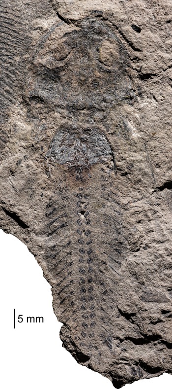
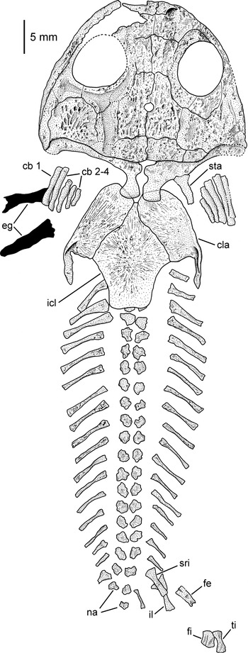
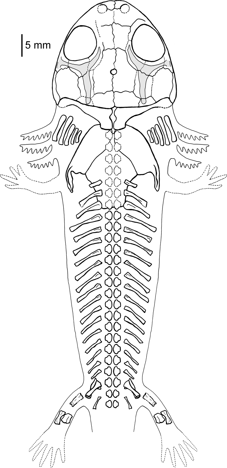
The only known specimen of Platycepsion wilkinsoni is an almost complete skeleton in dorsal view (MMF12572, Fig. 1) with the skull, hyobranchium, presacral vertebral column plus ribs and anteriormost portion of tail, parts of the pectoral and pelvic girdle, and the right hindlimb preserved (Figs. 1, 2). Additionally, soft-tissue preservation (external gills and presumably the skin) is present in the region of the branchial skeleton and the anterior part of the trunk.
Photograph of the complete specimen of Platycepsion wilkinsoni (MMF12572) from the Terrigal (Gosford) Formation, Narrabeen Group, latest Early Triassic or earliest Middle Triassic (Olenekian–Anisian), Gosford, New South Wales, Australia.

Repository and institutional abbreviation
MMF, Mining Museum of the New South Wales Geological Survey, Sydney, Australia.
Systematic paleontology
Temnospondyli Zittel, Reference Zittel1888
Stereospondyli Fraas, Reference Fraas1889
Brachyopoidea Lydekker, Reference Lydekker1885
Brachyopidae Lydekker, Reference Lydekker1885
Platycepsion Kuhn, Reference Kuhn1961
Type species
Platycepsion wilkinsoni (Stephens, Reference Stephens1887).
Diagnosis
As for the type species by monotypy.
Holotype
MMF12572, imprint of a skull (23.2 mm long) in dorsal view plus numerous parts of the postcranium (Fig. 1).
Interpretative drawing of the complete specimen of Platycepsion wilkinsoni (MMF12572). cb = ceratobranchial; cla = clavicula; eg = external gills; fe = femur; fi = fibula; icl = interclavicula; il = ilium; na = neural arches; sri = sacral rib; sta = stapes; ti = tibia.

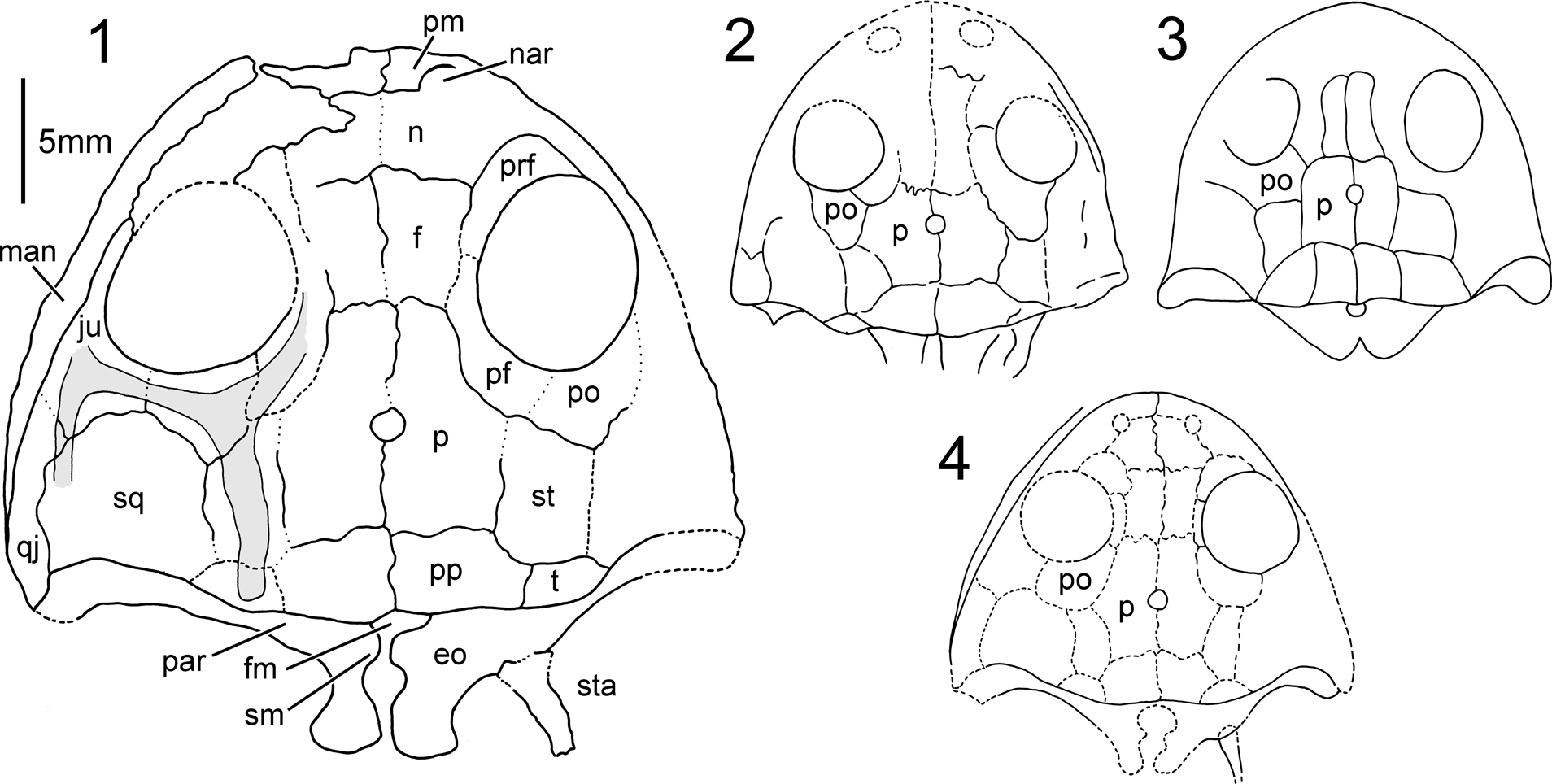
Interpretative drawings of the skull of Platycepsion wilkinsoni (MMF12572): (1) present study, lateral line sulci shown in gray; the sutures that can be traced with the highest degree of certainty are drawn with continuous lines, ambiguous sutures are drawn with dashed lines, and hypothetical sutures are drawn with dotted lines; (2) redrawn from Watson (Reference Watson1956, fig. 11); (3) redrawn from Cosgriff (Reference Cosgriff1969, fig. 3); (4) redrawn from Warren and Marsicano (Reference Warren and Marsicano1998, fig. 2b) with lateral lines omitted. eo = exoccipital; f = frontal; fm = foramen magnum; ju = jugal; man = mandible; n = nasal; nar = narial opening; p = parietal; par = paroccipital process of exoccipital; pf = postfrontal; pm = premaxilla; po = postorbital; pp = postparietal; prf = prefrontal; qj = quadratojugal; sm = submedullar process of exoccipital; sq = squamosal; st = supratemporal; sta = stapes; t = tabular.

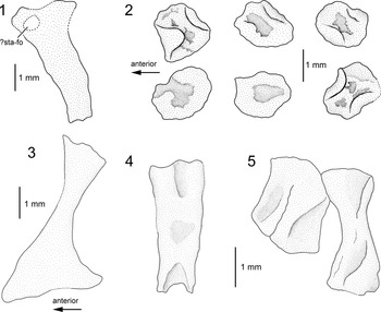
Platycepsion wilkinsoni (MMF12572), interpretative drawings of skeletal elements: (1) stapes; (2) three consecutive vertebrae, only the paired neural arches are ossified; (3) ilium; (4) femur; (5) tibia (right) and fibula. ?sta-fo = possible stapedial foramen.

Diagnosis
Autapomorphies: (1) posterior process of interclavicle with slightly concave posterior margin; (2) probably five pairs of ossified ceratobranchials.
Occurrence
Terrigal (Gosford) Formation, Narrabeen Group, latest Early Triassic or earliest Middle Triassic (Olenekian–Anisian) (Warren and Marsicano, Reference Warren and Marsicano1998; Schoch and Milner, Reference Schoch, Milner and Seus2014); Gosford, New South Wales, Australia.
Description
Nearly the complete dermal skull roof and parts of the occipital region are preserved (Fig. 3.1). The dermal skull roof is broad parabolic in outline and ~23.2 mm long as measured from the tip of the premaxillae to the posterior end of the postparietals. It is ~1.3 times broader than long and has a strikingly short preorbital region, which accounts for only 0.6 times the length of the postorbital skull table. The complete skull is two-dimensionally flattened, but the narrow pectoral girdle indicates that it may have been more slender, and consequently quite deep posteriorly. The quadrate condyles, whose position is inferred based on the morphology of the quadratojugals, are situated well anterior to the exoccipital condyles and anterior to the posterior end of the postparietals. The orbits are nearly circular (the right orbit is probably slightly medio-laterally compressed; as preserved, the maximum width of the right orbit accounts for 84% of the width of the left one) and proportionally large, their sagittal length accounting for more than one-third of the length of the skull. The circular pineal foramen is situated a short distance posterior to the posterior rim of the orbits, and located at midlength of the interparietal suture. The right naris is partially preserved. Its shape indicates that the nares were proportionally large and round. The posterolateral corner of the skull table is formed by a blunt tabular without any evidence of a tabular horn. The posterior margin of the squamosal and the posterolateral edge of the tabular form a shallow embayment whose degree of concavity is most pronounced at the lateral margin of the squamosal where it contacts the quadratojugal. The skull roofing bones are well ossified and bear—where preserved—a well-developed dermal sculpture of pits, ridges, and furrows. This is particularly well visible on the left squamosal with a region of pits in the posterior part of the bone from which deep furrows and pronounced ridges radiate anteriorly, laterally, and medially. It is striking that the lateral line sulci (at least on the postorbital skull roof) are deeply depressed and broad. The supraorbital sulcus is visible on the postfrontal, but its course cannot be traced in an anterior direction because of poor preservation. It continues as the postorbital part of the infraorbital sulcus on the postorbital and the posterior part of the jugal. On the postorbital, it gives rise to the otic part of the infraorbital sulcus that extends on the supratemporal and terminates on the tabular. On the jugal, the infraorbital sulcus branches in an anterior portion, which cannot be traced farther, and in a posterior portion that extends as the jugal sulcus on the lateral part of the squamosal (and possibly a small portion of the quadratojugal).
Skull roof
Most sutures between the dermal skull roofing bones are extremely difficult to trace, and thus their configuration remains hypothetical and depends to a large extent on the subjective impression of the researcher (Fig. 3). Nevertheless, we agree in most aspects with the interpretation of the sutures in the detailed study of Warren and Marsicano (Reference Warren and Marsicano1998) with one possible exception (see below). Where the sutures are traceable, they do not seem to be serrated or interdigitating. In Figure 3.1, the sutures that can be traced with the highest degree of certainty are drawn with continuous lines, ambiguous sutures are drawn with dashed lines, and hypothetical sutures are drawn with dotted lines. The bones whose outline can be determined with sufficient confidence are the frontal, parietal and postparietal, supratemporal, tabular, squamosal, and quadratojugal. The premaxilla is the shortest bone of the midline series of the skull and forms the anteromedial margin of the narial opening. There is no evidence of an alary process. Corresponding to the strikingly short preorbital region, the nasal is much abbreviated and interpreted here as wider than long. The frontal is >1.5 times longer than wide and is probably excluded from the orbital margin by a prefontal-postfrontal contact. The broadly L-shaped parietal is the largest bone of the skull roof. It is almost twice as long as wide, >1.5 times longer than the frontal, and >2.5 times longer than the nasal. The postparietals are rectangular in outline, much wider than long, and form a slightly concave anterolateral suture with the supratemporal. The boundaries of the supratemporal are hard to determine, but seem to be delineated by an elongate bone that probably wedges slightly between postfrontal and postorbital (see below). Posteriorly, it sutures with the small, blunt tabular that does not form a tabular process. The tabular forms a short sutural contact with the squamosal, and thus excludes the supratemporal from the shallow squamosal embayment. The squamosal is broadly lunate in shape and posteriorly concave. Its descending lamina, or flange, appears to be relatively deep, but no statements concerning its exact extent or the presence or absence of a palatoquadrate fissure (sensu Warren and Black, Reference Warren and Black1985) are possible. The quadratojugal is a slender bone that appears to extend slightly more anterior than the squamosal. The orbits are framed anteromedially by the prefrontal, posteromedially by a similar-sized postfrontal, the triangular postorbital posteriorly, and posterolaterally and laterally by the jugal, which is extremely slender lateral to the orbits. There is no evidence of a lacrimal. The boundaries of postfrontal and postorbital are difficult to discern.
We favor the interpretation that the postfrontal extends posteriorly, contacts the supratemporal, and precludes a postorbital-parietal contact. However, this stands in contrast to the interpretation of Watson (Reference Watson1956), Cosgriff (Reference Cosgriff1969), and Warren and Marsicano (Reference Warren and Marsicano1998), who reconstructed the postorbital as extending anteromedially at the expense of the postfrontal length, thus facilitating a postorbital-parietal contact (Fig. 3.2–3.4). This configuration is unknown in other brachyopids and uncommon in temnospondyls except for dvinosaurians such as Erpetosaurus radiatus (Cope, Reference Cope1874) (Milner and Sequeira, Reference Milner and Sequeira2011), Dvinosaurus primus Amalitzky, Reference Amalitzky1921 (Shishkin, Reference Shishkin1973), Tupilakosaurus wetlugensis Shishkin, Reference Shishkin1961 (Shishkin, Reference Shishkin1973), Thabanchuia oomieae Warren, Reference Warren1998 (Warren, Reference Warren1998), Kourerpeton bradyi Olson and Lammers, Reference Olson, Lammers and Churcher1976 (Olson and Lammers, Reference Olson, Lammers and Churcher1976), and Slaugenhopia texensis Olson, Reference Olson1962 (Milner and Sequeira, Reference Milner and Sequeira2004). We do not rule out that the interpretation of Watson (Reference Watson1956), Cosgriff (Reference Cosgriff1969), and Warren and Marsicano (Reference Warren and Marsicano1998) is correct, and given the poor state of preservation we do not want to commit ourselves to our interpretation. However, given the available data, we regard our more conservative interpretation as the more probable one.
Mandible
An imprint of an elongate bone that narrows posteriorly and has a rounded hind margin is preserved posterior to the position of the left quadrate condyle. Morphology and location of the bone strongly suggests that it represents the postglenoid area of the left mandibular ramus, as already suggested by Watson (Reference Watson1956) and Warren and Marsicano (Reference Warren and Marsicano1998). No further details are discernible.
Endocranium
From the endocranium, only the exoccipitals are exposed. The exoccipitals are well ossified, proportionally large, and their elongate condyles extend far posteriorly, probably slightly exaggerated by postmortem compression (Fig. 3.1). As in most stereospondyls (e.g., Bystrow and Efremov, Reference Bystrow and Efremov1940), there is no evidence of an ossified basioccipital. The foramen magnum is partially visible posterior to the postparietals and bordered ventrally by the submedullar processes. The stout vertical column of the exoccipital sends out a paroccipital process towards the tabular, but no details of this process can be discerned. No foramina for nerves or blood vessels are preserved. The presence or absence of the quadrate and other endocranial ossifications cannot be determined.
Ceratobranchials
One striking feature of P. wilkinsoni is the presence of ossified branchial arches on the left and right sides between skull and pectoral girdle. Stephens (Reference Stephens1887, pl. 22) illustrated three arches on either side and described them as “‘rows of ossicles’ more or less united in continuous lines” (Stephens, Reference Stephens1887, p. 1185). Watson (Reference Watson1956) determined four pairs of ceratobranchials and described them as long bony rods with a possible short ceratohyal anterior to the right first ceratobranchial. He mentioned soft-tissue preservation associated with the arches, but he did not mention or illustrate any gills proper. Cosgriff (Reference Cosgriff1969, fig. 3) illustrated three pairs of branchial arches and Warren and Marsicano (Reference Warren and Marsicano1998, fig. 2) illustrated four pairs on the left and five on the right, but neither study described them.
The present work reveals that five arches are present on both sides. They are well ossified, slightly bent medially, and more robust than the ribs. The second arch is the longest and the following ones become successively shorter. The first arch is approximately the length of the third one. We tentatively interpret all five pairs as ceratobranchials rather than one anterior pair of ceratohyals and subsequently following four pairs of ceratobranchials. This interpretation is based on the following reason. If the anteriormost arch would represent a ceratohyal, we would expect a different morphology compared to the other arches, as commonly occurs in early tetrapods (Witzmann, Reference Witzmann2013, and references therein). However, the morphology of the first pair does not differ from that of the following pairs. If this unorthodox interpretation is correct, then Platycepsion wilkinsoni is unique among early tetrapods in possessing five pairs of ceratobranchials. Five pairs are probably plesiomorphic for osteichthyans (Carvalho et al., Reference Carvalho, Bockmann and de Carvalho2013) and are present in most actinopterygians and in sarcopterygian fishes such as porolepiforms (Kanyukin, Reference Kanyukin2006) and coelacanths (Forey, Reference Forey1998). The fifth arch, however, was lost in more-derived sarcopterygians (Carvalho et al., Reference Carvalho, Bockmann and de Carvalho2013), and, accordingly, only four pairs of ceratobranchials are known in tetrapodomorph fishes such as Eusthenopteron foordi Whiteaves, Reference Whiteaves1881, and Tiktaalik roseae Daeschler, Shubin, and Jenkins, Reference Daeschler, Shubin and Jenkins2006 (Jarvik, Reference Jarvik1954; Downs et al., Reference Downs, Daeschler, Jenkins and Shubin2008). In early tetrapods and larval lissamphibians, the number of ceratobranchials is restricted to four or three pairs (Witzmann, Reference Witzmann2013). Given that Platycepsion wilkinsoni really had five ceratobranchials, the fifth was more likely a novelty than an atavism.
Branchial denticles and ventral grooves for gill arteries cannot be determined on the ceratobranchials. On the left side, two black, coaly imprints are visible lateral and posterior to the arches. Most plausibly, they can be interpreted as soft-tissue preservation of external gill rami. No further elements of the hyobranchial skeleton such as basibranchial or hypobranchials are preserved.
Stapes
A rod-like bone is preserved on the right side between clavicle and branchial arches. It is stouter than the latter and reaches about half the size of the presumed second ceratobranchial. Proximally, it abuts against the exoccipital and its longitudinal axis is directed posteriorly and slightly laterally. Watson (Reference Watson1956) interpreted this bone as the paroccipital process of the tabular. It was neither illustrated nor described by Cosgriff (Reference Cosgriff1969). Warren and Marsicano (Reference Warren and Marsicano1998, fig. 2) illustrated this bone, but neither mentioned nor labeled it. The position lateral to the exoccipitals and its morphology strongly suggest that this element is a stapes (Figs. 3.1, 4.1). It has a broad proximal base, possibly consisting of two slightly separated heads, and narrows to a straight shaft. The broader head represents the footplate, whereas the smaller head is the ventral process. The element as a whole most closely resembles the stapes of Micromelerpeton credneri Bulman and Whittard, Reference Bulman and Whittard1926, in outline and degree of ossification (Boy, Reference Boy1995). There appears to be a stapedial foramen near the base, but due to poor preservation it cannot be ruled out that this is an artefact. In its distal third, the bone narrows again and is slightly bent. On the convex side, a weakly developed quadrate process might be indicated. The distal end is irregular and suggests continuation in cartilage.
Postcranium
An interpretative drawing of the complete postcranium has so far only been provided by Stephens (Reference Stephens1887, pl. 2), whereas in Watson (Reference Watson1956, fig. 11), Cosgriff (Reference Cosgriff1969, fig. 3), and Warren and Marsicano (Reference Warren and Marsicano1998, fig. 2), graphical interpretations of the postcranium are restricted to the pectoral girdle.
Pectoral girdle
From the pectoral girdle, only the interclavicle and the clavicles are preserved, whereas the scapulocoracoid and the cleithrum are absent (Fig. 2). Considering the generally poor state of ossification of the postcranium, we assume that the scapulocoracoid was still completely cartilaginous, whereas the cleithrum might have been ossified but was a feeble element that is not preserved. We completely agree with the illustration of the dermal pectoral bones and their articulation provided by Warren and Marsicano (Reference Warren and Marsicano1998, fig. 2). The clavicular blades do not meet anteriorly and extend farther anteriorly than the anterior margin of the interclavicle. The ventral dermal sculpture is not directly visible because of preservation in dorsal aspect. However, due to erosion of the most-superficial layers of bone, radiating, mineral-filled canals within the bones are visible. They suggest a radial sculptural pattern of ridges and furrows on the ventral surface (see Witzmann et al., Reference Witzmann, Scholz, Müller and Kardjilov2010). The ossification center of the interclavicle, as indicated by the area where the radiating canals intersect, is located slightly posterior to the bone's midlength. Posteriorly, the interclavicle extends in a short process that has a slightly concave posterior end.
Vertebrae
From the vertebrae, only the paired neural arches are present without any evidence of ossified vertebral centra. If ossified centra were present in the living animal, they would be visible directly below or between the neural arches since all skeletal elements are preserved two-dimensionally in the same plane. Furthermore, as already reported by Cosgriff (Reference Cosgriff1969, p. 68), “small excavations were made along the vertebral column” but no remnants of vertebral centra were found. These “small excavations” are still visible on the specimen. The neural arches lack neural spines and transverse processes. Rudimentary zygapophyses are indicated in some neural arches (Fig. 4.2). In some more three-dimensionally preserved neural arches, the anterior and posterior margins are inwardly curved and wrap around the medial surface where they probably enclosed a cartilaginous core. These poorly differentiated neural arches closely resemble early larvae of branchiosaurids (Boy, Reference Boy1974) and small larvae of stereospondylomorphs such as Sclerocephalus haeuseri (Schoch, Reference Schoch2003) or Archegosaurus decheni (Witzmann and Schoch, Reference Witzmann and Schoch2006b). The sacral vertebra can be identified based on the presence of a sacral rib (see below). Fifteen pairs of presacral neural arches are exposed posterior to the interclavicle, and one pair is located at the posterior margin of the interclavicle. We estimate that the anteriormost six pairs of neural arches are not preserved. Thus, P. wilkinsoni probably had 22 presacral vertebrae in total. The first two postsacral vertebrae (three neural arches) are preserved, whereas the remainder of the tail is missing due to breakage of the slab. The neural arches of the presacral and sacral region have all roughly the same size, but the postsacral neural arches are reduced in size.
Ribs
An almost complete series of articulated ribs from (presumed) vertebra 5 to vertebra 24 (the first postsacral vertebra) are preserved in P. wilkinsoni (Fig. 2). As is common in temnospondyl larvae, the ribs are relatively simple rods with no indication of uncinate processes. In P. wilkinsoni, the fourth rib is straight and proportionally short (less than the length of two vertebral segments). The length of the subsequently following ribs increases until rib eight, which is the longest rib (as long as three vertebral segments). Ribs 9–14 are only slightly smaller, and ribs 15–22 decrease from a length of ~2.5 vertebral segments to a length of ~1.5 segments. Ribs 6–18 are slightly curved. The proximal rib head is expanded (most pronounced in ribs 6–8, which are also the most robust ribs) and not differentiated into tuberculum and capitulum. The distal ends also are expanded in ribs 6–16. The right sacral rib is completely preserved, whereas only the proximal head is preserved on the left side. The length of the sacral rib measures ~2 vertebral segments. It is much more robust than the neighboring ribs and has a distinctly broadened proximal and slightly widened distal end. The first postsacral rib is slightly longer than the last presacral rib, but is more slender.
Pelvic girdle
From the pelvic girdle, only the ilium is ossified in P. wilkinsoni, whereas ischium and pubis remained cartilaginous (Figs. 2, 4.3). It is probably exposed in medial view so that the dorsal part of the acetabulum is not visible. The ilium is already well differentiated and consists of a triangular base and a posterodorsally directed slender shaft, which is expanded dorsally. The dorsal margin is sloping posteroventrally. Comparison with other brachyopids is not possible because only a small distal fragment of an ilium is known from Batrachosuchus sp. (Warren and Marsicano, Reference Warren and Marsicano2000); otherwise the brachyopid ilium is unknown. The ilium, however, is known in some chigutisaurids and differs from that in P. wilkinsoni either in being much stouter with a robust shaft, as in Siderops kehli Warren and Hutchinson, Reference Warren and Hutchinson1983 (see Warren and Hutchinson, Reference Warren and Hutchinson1983, fig. 30), or in having a slender shaft without dorsal expansion, as in Pelorocephalus mendozensis Cabrera, Reference Cabrera1944 (Marsicano, Reference Marsicano1993, fig. 10). Most similar is the ilium of Compsocerops cosgriffi Sengupta, Reference Sengupta1995 (see Sengupta, Reference Sengupta1995, fig. 14) in having a slender (albeit more robust) shaft and a widened dorsal end. In plagiosaurids, the rod-like ilium is known in Gerrothorax pulcherrimus Fraas, Reference Fraas1913 (Hellrung, Reference Hellrung2003) where it is conspicuously shortened dorso-ventrally, corresponding to the flattened body of the animal.
Limbs
Only the right hindlimb without foot is preserved (Figs. 2, 4.4, 4.5). The femur is elongate rectangular, extremely poorly ossified, and undifferentiated (Fig. 4.4). It is strikingly short and has the same length as the tibia. Its rudimentary appearance closely resembles the femur of early larval Sclerocephalus haeuseri (Schoch, Reference Schoch2003). It is not possible to tell if the femur (and thus the hind limb) of P. wilkinsoni might have been proportionally short also in adults. The only other described brachyopid femur belongs to Banksiops townrowi (Cosgriff, Reference Cosgriff1974) and does not differ from other temnospondyl femurs except for its slender shaft (Cosgriff, Reference Cosgriff1974). Among chigutisaurids, the femora of Siderops kehli (Warren and Hutchinson, Reference Warren and Hutchinson1983), Pelorocephalus mendozensis (Bonaparte, Reference Bonaparte1978) and of an undetermined chigutisaurid (Marsicano, Reference Marsicano1993) have been described as likewise slender elements, and the same is true for the femur of the plagiosaurid Gerrothorax pulcherrimus (Nilsson, Reference Nilsson1946). Tibia and fibula have a strikingly different morphology (Fig. 4.5). The tibia is ~1.4 times longer than the fibula and has a slender shaft with broadened proximal and distal ends. It is asymmetrical in shape, with the medial margin showing a larger degree of concavity than the lateral one. The short, flattened fibula is only slightly longer than wide and is almost 1.5 times broader than the tibia. Proximal and distal ends are of roughly equal size, and the stout, robust shaft is slightly concave on the medial and lateral sides. A medial broad ridge extends proximodistally. The fibular morphology of P. wilkinsoni is highly unusual among temnospondyls. No fibulae are known in other brachyopids, but have been described in an undetermined chigutisaurid (Marsicano, Reference Marsicano1993) and the chigutisaurid Koolasuchus cleelandi Warren, Rich, and Rich, Reference Warren, Rich and Rich1997, in which they have the more characteristic temnospondyl shape with the distal head more expanded than the proximal one and separated by a slender shaft. The fibular morphology of P. wilkinsoni most closely resembles that of the plagiosaurid Gerrothorax pulcherrimus (Nilsson, Reference Nilsson1946, fig. 7), which is a similarly flattened bone that is much wider than the rod-like tibia. In contrast to P. wilkinsoni, it is only slightly shorter than the tibia.
Remarks
None.
Discussion
Assignment to Brachyopidae
We follow Watson (Reference Watson1956), Cosgriff (Reference Cosgriff1969), and Warren and Marsicano (Reference Warren and Marsicano1998, Reference Warren and Marsicano2000) in assigning Platycepsion wilkinsoni to the Brachyopidae. Warren and Marsicano (Reference Warren and Marsicano1998, Reference Warren and Marsicano2000) listed the following derived characters shared by P. wilkinsoni and the Brachyopidae: (1) deep skull with exoccipital condyles extending far posterior; (2) absence of tabular horns; (3) occipital portion of squamosal convex (although we cannot follow point [3] because we do not see a convex portion of the squamosal); (4) postglenoid area of mandible elongate; (5) narrow pectoral girdle. Point (5) deserves the following comment. In our view, a narrow pectoral girdle probably is not a characteristic of brachyopids or brachyopoids. Warren and Marsicano (Reference Warren and Marsicano1998) emphasized that the pectoral girdle of Platycepsion wilkinsoni is strikingly narrower than the width of the skull and pointed to similar proportions in the chigutisaurid Siderops kehli (Warren and Hutchinson, Reference Warren and Hutchinson1983). These authors thus hypothesized that a pectoral girdle much narrower than the skull could be a derived brachyopoid characteristic, but admitted that these proportions have been observed only in the two mentioned taxa. However, the condition in Siderops kehli is probably not comparable with that in P. wilkinsoni. Siderops kehli has a well-ossified skull, which is extremely broad also when three-dimensionally reconstructed (Warren and Hutchinson, Reference Warren and Hutchinson1983). In contrast, the wide skull of P. wilkinsoni is the result of complete flattening in one layer. In life, this skull was definitely deeper (Figs. 5, 6). More generally, a narrow pectoral girdle is a plesiomorphic character among temnospondyls, as exemplified by edopoids, Dendrysekos helogenes Steen, Reference Steen1934, zatracheids, and trematosaurids (Schoch and Milner, Reference Schoch, Milner and Wellnhofer2000, 2014; Witzmann and Schoch, Reference Witzmann and Schoch2006a), whereas the pectoral girdle is at least the width of the skull in plagiosaurids such as Gerrothorax pulcherrimus (Hellrung, Reference Hellrung2003) and probably Plagiosaurus depressus Jaekel, Reference Jaekel1914 (Nilsson, Reference Nilsson1937). An additional character—the wide and blunt posterior end of the interclavicle—is shared among chigutisaurids, brachyopids, and plagiosaurids. The interclavicle of P. wilkinsoni is consistent especially with that of Siderops kehli in outline, except for the concave posterior end.
Platycepsion wilkinsoni (MMF12572), reconstruction of skeleton with body contour and external gills.

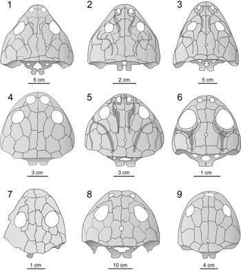
Brachyopid skulls in dorsal view: (1) Brachyops laticeps Owen in Anonymous, 1854; (2) Xenobrachyops allos (Howie, Reference Howie1972); (3) Bothriceps australis; (4) Bathignathus watsoni; (5) Batrachosuchus browni; (6) Platycepsion wilkinsoni; (7) Notobrachyops picketti; (8) Vigilius wellesi; (9) Sinobrachyops placenticephalus Dong, Reference Dong1985. Platycepsion wilkinsoni is based on the present study, Notobrachyops picketti is based on Warren and Marsicano (Reference Warren and Marsicano1998), and all others are redrawn from Schoch and Milner (Reference Schoch, Milner and Seus2014) and references cited therein.

Derived characters (1) and (2) can also be found in tupilakosaurid dvinosaurians, but the elongate postglenoid process that is visible on the left mandibular ramus clearly precludes tupilakosaurid affinities. This is further supported by the fact that there is no evidence of a dorsally exposed palatine, which is a diagnostic character of tupilakosaurids (Schoch and Milner, Reference Schoch, Milner and Seus2014). The basioccipital, which makes a major contribution to the single occipital condyle in dvinosaurs, is absent and a wide gap remains between the exoccipitals. Finally, the short postorbital in P. wilkinsoni differs from the much expanded postorbital of tupilakosaurids. An additional and very distinctive brachyopid character of P. wilkinsoni is the unusually wide lateral line sulci (not present in dvinosaurs), which otherwise are known only from the genera Mastodonsaurus (Schoch, Reference Schoch1999, fig. 9) and Buettneria (Sawin, Reference Sawin1945, fig. 3), where outgroup comparison shows that they evolved in parallel. Despite the larval state of P. wilkinsoni, this feature is as pronounced as in Bathignathus watsoni (Haughton, Reference Haughton1925).
Phylogenetic position within Brachyopidae
Platycepsion wilkinsoni is consistent with many brachyopids in the parabolic skull outline, the position of the orbits, and proportions of the preorbital and postorbital region (Fig. 6). The wide lateral line sulci are shared with Bathignathus watsoni and Batrachosuchus browni Broom, Reference Broom1903, but the highly apomorphic proportions of these taxa, such as the postfrontal (posteromedially extended and medially convex), slender parietal, and much wider supratemporal, are not shared by P. wilkinsoni, which instead has a more plesiomorphic condition similar to that of Bothriceps australis Huxley, Reference Huxley1859. We conclude that P. wilkinsoni was most likely related to a clade that was recovered by Warren and Marsicano (Reference Warren and Marsicano2000) and includes Vigilius wellesi Warren and Marsicano, Reference Warren and Marsicano2000, Bathignathus watsoni, and Batrachosuchus browni. Any further, more exact placement within the tree is hampered by the larval morphology.
Validity of the taxon Platycepsion wilkinsoni
Warren and Marsicano (Reference Warren and Marsicano1998, Reference Warren and Marsicano2000) pointed out that the contact between parietal and postorbital was the only autapomorphy of Platycepsion wilkinsoni. We do not see evidence for this “dvinosaurian” bone configuration and prefer the more conservative interpretation that postorbital and parietal are separated by the postfrontal. The poor preservation precludes a definitive statement and weakens this character as an autapomorphy of Platycepsion wilkinsoni.
A unique character among brachyopids (and other temnospondyls) forms the shape of the interclavicle. Cosgriff (Reference Cosgriff1969) stressed that in contrast to most temnospondyls, the posterior process of the interclavicle in P. wilkinsoni does not have a rounded posterior end but is rather straight. Indeed, as described above, the posterior end is even slightly concave. The only other brachyopid in which a complete interclavicle is known is Blinasaurus townrowi, whose interclavicle has a rounded posterior end (Cosgriff, Reference Cosgriff1974, fig. 26). Cosgriff (Reference Cosgriff1974) even consulted the different posterior ends of the interclavicles to justify that B. townrowi and Platycepsion wilkinsoni (referred by him to as Blinasaurus wilkinsoni) are different species. The interclavicles known in chigutisaurids have a similar diamond shape as in Platycepsion wilkinsoni and B. townrowi, but in contrast to P. wilkinsoni they have a rounded or pointed posterior end (Warren and Hutchinson, Reference Warren and Hutchinson1983). A possible exception is the chigutisaurid Koolasuchus cleelandi whose interclavicle might have a straight or faintly convex posterior end (Warren et al., Reference Warren, Rich and Rich1997, fig. 9). In plagiosaurids, the interclavicle is roughly triangular in shape, with the apex pointing anteriorly and the posterior border being straight (Nilsson, Reference Nilsson1937; Hellrung, Reference Hellrung2003). To our best knowledge, a concave posterior margin of the interclavicle appears to be a unique character among temnospondyls. We suggest this character as a possible autapomorphy of P. wilkinsoni, although we cannot rule out that the morphology of the interclavicle might have changed during further ontogeny. A further autapomorphic character of P. wilkinsoni might be the presence of five pairs of ceratobranchials. However, this character is weakened by the fact that the hyobranchial morphology is poorly known in other brachyopids.
Finally, the morphology of the skull as a whole differs from all other brachyopids and chigutisaurids in the large size of the orbits, the short postfrontal and postorbital, and the relatively small supratemporal, especially compared with the parietal. These features are probably larval traits and it is impossible to deduce the adult morphology of the taxon, but at present they give a distinctive cranial morphology that further distinguishes P. wilkinsoni, especially, from the taxa that have similar skull outlines—Bathignathus watsoni and Batrachosuchus browni. This said, we emphasize that comparison with such large specimens is necessarily limited because size differences are enormous. Only plagiosaurids have proportionally larger orbits, but their skull morphology is highly derived, with either having huge combined orbitotemporal fenestrae (Plagiosuchus pustuliferus [Fraas, Reference Fraas1896]) or skulls being more than twice wider than long (Plagiosterninae and Plagiosaurinae) (Schoch and Milner, Reference Schoch, Milner and Seus2014).
Interpretation of the ontogeny of Platycepsion wilkinsoni
The present study demonstrates for the first time soft-tissue preservation of external gills in P. wilkinsoni. External gills are a larval specialization in temnospondyls and other tetrapods (see discussions in Schoch and Witzmann, Reference Schoch and Witzmann2011; Witzmann, Reference Witzmann2013). Therefore, P. wilkinsoni is not only immature, but indeed a larval form. The alternative interpretation, that P. wilkinsoni is a paedomorphic adult that retained its larval external gills (see Watson, Reference Watson1956), is unlikely for the following reasons. The extremely short snout and the large orbits and pineal opening stand in contrast to an interpretation as a small adult or juvenile. Only adult plagiosaurids have proportionally shorter snouts, but their skulls are highly aberrant with huge orbits (Schoch and Milner, Reference Schoch, Milner and Seus2014). What is even more important is the extremely low degree of ossification and differentiation of the postcranial skeleton. Adult temnospondyls with paedomorphic skull proportions (e.g., dvinosaurians or lapillopsids and lydekkerinids among stereospondyls) have well-ossified postcranial skeletons throughout (Schoch and Milner, Reference Schoch, Milner and Wellnhofer2000, Reference Schoch, Milner and Seus2014). Thus, the interpretation of the specimen as a larva appears the most plausible for us.
Platycepsion wilkinsoni is the only unambiguously known larva of a stereospondyl, despite the fact that growth series are known from certain Early Triassic stereospondyls such as Warrenisuchus aliciae (Warren and Hutchinson, Reference Warren and Hutchinson1988) (see, however, the comment of Steyer [Reference Steyer2003] that this series is not represented by a single species), Edingerella (Watsonisuchus) madagascariensis (Lehman, Reference Lehman1961) (Steyer, Reference Steyer2003), and Benthosuchus sushkini (Efremov, Reference Efremov1929) (Bystrow and Efremov, Reference Bystrow and Efremov1940). Even their smallest specimens lack unambiguous larval characters.
It is worth mentioning here that the ~40 mm long, poorly preserved skull of Notobrachyops picketti Cosgriff, Reference Cosgriff1973, has been assigned to brachyopids with reservation (Warren and Marsicano, Reference Warren and Marsicano1998, Reference Warren and Marsicano2000). If this assignment is correct, then N. picketti is the second smallest brachyopid after P. wilkinsoni. However, the proportionally long preorbital region, the rather small orbits, pineal opening and, to a lesser degree, the absence of lateral line sulci support the interpretation of Warren and Marsicano (Reference Warren and Marsicano1998) that the specimen is an adult. It thus provides no ontogenetic data and cannot be compared with P. wilkinsoni.
In the following, a short summary of the general larval ontogeny in temnospondyls is given. It is then compared with the degree of ossification and differentiation of the skeleton in P. wilkinsoni to derive information about its developmental stage and, as far as possible, the sequence of ossification.
General features of Paleozoic temnospondyl larvae.—A great number of well-preserved specimens enabled detailed study of the formation of skull and postcranium in Permo-Carboniferous larval temnospondyls (e.g., Boy, Reference Boy1974; Milner, Reference Milner1982; Schoch, Reference Schoch1992, Reference Schoch2002, Reference Schoch2003, Reference Schoch2004; Werneburg, Reference Werneburg1992, Reference Werneburg2002, Reference Werneburg2021; Witzmann and Pfretzschner, Reference Witzmann and Pfretzschner2003; Witzmann, Reference Witzmann2005, Reference Witzmann2006; Fröbisch et al., Reference Fröbisch, Carroll and Schoch2007, Reference Fröbisch, Olori, Schoch and Witzmann2010). These studies indicate that rapid ossification of the dermal skull roof and palate early in ontogeny, contrasted with a much slower pace in postcranial development, is plesiomorphic for temnospondyls. A more derived early ontogeny is found in the Branchiosauridae, a group of larval or paedomorphic dissorophoids in which formation of the dermal skull bones was delayed, whereas the postcranial elements, especially the limbs, ossified and differentiated earlier (Boy, Reference Boy1974; Schoch, Reference Schoch1992, Reference Schoch2004).
The ossification sequence in skull and postcranium of Paleozoic temnospondyls generally proceeded in the following pattern. Endocranial bones started to ossify after formation of the skull roof, the first elements to ossify being usually the exoccipitals and quadrates. Postcranial ossification commenced with undifferentiated, paired neural arches in the anterior trunk region and ossified in an anterior to posterior direction, followed by successive formation of the ribs, the humerus, and the dermal bones of the pectoral girdle. The endochondral scapulocoracoid ossified much later (first the scapula and, much later, the coracoid), and the ilium was the first element of the pelvic girdle that started ossification already in small larvae, followed by the ischium and, much later, the pubis. The latter remained cartilaginous in many aquatic forms. Initial ossification of the femur occurred after the humerus and the dermal pectoral girdle had started to ossify (Schoch, Reference Schoch1992; Witzmann and Pfretzschner, Reference Witzmann and Pfretzschner2003).
Developmental stage of P. wilkinsoni and comparison with other larval temnospondyls.—The dermal skull roof of Platycepsion wilkinsoni is well ossified and bears distinct ornament with broad lateral lines, and the exoccipitals not only have started to ossify, but are already well differentiated with conspicuous occipital condyles and paroccipital processes. Similarly, the viscerocranium is in an advanced stage of development with massive branchial arches and a well-differentiated stapes. The ossified branchial arches are no larval character. Their presence in adult brachyopids (Warren et al., Reference Warren, Rozefelds and Bull2011) indicates that they also were retained in P. wilkinsoni in the adult stage.
The stapes is of special interest because this element has been rarely reported in brachyopids. In the brachyopid Batrachosuchus watsoni and the plagiosaurids Gerrothorax pulcherrimus and Megalophthalma ockerti Schoch, Milner, and Witzmann, Reference Schoch, Milner and Seus2014 (Watson, Reference Watson1956; Witzmann et al., Reference Witzmann, Schoch, Hilger and Kardjilov2012; Schoch et al., Reference Schoch, Milner and Witzmann2014), it forms a highly aberrant, blade-like element. In contrast, the probably closely related chigutisaurids had a rather plesiomorphic stapes, with a long rod-like shaft (Warren and Hutchinson, Reference Warren and Hutchinson1983). In Platycepsion wilkinsoni, the stapes was still in an immature stage (as indicated by its consistency with the stapes of Micromelerpeton credneri), but clearly represents the rod-like type found in chigutisaurids and most other stereospondyls, which is also widespread in Paleozoic taxa (Schoch, Reference Schoch2017). These data suggest that the blade-like stapes of brachyopids and plagiosaurids was derived, and not paedomorphic as suggested by Schoch (Reference Schoch2017), but it remains unclear what the stapes of adult P. wilkinsoni looked like.
In contrast to the skull and viscerocranium, the postcranial skeleton was in a much less advanced stage. Still completely unossified are the scapulocoracoid, ischium and pubis, and the vertebral centra. This corresponds to an early larva of an eryopiform (Sclerocephalus haeuseri or Onchiodon labyrinthicus Geinitz, Reference Geinitz1861). The femur is short and undifferentiated. The neural arches are poorly developed and still paired, and the centra are completely unossified. Vertebral development, starting with paired neural arches and subsequent ossification of centra, is plesiomorphic for temnospondyls and probably for early tetrapods in general (Danto et al., Reference Danto, Witzmann and Fröbisch2016). Because brachyopids have disc- or spool-shaped centra as adults (Warren et al., Reference Warren, Rozefelds and Bull2011), P. wilkinsoni shows that the plesiomorphic pattern of development was conserved also in Mesozoic stereospondyls, irrespective of whether the adult vertebral morphology was rhachitomous (horseshoe-shaped intercentra), stereospondylous (disc-like intercentra), or diplospondylous (disc-like intercentra and pleurocentra). The ribs are moderately short and straight, similar to those of larval dvinosaurians, eryopids, and Micromelerpeton credneri, but much longer than in branchiosaurids. It is not clear whether adult brachyopids developed uncinate processes. The only other described brachyopid with ribs is an adult specimen of Bothriceps australis, in which slender, almost straight ribs without uncinate processes have been described (Warren et al., Reference Warren, Rozefelds and Bull2011). However, these ribs are derived from the posterior trunk region, whereas ribs with uncinate processes are usually present in the “thoracic” region (e.g., Witzmann and Schoch, Reference Witzmann and Schoch2006b). At least the closely related chigutisaurids developed distinct uncinate processes as adults, as described by Warren and Hutchinson (Reference Warren and Hutchinson1983) and Marsicano (Reference Marsicano1993), whereas uncinate processes are unknown in plagiosaurids (Nilsson, Reference Nilsson1937, Reference Nilsson1946; Hellrung, Reference Hellrung2003).
In its overall degree of ossification, P. wilkinsoni resembles early larvae of Permo-Carboniferous eryopiforms such as the eryopid Onchiodon labyrinthicus (at a skull length between 16–20 mm; Witzmann, Reference Witzmann2005), the basal stereospondylomorph Sclerocephalus haeuseri (at a skull length of ~20 mm; Schoch, Reference Schoch2003; Witzmann, Reference Witzmann2006), and the more derived stereospondylomorph Archegosaurus decheni (at a skull length between 30–40 mm; Witzmann, Reference Witzmann2006). However, P. wilkinsoni differs from Paleozoic temnospondyl larvae in a more pronounced dermal ornament and distinct lateral lines, and in the earlier ossification and differentiation of the occiput. This earlier development of the skull might be a stereospondyl characteristic, as indicated by early juveniles of Warrenisuchus aliciae (Warren and Hutchinson, Reference Warren and Hutchinson1988) and Benthosuchus sushkini (Bystrow and Efremov, Reference Bystrow and Efremov1940), which likewise already possess well-ossified exoccipitals and well-developed dermal ornament.
Degree of ossification and implications for ossification sequence.—Based on the degree of ossification of the different skeletal elements in Platycepsion wilkinsoni, it is possible to detect similarities and differences in a small part of the ossification sequence between P. wilkinsoni and Paleozoic temnospondyls. In the dissorophoid Micromelerpeton credneri, the ilium started to ossify prior to the scapulocoracoid, which in turn ossified earlier than the stapes, and which was followed later by ossification of the exoccipitals (Boy, Reference Boy1995; Witzmann and Pfretzschner, Reference Witzmann and Pfretzschner2003). The same sequence was probably present in the eryopid Onchiodon labyrinthicus (although ilium and scapula are already ossified in the smallest available specimen; Witzmann, Reference Witzmann2005) and was documented in the basal stereospondylomorph Sclerocephalus haeuseri (Witzmann, Reference Witzmann2006; Schoch and Witzmann, Reference Schoch and Witzmann2009a). In the more derived and more aquatic stereospondylomorph Archegosaurus decheni, the ossification sequence had partially changed: the scapulocoracoid started to ossify after an ossified stapes had formed. In P. wilkinsoni, the ossification of the scapulocoracoid is not only delayed with respect to the formation of the stapes, but also with respect to the exoccipitals, which ossified prior to the scapulocoracoid. This heterochronic shift might be correlated with the strict aquatic lifestyle of both adult Archegosaurus decheni and brachyopids throughout their life histories, in which the limbs were hardly used for terrestrial locomotion.
Paleobiological implications
The striking contrast between the strongly ossified skull and hyobranchium on the one hand and the feebly ossified and poorly developed postcranium, including the tiny limbs, on the other hand might give some interesting hints concerning the lifestyle of P. wilkinsoni. The branchial arches (which are more strongly developed than the ribs) and the well-differentiated exoccipitals indicate that Platycepsion wilkinsoni relied on strong suction feeding similar to plagiosaurid temnospondyls (Witzmann and Schoch, Reference Witzmann and Schoch2013). Posteroventral rotation of the robust hyobranchial apparatus expanded the buccal cavity and generated powerful suction of prey into the mouth. The morphologically already advanced exoccipital condyles probably facilitated rapid mouth opening by skull elevation, which has been shown to occur in plagiosaurid feeding (Jenkins et al., Reference Jenkins, Shubin, Gatesy and Warren2008). In contrast to most actinopterygians and Latimeria chalumnae Smith, Reference Smith1939, suction feeding was akinetic in P. wilkinsoni because all skull roofing bones were already tightly sutured. This mode of feeding was probably retained in P. wilkinsoni throughout its life history. It suggests that brachyopids had a uniphasic trophic life history, even though a change in prey items from invertebrates and small fishes to larger aquatic vertebrates was likely.
The poor degree of postcranial ossification of P. wilkinsoni might indicate that it was a lurking predator, at least in this growth stage, waiting for bypassing prey. The external gills were probably the main site of gas exchange in the larva, conceivably supplemented by the developing lungs when air breathing was necessary (e.g., depletion of oxygen in the water) and possibly by skin breathing. Ventral grooves for branchial arteries are not preserved on the gill arches in Platycepsion wilkinsoni, whereas adult brachyopids have ventrally grooved gill arches (Warren et al., Reference Warren, Rozefelds and Bull2011), indicating the presence of internal gills (Schoch and Witzmann, Reference Schoch and Witzmann2011). We conclude that internal gills were not yet developed in Platycepsion wilkinsoni, but developed later at the end of the larval stage or thereafter. This is analogous to the situation in extant lepidosirenid lungfishes, which possess larval external and adult internal gills plus lungs (Graham, Reference Graham1997).
Conclusions
Although brachyopid stereospondyls are a highly derived group of Mesozoic temnospondyls, the morphology and developmental stage of P. wilkinsoni indicate that they conserved the plesiomorphic ontogenetic pattern seen in most Paleozoic temnospondyls. Platycepsion wilkinsoni, and thus probably all other brachyopids, underwent a larval stage with an early ossification of the dermal skull roof, and delayed ossification of the postcranium. They are derived in the even faster ossification and differentiation of the skull compared to Paleozoic forms, as indicated by the strongly developed ornament, the widened lateral lines, and the well-ossified exoccipitals and stapes. A more rapid development of the skull in stereospondyls with respect to Paleozoic forms has already been suggested by the few taxa from which extensive size series exist, such as Warrenisuchus aliciae (Warren and Hutchinson, Reference Warren and Hutchinson1988) and Benthosuchus sushkini (Bystrow and Efremov, Reference Bystrow and Efremov1940). The low degree of ossification of the postcranium, its slow development, and probably the altered ossification sequence indicate an aquatic mode of life, whereas the strongly ossified skull and hyobranchial apparatus suggest an emphasis on powerful suction feeding. However, this does not necessarily mean that the postcranial skeleton developed similarly slowly in all non-brachyopid stereospondyls. Although postcranial ontogeny in stereospondyls is still poorly known, there is increasing evidence for a faster pace of postcranial development in certain taxa. For example, the limb bones of the smallest specimens of the capitosauroid Warrenisuchus aliciae and the trematosauroids Trematolestes hagdorni Schoch, Reference Schoch2006, and Callistomordax kugleri Schoch, Reference Schoch2008, are already elongate and slender, much like in adults (personal observations), indicating that these stereospondyls reached their adult morphology earlier in ontogeny than Paleozoic forms. Faster postcranial development during ontogeny can be assumed especially for those stereospondyls that had at least limited terrestrial capabilities as adults. For example, the small stereospondyl Lapillopsis nana Warren and Hutchinson, Reference Warren and Hutchinson1990, which had a terrestrial lifestyle as an adult, shows early ossification and differentiation of the postcranial skeleton, especially in the limbs and pectoral girdle (Yates, Reference Yates1999).
Acknowledgments
We are indebted to B. Gee and an anonymous referee for their thoughtful comments that significantly improved the manuscript. Y.Y. Zhen (Geological Survey of New South Wales, Londonderry) is thanked for access to the collection under his care and for providing the photograph in Figure 1. FW thanks the Australian government for financial support by an Endeavour Research Leadership Award.
Competing interests
The authors declare none.