INTRODUCTION
Members of the Actiniidae family, one of the largest in the order Actiniaria, are among the best-known sea anemones. Nevertheless, distinguishing genera and species in Actiniidae is often very difficult due to great character variability (Daly, Reference Daly2003, Reference Daly2004) and because species are often defined by the absence of characteristics (Daly et al., Reference Daly, Chaudhuri, Gusmão and Rodríguez2008). Two genera in this family, Bunodosoma Verrill, 1899 and Phymactis Milne-Edwards, 1857, are closely related and clearly differ from other actinids by the presence of typical non-adhesive vesicles on the column and acrorhagi with holotrichs in the fosse (Carlgren, Reference Carlgren1899, Reference Carlgren1924, Reference Carlgren1949; Belém, Reference Belém1988; Haussermänn, Reference Haussermänn2004).
The genus Bunodosoma has 13 described species, mostly in tropical and subtropical waters, occurring on both coasts of the Atlantic Ocean and on the Pacific coast of the Americas. On the Atlantic coast of South America, only three Bunodosoma species are found: B. cangicum Corrêa, 1973, occurs along the Brazilian coast (Belém & Monteiro, Reference Belém and Monteiro1981; Gomes et al., Reference Gomes, Belém and Schlenz1998) and in Uruguay (Zamponi et al., Reference Zamponi, Belém, Schlenz and Acuña1998a); B. caissarum Corrêa in Belém, Reference Belém1988, is an endemic Brazilian species that occurs in the south-east and south Brazil and some oceanic islands (Zamponi et al., Reference Zamponi, Belém, Schlenz and Acuña1998a); B. granuliferum (Le Sueur, 1817), has been recorded in the Caribbean and Brazilian north-east (Paranhos et al., Reference Paranhos, Pinto, Silva, Amaral and Farias1999) and south-east (Grohmann, Reference Grohmann1998) regions.
In a recent revision of the genus Phymactis Haussermänn (Reference Haussermänn2004) recognized two valid species: P. papillosa (Lesson, 1830) and P. sanctahelenae (Lesson, 1830). Other species of the genus were considered with unknown status and P. polydactyla (Hutton, 1879), recorded only from New Zealand (Fautin, Reference Fautin2011), was mentioned as belonging to the genus Bunodosoma. Phymactis sanctaehelenae has only been recorded in St Helena Island and it does not seem to be found in the Pacific Ocean (Carlgren, Reference Carlgren1949). Phymactis papillosa (=P. clematis), the genus type species, was originally described from the Chilean coast (Valparaíso region). Later, the species was recorded on other localities in the Pacific Ocean, from the Gulf of California to Tierra del Fuego (Patagonia), including Juan Fernandez Archipelago, Pascua Island and the Galapagos Islands (Carlgren, Reference Carlgren and Skottsberg1922, Reference Carlgren1951, Reference Carlgren1959; Carter-Verdeilhan, Reference Carter-Verdeilhan1965; Sebens & Paine, Reference Sebens and Paine1978; Brattstrom & Johanssen, Reference Brattstrom and Johanssen1983; Rivadeneira & Oliva, Reference Rivadeneira and Oliva2001; Haussermänn, Reference Haussermänn2004; Fautin et al., Reference Fautin, Hickman, Daly and Molodtsova2007; Garese et al., Reference Garese, Guzmán and Acuña2009).
Phymactis papillosa has also been recorded in Argentina (as P. clematis), where it is a very common sea anemone species distributed along the coast of Mar del Plata (Zamponi, Reference Zamponi1977; Acuña & Zamponi, Reference Acuña and Zamponi1996; Zamponi & Perez, Reference Zamponi and Perez1996; Oliveira et al., Reference Olivera, Patronelli and Zamponi2009). However, the specific status of these populations has been questioned (Haussermänn, Reference Haussermänn2004).
Molecular markers have been widely used to investigate taxonomic problems in the Actiniaria because of their independence from morphological characters (Carter & Thorpe, Reference Carter and Thorpe1981; Bucklin & Hedgecock, Reference Bucklin and Hedgecock1982; Solé-Cava et al., Reference Solé-Cava, Thorpe and Kaye1985, 1994; Billingham & Ayre, Reference Billingham and Ayre1996; McFadden et al., Reference McFadden, Grosberg, Cameron, Karlton and Secord1997; McManus et al., Reference McManus, Place and Zamer1997; Monteiro et al., Reference Monteiro, Solé-Cava and Thorpe1997; Manchenko et al., Reference Manchenko, Dautova and Latypov2000; Schama et al., Reference Schama, Solé-Cava and Thorpe2005; Stoletzki & Schierwater, Reference Stoletzki and Schierwater2005; Acuña et al., Reference Acuña, Excoffon, McKinstry and Martinez2007; Gusmão, Reference Gusmão2010). Therefore, they are particularly interesting when trying to resolve disputes regarding species limits. In many marine invertebrates it has been demonstrated that much of the assumed intraspecific morphological variability in fact represents differences between species (Knowlton, Reference Knowlton2000).
The aim of the present work is to analyse the relationship among the different Bunodosoma species occurring in South America and to determine the specific status of P. papillosa from Argentina. We used molecular (allozyme electrophoresis and DNA sequencing of the ribosomal internal transcribed spacers (ITS)) and morphological data to compare P. papillosa (identified as P. clematis) from Argentina with P. papillosa from Chile and with other South American Bunodosoma species. Actiniid species Anthopleura cascaia Corrêa in Dube, 1977 and Actinia bermudensis (McMurrich, 1889) were used as outgroups.
MATERIALS AND METHODS
Sample collection
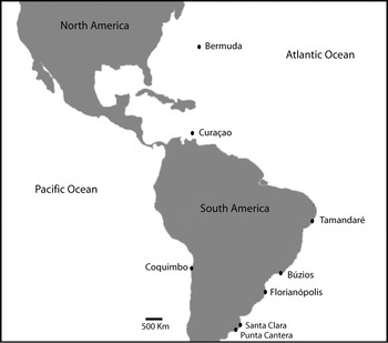
Bunodosoma cangicum, B. caissarum and Anthopleura cascaia specimens were collected in Búzios (south-east Brazil, 22°57′S 43°10′W). Bunodosoma cangicum specimens were also collected in Tamandaré (north-east Brazil, 03°45′S 38°36′W). Bunodosoma granuliferum, the genus type species, was collected in Curaçao (Boca Sami, 12°06′N 68°55′W). Phymactis papillosa specimens from Argentina were collected at Santa Clara del Mar (37°50′S 57°29′W) and Mar del Plata (38°05′S 57°32′W) in three localities (Punta Cantera Beach, Acantilados and Escollera Norte). Samples of P. papillosa var. rubra-viridis Haussermänn, Reference Haussermänn2004 from the Chilean coast were collected at Coquimbo (29°57′S 71°19′W). Actinia bermudensis samples were collected in Florianopolis (27°26′S 48°34′W), south of Brazil and in Bermuda (32°18′N 64°44′W). To minimize the possible collection of clone mates, all individuals were sampled at least 1 m apart. The anemones were wrapped in damp paper towels and transported to the laboratory, where they were processed for each analysis. For the molecular work (both allozymes and DNA extraction) samples were stored in liquid nitrogen until required for analysis. Figure 1 shows the collecting sites.
Collection sites for all species analysed in the study.

Morphological analysis
Specimens of P. papillosa, B. cangicum and B. caissarum were observed in situ and also in aquaria. Collected specimens were anaesthetized in a 7% magnesium chloride solution and then fixed and preserved in 4% formaldehyde. For the new species described, type and voucher specimens have been deposited at the Museo Argentino de Ciencias Naturales ‘Bernardino Rivadavia’ (MACN), in the Actiniarian Collection of Universidad Nacional de Mar del Plata (UNMdP), Argentina and in the Museu Nacional do Rio de Janeiro (MNRJ), Brazil. Some specimens were also deposited at the Cnidarian Collection of Anthozoan Research Group (GPA) at the Universidade Federal Rural de Pernambuco (UFRPE), Brazil. For purposes of comparison, several specimens collected and identified by Zamponi and deposited in the Cnidaria Collection of UNMdP were also examined.
Measurements of pedal disc width and column height were made on preserved material. Longitudinal and transverse sections, 8–10 µm thick, were made from paraffin embedded specimens. Sections were stained with haematoxylin–eosin and Mallory triple stain methods (Kiernan, Reference Kiernan1990). Cnidae measurements were taken from undischarged capsules in squash preparations mounted with fresh water and without stain at 1000X magnification. Relative frequencies of nematocyst types are subjective estimates based on all the cnidae observed on slides. The nomenclature of cnidae follows that of Schmidt (Reference Schmidt1969, Reference Schmidt1972, Reference Schmidt1974).
Allozyme analysis
Frozen tissue samples were homogenized with 100 µl of distilled water and analysed by horizontal 12.5% starch gel electrophoresis, using a Tris Citrate pH 8.0 buffer (Ward & Beardmore, Reference Ward and Beardmore1977). A total of 15 enzymes, coding for 19 loci were used: acid phosphatase (ACP, # E.C. 3.1.3.2); catalase (CAT, # E.C. 1.11.1.6); α-esterase (α-EST, # E.C. 3.1.1.1); glucose-6-phosphate isomerase (GPI, # E.C. 5.3.1.9); glutamate dehydrogenase (GDH, # E.C. 1.4.1.3); glutamate-oxaloacetate transaminase (GOT, # E.C. 2.6.1.1); hexokinase (HK, # E.C. 2.7.1.1); isocitrate dehydrogenase (IDH, # E.C. 1.1.1.42); malate dehydrogenase (MDH, # E.C. 1.1.1.37); malate dehydrogenase NADP (ME, # E.C. 1.1.1.40); mannose-6-phosphate isomerase (MPI, # E.C. 5.3.1.8); octopine dehydrogenase (ONDH, # E.C. 1.5.1.11); α-prolyl-phenylalanine peptidase (PEP, # E.C. 3.4.11); phophoglucomutase (PGM, # E.C. 5.4.2.2); and phosphogluconate dehydrogenase (PGD, # E.C. 1.1.1.4.4). Enzymes were stained according to Manchenko (Reference Manchenko1994).
DNA extraction and amplification
For the DNA extractions, 10 mg of frozen tissue was homogenized in a microcentrifuge tube in 500 µl of CTAB extraction buffer (CTAB 2%, EDTA 20 mM, 2-mercaptoethanol 0.2% v/v, Tris 100 mM, NaCl 1.4M, proteinase K 30 µg) following the protocol of Damato & Corach (Reference Damato and Corach1996) modified with a precipitation with 3M sodium acetate and 100% ethanol at –20oC. The pellet was re-suspended in ultrapure water. The two ribosomal internal transcribed spacers (ITS1 and ITS2), together with the 5.8S rDNA were directly amplified using the polymerase chain reaction (PCR). Each 20 µl PCR reaction consisted of 20 ng of DNA template, 1 unit of Taq DNA polymerase, 0.8 µM of each primer, 0.2 mM dNTPs, 2 mM MgCl2 and 1 mg/ml BSA, in 1X PCR buffer. The primers used were the 18SF (5′TCA TTT AGA GGA AGT AAA AGT CG 3′) and 28SR (5′GTT AGT TTC TTT TCC TCC GCT T 3′) designed by Lôbo-Hajdu et al. (Reference Lôbo-Hajdu, Guimarães, Mendes, Lamarão, Vieiralves, Mansure and Albano2004). Cycling conditions were 4 minutes at 94oC, followed by 35 cycles of 1 minute at 92oC, 1 minute at 42oC and 1 minute at 72oC, with a final extension step of 5 minutes at 72oC. PCR products were treated with 0.5 units of exonuclease and 2 units of shrimp alkaline phosphatase and sequenced by the dideoxy termination method on an ABI3500 automatic sequencer. Although multiple copies of the ribosomal genes are usually present in the genome, it has been shown that concerted evolution is a major force maintaining the uniformity of paralogues and therefore direct sequencing of PCR products usually does not interfere with phylogenetic analyses (Hillis et al., Reference Hillis, Moritz, Porter and Baker1991).
Data analyses
Allozyme data were analysed using the program BIOSYS–2 (Swofford & Selander, Reference Swofford and Selander1981). Allele frequencies were calculated and all loci were tested for Hardy–Weinberg equilibrium with an exact test (Haldane, Reference Haldane1954) with Bonferroni correction for multiple tests (Lessios, Reference Lessios1992). Unbiased estimates of heterozygosity and pairwise genetic identity and distance (Nei, Reference Nei1978) were estimated for all populations analysed. The genetic distances were then used to build an Unweighted Pair Group Method with Arithmetic Mean dendrogram (Sneath & Sokal, Reference Sneath and Sokal1973) with 2000 bootstrap replicates using the program TFPGA v1.3 (Miller, Reference Miller1997; http://www.marksgeneticsoftware.net/tfpga.htm). An exact test for population differentiation as described in Raymond & Rousset (Reference Raymond and Rousset1995) was performed also using TFPGA v1.3. A factorial correspondence analysis (FCA) was performed with the program GENETIX 4.05 (Belkhir et al., Reference Belkhir, Borsa, Chikhi, Raufaste and Bonhomme2002). This type of analysis is especially useful for estimating associations between multiple independent qualitative variables, where no a priori hypothesis is present (Valentin, Reference Valentin2000).
Sequences of the two internal transcribed spacers (ITS1 and ITS2) were aligned using Clustal W (Thompson et al., Reference Thompson, Higgins and Gibson1994), followed by eye inspection. Neighbour-joining (NJ) and maximum likelihood (ML) methods were used for reconstructing the group's phylogeny (Felsenstein, Reference Felsenstein1981; Saitou & Nei, Reference Saitou and Nei1987). In this analysis only the species Actinia bermudensis was used as outgroup.
The model of evolution was estimated using the log-likelihood score as implemented in the programs Modeltest version 3.5 (Posada & Crandall, Reference Posada and Crandall1998). The program PAUP* 4.0b10 (Swofford, Reference Swofford2000) was used for the ML analysis and the program MEGA 3 (Kumar et al., Reference Kumar, Tamura and Nei2004) was used for the NJ analysis.
For the ML approach a full heuristic search with tree bisection reconnection (TBR) and starting tree obtained via NJ was used. For the NJ analyses the Jukes–Cantor distance (Jukes & Cantor, Reference Jukes, Cantor and Munro1969) was used. Bootstrap analyses were carried out for both NJ and ML methods, using 1000 replicates. The resulting trees were drawn using the program Figtree v1.3.1 (Rambaut, 2006–2009; http://tree.bio.ed.ac.uk/).
RESULTS
Allozymes
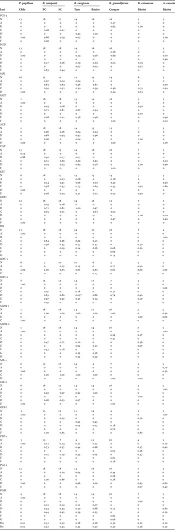
Gene frequencies and sample sizes for all studied loci are given in Table 1. Significant deviations from Hardy–Weinberg expectations (heterozygote deficiencies) were only found for the PGD locus in the Punta Cantera population. Heterozygote deficiencies are common in marine invertebrates and could be due to a variety of different factors such as gel scoring errors, null alleles, aneuploidy, effects of selection or the Wahlund effect (Zouros & Foltz, Reference Zouros and Foltz1984; Hare et al., Reference Hare, Karl and Avise1996). Heterozygosity levels (H) were high in most populations (Table 1) but were well within the range of those usually observed in sea anemone species (Solé-Cava & Thorpe, Reference Solé-Cava and Thorpe1989; Russo et al., Reference Russo, Solé-Cava and Thorpe1994).
Bunodosoma spp., Phymactis papillosa and Anthopleura cascaia. Gene frequencies of the populations studied. N, number of individuals analysed; Hobs and Hexp, direct count and Hardy–Weinberg expected mean heterozygosities per locus, respectively; PC, Punta Cantera Beach; SC, Santa Clara del Mar; Tam, Tamandaré.

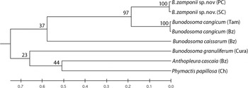
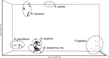
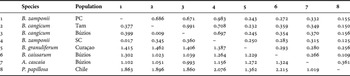
Genetic distance found between Phymactis papillosa from Chile and the putative P. papillosa from Argentina was very large, at a level typically found between different genera (Thorpe & Solé-Cava, Reference Thorpe and Solé-Cava1994; Vianna et al., Reference Vianna, Schama and Russo2003). The smallest gene divergence was found between the two Bunodosoma cangicum populations (D = 0.009; Table 2). The Santa Clara and Punta Cantera P. cf. papillosa populations also presented little gene divergence (D= 0.017). Within the Bunodosoma genus, B. granuliferum was found to be the most divergent species (D ranges from 1.024 to 1.363). The samples of the putative P. papillosa from Argentina clustered with the Bunodosoma species analysed clearly indicating that P. cf. papillosa from Argentina is not conspecific with P. papillosa from Chile (Figures 2 & 3). The FCA graphically shows the differentiation found among the species studied, indicating a close relationship between B. cangicum and P. cf. papillosa from Argentina (Figure 3). However, the genetic distance values between those two species (D ranging from 0.345 to 0.399) were within the range usually found between different species of the same genus (Thorpe & Solé-Cava, Reference Thorpe and Solé-Cava1994; Vianna et al., Reference Vianna, Schama and Russo2003). Furthermore, those two species showed a high degree of gene frequency differentiation across loci (Fisher's exact test; P < 0.0001), confirming their genetic distinctiveness.
Unweighted Pair Group Method with Arithmetic Mean dendrogram of allozyme unbiased genetic distances (Nei, Reference Nei1978) between the populations studied. Bootstrap values for 2000 replicates on branches.

Three-dimensional representation of a factorial correspondence analysis based on 19 allozyme loci.

Bunodosoma spp., Anthopleura cascaia and Phymactis papillosa unbiased genetic identities (above diagonal) and distances (below diagonal) between pairwise populations (Nei, Reference Nei1978). PC, Punta Cantera Beach; SC, Santa Clara del Mar; Tam, Tamandaré.

DNA sequencing analysis
The ITS1/5.8S/ITS2 region was amplified in all samples and ranged in length from 665 to 740 base pairs. The alignment was made with the three regions together. The two ITS regions were used as different data sets in order to avoid heterogeneity among sites, although the results of a preliminary NJ combined analysis were exactly the same (i.e. same tree topology and bootstrap confidence, results not shown). Average nucleotide composition was 20.4% T, 29.2% C, 24.0% A and 26.5% G. Base composition did not vary significantly between species. The complete ITS1, 5.8S and ITS2 sequences were deposited in GenBank with accession numbers JN118557–JN118569.
A partition homogeneity test (Farris et al., Reference Farris, Kallersjo, Kluge and Bult1995) as implemented in PAUP* was performed to verify if the two ITS regions would give significantly different results. The test result indicates no evidence that the two regions were incongruent, so the analyses were subsequently done with the combined data sets. The evolutionary model chosen by Modeltest was the Jukes–Cantor model with a gamma distribution of among site variation (JC + G).
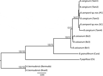
Both phylogenetic methods gave similar results (same tree topology) and the relationships found between the studied species were the same as those found in the analyses of allozyme loci. Once again, the putative Phymactis papillosa samples from Argentina were genetically more similar to Bunodosoma than to P. papillosa from Chile (Figure 4). High divergence levels (16–20%) were observed among the genera studied further emphasizing the separation of the two closely related genera Bunodosoma and Phymactis (18% mean Jukes–Cantor distance). The mean intraspecific variability levels observed for Bunodosoma ITS sequences (0.25%), although expected for Anthozoans (Forsman et al., Reference Forsman, Guzman, Chen, Fox and Wellington2005, Reference Forsman, Barshis, Hunter and Toonen2009; Fukami et al., Reference Fukami, Chen, Budd, Collins, Wallace, Chuang, Chen, Dai, Iwao, Sheppard and Knowlton2008), were smaller than those observed in other Actiniaria populations (Stoletzki & Schierwater, Reference Stoletzki and Schierwater2005; Acuña et al., Reference Acuña, Excoffon, McKinstry and Martinez2007; Gusmão, Reference Gusmão2010). This lower evolutionary rate may explain the lack of significant differences between B. cangicum and the putative P. papillosa from Argentina, which could be clearly separated by allozyme data.
Phylogenetic tree constructed by the neighbour-joining (NJ) method with Jukes–Cantor distances and pairwise deletion. Numbers on branch are: 1000 bootstrap replicates NJ/1000 bootstrap replicates maximum likelihood.

The joint results of the allozyme and DNA sequence data clearly show that the common intertidal anemone from Argentina identified as P. clematis by Zamponi (Reference Zamponi1977) does not belong to that species but, instead, is a species of the genus Bunodosoma. The high differentiation observed in the allozyme analyses, together with the morphological diagnostic characteristics observed lead us to conclude that the anemones formerly named P. clematis in Argentina belong to a new species of Bunodosoma, that we describe below.
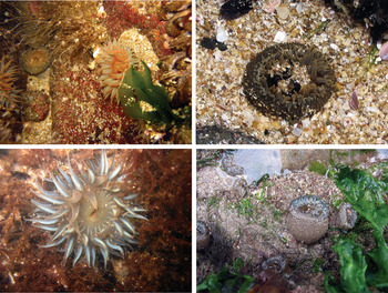
External morphology of Bunodosoma zamponii sp. nov. on the intertidal zone of Mar del Plata. Photograph by Gabriel Genzano.

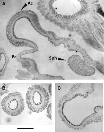
Internal anatomy of Bunodosoma zamponii sp. nov.: (A) marginal sphincter muscle circumscribed, palmated (Sph), acrorhagus (Ac) and ectoderm (Ec); (B) cross-section through tentacles; (C) cross-section through a vesicle. Scale bars: A, 700 µm; B, 1.1 mm; C, 0.8 mm.

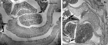
Internal anatomy, cross-section showing mesenteries with retractor muscles (R), gonads (G) and ectoderm (Ec). (A) Bunodosoma zamponii sp. nov.; (B) B. cangicum. Scale bar: 200 µm.

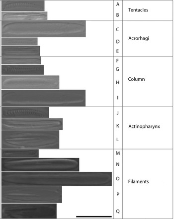
Cnidae of Bunodosoma zamponii sp. nov.: (A) spirocyst; (B) b-rhabdoid; (C) holotrich; (D) spirocyst; (E) b-rhabdoid; (F) b-rhabdoid; (G) spirocyst; (H) holotrich; (I) holotrich; (J) spirocyst; (K) b-rhabdoid; (L) p-rhabdoid; (M) b-rhabdoid 1; (N) b-rhabdoid 2; (O) b-rhabdoid 3; (P) p-rhabdoid A; (Q) p-rhabdoid B1a. Scale bar: 10 µm.

Phymactis clematis; Zamponi, Reference Zamponi1977: 139,141 (Argentina); Pollero, Reference Pollero1983; Patronelli et al., Reference Patronelli, Zamponi, Bustos and Vega1987; Zamponi, Reference Zamponi1989, Reference Zamponi1993, Reference Zamponi2000, Reference Zamponi2005; Excoffon & Zamponi, Reference Excoffon and Zamponi1991; Acuña & Zamponi, Reference Acuña and Zamponi1995, Reference Acuña and Zamponi1996, Reference Acuña and Zamponi1997; Acuña et al., Reference Acuña, Zamponi and Perez1996; Zamponi & Perez, Reference Zamponi and Perez1996, Genzano et al., Reference Genzano, Acuña, Excoffon and Perez1996; Acuña, Reference Acuña1997; Zamponi et al., Reference Zamponi, Belém, Schlenz and Acuña1998a, Reference Zamponi, Genzano, Acuña and Excoffonb; Gomes et al., Reference Gomes, Belém and Schlenz1998; Excoffon et al., Reference Excoffon, Genzano and Zamponi1999; Patronelli et al., Reference Patronelli, Olivera and Zamponi2005, Reference Patronelli, Olivera, Zamponi and Crupkin2008; Olivera et al., Reference Olivera, Patronelli and Zamponi2009; not Phymactis clematis (Drayton in Dana, 1846: 130).
TYPE MATERIAL
Holotype: MACN (In-35365), Atlantic Ocean, Mar del Plata (38°05′S 57°32′W), Punta Cantera, intertidal, Coll. P.B. Gomes, 16 September 1999, preserved in formalin.
Paratypes: UNMdP (C.A. 27), three specimens, MNRJ (6274), one specimen, both samples collected at the same time and place as the holotype.
ADDITIONAL MATERIAL
UFPE (GPA 092), two specimens, MNRJ (6275), one specimen, Atlantic Ocean, Mar del Plata (38°05′S 57°32′W), Acantilados, intertidal, Coll. P.B. Gomes, 24 November 1999, preserved in formalin; UFPE (GPA 093), one specimen, Atlantic Ocean, Mar del Plata (38°05′S 57°32′W), Escollera Norte, intertidal, Coll. A.C. Excoffon, 14 November 1999, preserved in formalin.
DIAGNOSIS
Actiniidae with rows of non-adhesive vesicles on the column from margin to limbus. Margin with marginal projections each one with a single holotrichous acrorhagus on the oral surface and simple or compound vesicles on the adoral surface. Projections arranged in two alternate crowns of slightly different size. Fosse present. Column orange to cream with dark grey vesicles, or dark orange with olive green vesicles. Tentacles arranged in five cycles, approximately 96 total, brownish or crimson red. Size and coloration distinguish live specimens of B. zamponii from other Bunodosoma species, except B. cangicum from which it only differs on the arrangement of the mesenteries.
DESCRIPTION
Column
Cylindrical with a great capacity of elongation. Live specimens vary from 3 to 6 cm long, width of the base between 1.4 and 3.8 cm. Contracted specimens often dome-shaped. Fosse deep. Margin denticulate, with vesicle-covered marginal projections arranged in two alternate crowns of slightly different size; each projection bears a single holotrichous acrorhagus on the oral surface. Acrorhagi simple, rounded, of cream or opaque white colour, arranged in two alternate cycles with 48 acrorhagi each (Figure 5). External cycle endocoelic and large (diameter 1.1 cm), internal cycle exocoelic and small (0.75 cm of diameter). In some specimens, the second cycle may be absent or incomplete. Column covered from margin to just above the limbus with endocoelic and exocoelic non-adhesive vesicles, mostly with no clear arrangement due to the state of contraction of the specimens, but in some specimens, especially near the margin, a clear arrangement can be observed forming approximately 96 longitudinal rows. Vesicles rounded, sometimes compound only near the margin and on marginal projections, without nematocyst batteries. Column orange to cream with dark grey vesicles or dark orange with olive green vesicles. Adherent base roughly circular, bigger in diameter than the column, cream with orange or pale brown radial lines.
Oral disc and tentacles
Number of tentacles from 96 to about 102, in 5 cycles. The outermost cycle, exocoelic, alternating with the largest row of acrorhagi. Tentacles of the outermost cycle are the same length as innermost tentacles (from 1.5 to 2.5 cm long). Tentacles pale or dark brownish or crimson red typically without marks. Oral disc brownish or pale cream, mesentery insertions visible as orange or cream lines. Oral disc diameter from 2 to 5 cm, central mouth, rounded, atop an oral cone. Actinopharynx creamy white.
Internal anatomy
Actinopharynx extends half to three-quarters of the length of the column, with folds. Two siphonoglyphs, extending below the end of the actinopharynx, each one attached to a pair of directive mesenteries. Equal number of mesenteries distally and proximally (46 to 52 pairs, usually 48). Mesenteries hexamerously arranged in four cycles, all perfect. The last cycle is only attached to the actinopharynx just below the oral disc. Near the aboral end, all mesenteries join together, forming a basal node. Gonochoric. All mesenteries, except the directives, are fertile. Gametogenic tissue may be poorly developed or absent in mesenteries of the last cycle. In younger mesenteries, the gametogenic tissue is positioned more distally. The male gametogenic tissue appears rounded and pale white or yellow in fixed specimens. The female tissue, when poorly developed, is similar to that of the male. Well-developed, female gametogenic tissue is grey and not as round.
Marginal sphincter strong, circumscribed, palmate (Figure 6A), rounded or oval. Endodermal circular musculature well developed at mid-column. Vesicles in the column show no histological differentiation (Figure 6C). Parietobasilar muscles diffuse, with a short pennon (Figure 7A). Retractor muscles circumscribed to diffuse strong (Figure 7A). Tentacles and oral disc with ectodermal longitudinal musculature (Figure 6B). As in other species of the genus, tissues without zooxanthellae.
Cnidom
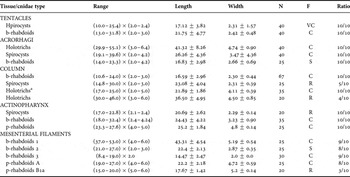
Spirocysts, b-rhabdoids, p-rhabdoids A, p-rhabdoids B1a, holotrichs (Figure 8). See Table 3 for sizes and distribution.
Size and distribution of Bunodosoma zamponii sp. nov. cnidae. All measurements are in µm; range: length × width; mean ± standard deviation; N, total number of capsules measured; F, frequency; VC, very common; C, common; S, scarce; R, rare; ratio, ratio of number of specimens in which each cnidae was found to number of specimens examined.

a, very common in the middle column, absent or sporadic near the oral and pedal disc.
Distribution and natural history
Atlantic Ocean, Argentina, from Santa Clara del Mar (37°50′S 57°32′W) to Mar del Plata (38°05′S 57°32′W). The species inhabits crevices, clefts and tide pools in the intertidal and subtidal zones. Bunodosoma zamponii sp. nov. is the most abundant species where it occurs (Zamponi et al., Reference Zamponi, Genzano, Acuña and Excoffon1998b). Much work has been done on the species (all still considering the species as Phymactis clematis) about its autoecology (Acuña & Zamponi, Reference Acuña and Zamponi1995; Acuña, Reference Acuña1997) and morphological variation (Zamponi & Perez, Reference Zamponi and Perez1996; Acuña & Zamponi, Reference Acuña and Zamponi1997). The species is polyphagous opportunistic (Acuña & Zamponi, Reference Acuña and Zamponi1996). It is gonochoric, with an oviparous–pelagic–planktotrophic pattern (Excoffon & Zamponi, Reference Excoffon and Zamponi1991). No fission scars or anatomical irregularities have been reported for the species. Many studies have been conducted about the morphological, functional and biochemical characterization of the sphincter of B. zamponii sp. nov. (Patronelli et al., Reference Patronelli, Olivera and Zamponi2005, Reference Patronelli, Olivera, Zamponi and Crupkin2008; Olivera et al., Reference Olivera, Patronelli and Zamponi2009).
ETYMOLOGY
Bunodosoma zamponii is named after Dr Mauricio O. Zamponi, in recognition of his numerous contributions to the study of cnidarians, especially in Argentina.
TAXONOMIC REMARKS
This species has a circumscribed sphincter, typical non-adhesive vesicles and old, strong fertile mesenteries typical of the genus Bunodosoma. The specimens of P. papillosa collected in Chile and used in the present study presented a diffuse sphincter and no reproductive tissue in the first and second mesentery cycles, which is similar to previous descriptions of the species (McMurrich, Reference McMurrich1904; Carlgren, Reference Carlgren1899, Reference Carlgren1920, Reference Carlgren1945, Reference Carlgren1959; Stotz, Reference Stotz1979; Haussermänn, Reference Haussermänn2004). The fact that the new species is the most common on the intertidal zone of Mar del Plata and that there are no other species that resemble the genus Phymactis in the area nor in the Cnidarian Collection of UNMdP lead us to believe that the records of P. papillosa (= P. clematis) from Argentina were misidentifications. Phymactis papillosa probably does not occur on the Atlantic coast of South America, as already suggested by Carlgren (Reference Carlgren1939).
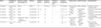
Bunodosoma zamponii sp. nov. differs from other species of the genus by a combination of characters such as retractor muscle type, number of mesenteries and tentacles, reproductive tissue distribution and cnidom. From the species of Bunodosoma that occur in the South Atlantic Ocean, B. zamponii sp. nov. resembles B. granuliferum and B. biscayense (Fischer, 1874) by having 96 mesenteries disposed in 4 cycles. However, these two species have the column marked with alternating dark and white longitudinal bands (Watzl, Reference Watzl1922; Pax, Reference Pax1924; den Hartog, Reference den Hartog1987), which are not present in B. zamponii sp. nov. (Figure 5). Furthermore, B. zamponii sp. nov. can be distinguished from those two species by cnidae differences and gonad distribution.
The cnidae distribution of the new species is very similar to that of other Bunodosoma species. Nevertheless, the presence of spirocysts in the filaments and column observed in B. zamponii sp. nov. differentiate this species from B. caissarum and B. cangicum.
Bunodosoma zamponii sp. nov. is very similar to B. cangicum, a species present along the coasts of Brazil and Uruguay. Their major morphological differences are the mesentery cycle number (four in Bunodosoma zamponii sp. nov., against three in B. cangicum) and retractor muscle type (strong and circumscribed in B. cangicum; Figure 7). Table 4 summarizes the major differences among the different species.
Bunodosoma spp. and Phymactis papillosa. Main morphological characters.

a, Haussermänn, 2004 and personal observation; b, Belém, 1988 and personal observation; c, Pax, 1924; d, den Hartog, 1987.
Den Hartog (Reference den Hartog1987) proposed modifications to the diagnosis of the genus Bunodosoma establishing that the number of mesentery cycles should be four or five and that the retractor muscle of the mesenteries should be diffuse (den Hartog, Reference den Hartog1987: 555–556). This diagnosis is not compatible with the species of Bunodosoma from South America. Bunodosoma zamponii sp. nov., B. caissarum and B. cangicum present a circumscribed or circumscribed–diffuse retractor muscle and the last species has only three mesentery cycles.
In Mar del Plata and Santa Clara del Mar B. zamponii sp. nov. shares the hard substrate with Aulactinia marplatensis Zamponi, Reference Zamponi1977 and Oulactis muscosa (Drayton in Dana, 1846). The new species differs from all others in the field by the coloration and the presence of non-adhesive vesicles on the column. The presence of acrorhagi also distinguishes B. zamponii sp. nov. from Aulactinia marplatensis.
DISCUSSION
Both genetic and morphological data clearly show that Phymactis papillosa (formerly cited as Phymactis clematis) specimens from Argentina are not only very distinct from P. papillosa from Chile, the species' type locality, but, more importantly, belong to a different genus (Bunodosoma).
Only Carlgren (Reference Carlgren1949) reported P. papillosa along the whole Chilean coast as far south as Tierra del Fuego. No other study to date has found this species south of the Golfo de Penas (48°S) (Haussermänn, Reference Haussermänn2004), indicating that a phylogeographical break might occur in that region. Surface ocean currents are recognized as one of the most important factors for the dispersal of benthic marine animals, since many larvae are planktonic and expected to have great dispersal potential. Nevertheless, how much dispersal really occurs is not always well known and studies describing species with long-lived larvae that have low dispersal and species with no planktonic development with high dispersal capability have been reported (Miller & Ayre, Reference Miller and Ayre2008). Phylogeographical breaks, where there is a discontinuity in the distribution of a species that coincides with a geographical feature, seem to play an important role for benthic marine animals (Hellberg, Reference Hellberg2009). Golfo de Penas has already been reported as a dispersal barrier for some marine animals (Lancellotti & Vásquez, Reference Lancellotti and Vásquez1999). It is possible that the cold and low salinity waters in that area (Försterra & Haussermänn, Reference Försterra and Haussermänn2003) could be responsible for the southward restriction in P. papillosa distribution.
The external features of the genera Phymactis and Bunodosoma are very similar, increasing the difficulty in making a distinction between them, nevertheless other, more subtle, morphological features differentiate the two genera (den Hartog, Reference den Hartog1987; Haussermänn, Reference Haussermänn2004). Species of Phymactis have the first and second mesentery cycles sterile, a diffuse sphincter and few holotrichs in the acrorhagi, with abundant long b-rhabdoids instead (Carlgren, Reference Carlgren1934, Reference Carlgren1949, Reference Carlgren1959; Stotz, Reference Stotz1979; Haussermänn, Reference Haussermänn2004). Bunodosoma members have a variety of degrees of circumscribed sphincter (den Hartog, Reference den Hartog1987; Haussermänn, Reference Haussermänn2004; Fautin et al., Reference Fautin, Hickman, Daly and Molodtsova2007). Morphological analyses carried out in this study confirm all these differences, since all Bunodosoma species studied differ from P. papillosa on these characters. The specimens from Argentina presented the same type of sphincter and cnidae distribution of other Bunodosoma species and also all strong mesenteries, except the directives, were fertile (see description above).
The low genetic differentiation found between the Santa Clara del Mar and Punta Cantera B. zamponii sp. nov. populations (D = 0.02; 50 km distance) and between the Tamandaré and Búzios B. cangicum populations (D = 0.01; 3500 km distance) are similar to those found between other species of the genus. Russo & Solé-Cava (Reference Russo and Solé-Cava1991) found a low divergence (D = 0.05) between B. caissarum populations that were 180 km distant. The same was observed for B. cavernata (Bosc, 1802) (D = 0.11; 2000 km) and B. granuliferum (D = 0.16; 1600 km) populations from the Gulf of Mexico and the Caribbean, respectively (McCommas & Lester, Reference McCommas and Lester1980; McCommas, Reference McCommas1982). These values indicate that Bunodosoma species have a high potential for long distance dispersal. Studies on the reproductive biology of B. caissarum showed that they bear long-lived planktotrophic larvae (Belém, Reference Belém1987). Although there are no studies on B. cangicum reproduction patterns or larval biology, the Argentinean B. zamponii sp. nov. and B. caissarum have the same kind of larvae which can live for five days in the plankton (Belém, Reference Belém1987; Excoffon & Zamponi, Reference Excoffon and Zamponi1991), enabling them for long distance dispersal.
Bunodosoma cangicum and Bunodosoma zamponii sp. nov. are genetically, morphologically and ecologically more similar to each other than either of them is to B. caissarum or B. granuliferum. This indicates that B. cangicum and Bunodosoma zamponii sp. nov. have probably diverged very recently. It is possible that, due to its hydrographical characteristics and the high sedimentation rate, the La Plata River might have played an important role in the isolation of these two species. In some cases habitat specificity can be more important than larval dispersion when it comes to gene flow among populations (Miller & Ayre, Reference Miller and Ayre2008; Ayre et al., Reference Ayre, Minchinton and Perrin2009). In fact, large estuaries such as the Amazon River delta are an important source of phylogeographical breaks for costal marine species (Rocha et al., Reference Rocha, Bass, Robertson and Bowen2002; Lima et al., Reference Lima, Freitas, Araujo and Solé-Cava2005; Currie & Small, Reference Currie and Small2006). Although the Amazon River estuary did not seem to be an important barrier to gene flow (Vianna et al., Reference Vianna, Schama and Russo2003) for the sea anemone species Actinia bermudensis, that species is known to withstand large salinity variations, inhabiting estuaries (Stephenson, Reference Stephenson1935; Corrêa, Reference Corrêa1964; Douek et al., Reference Douek, Barki, Gateño and Rinkevich2002) where Bunodosoma species are never found (Gomes, Reference Gomes, Tabarelli and Silva2002). So far few papers have analysed the role of the La Plata River as a gene flow barrier for marine animals but Zamponi et al. (Reference Zamponi, Belém, Schlenz and Acuña1998a) have already called attention to this estuary as a potential barrier to cnidarian dispersal, since the Brazilian and Argentinean coasts only share three species of sea anemones out of more than 50 presently recognized species in the two areas.
ACKNOWLEDGEMENTS
This paper is part of Renata Schama's PhD thesis at the Genetics Program of the Federal University of Rio de Janeiro. A.O. Debrot generously provided Bunodosoma granuliferum specimens from Curaçao. The authors would also like to thank E.S. Lellis for help with the collection of P. papillosa samples from Chile and R.M. Albano for revision of an earlier version of this manuscript. The authors are grateful to CNPq for funding that enabled us to undertake this work (Brazilian Science and Technology Ministry, R.S., grant number: 141719/2001–0).