Introduction
Radiation therapy, based on simulation computed tomography (CT), is a core activity in the treatment planning process. Contrast agents are frequently used during CT to characterise both healthy and tumour tissues more accurately. Reference Elawadi, Almohsen and Algendy1,Reference Wertz and Jäkel2 Treatment planning systems (TPS) convert the Hounsfield Unit (HU) values of different tissues or materials in the CT to electron or mass densities, depending on the algorithm used for planning. As contrast agents have elements with higher atomic numbers than most of the human tissues, they increase HUs, which could result in changes in calculated doses. Reference Davis, Palmer and Nisbet3 Therefore, if the dose calculation is performed in a contrast-enhanced CT, there is a concern of dosimetric deviation because the treatment is administered in the absence of contrast. Reference Elawadi, Almohsen and Algendy1,Reference Jing, Tian and Tang4
In order to avoid this problem, it is a common practice to obtain two CT scans during the same simulation session: one unenhanced and one enhanced. Reference Hanna, Slevin and Appelt5 However, it has been described that performing two CT scans during the simulation not only exposes the patient to additional radiation but also reduces the optimisation of resources in the radiation oncology department by extending the simulation time. Reference Liu, Vora, Suh, Liu, Schultheiss and Wong6 Consequently, waiting times for radiotherapy increase. This problem is even more serious in low-resource countries where access to radiotherapy treatment is already very limited.
Some authors have reported tolerances for changes in HU that could be set to achieve less than 1% dose change: ±20 HU for soft tissue and ±50 HU for lung. Reference Davis, Palmer and Nisbet3,Reference Bissonnette, Balter and Dong7 These limits could explain why some researchers have described that, even if contrast agents cause noticeable increase in HU values, these changes are clinically insignificant. Reference Elawadi, Almohsen and Algendy1,Reference Létourneau, Finlay and O’Sullivan8–Reference Komiyama, Ohira and Kanayama11 In spite of that, there are few studies assessing dosimetric differences in pelvis. Reference Elawadi, Almohsen and Algendy1,Reference Jing, Tian and Tang4,Reference Shibamoto, Naruse, Fukuma, Ayakawa, Sugie and Tomita12–Reference Heydarheydari, Farshchian and Haghparast16
The present study evaluates the effect of oral contrast on dose distribution calculation for rectal cancer patients, using three-dimensional conformal radiation therapy (3D-CRT) and volumetric modulated arc radiotherapy (VMAT) planning.
Methods
A prospective dosimetric study was conducted as a before-and-after study. This study was approved by the institutional Human Research Ethics Committee prior to conducting this study.
Patient selection
The sample size was calculated, accepting an alpha risk of 0·05 and a beta risk of 0·2 in a bilateral contrast, and 26 subjects were required to detect a difference in dose target coverage equal to or greater than 0·45 units, as reported in previous studies. Reference Joseph, Liu and Severin15 Consecutive patients, older than 18 years, admitted to our radiotherapy unit with a diagnosis of non-metastatic rectal cancer were enrolled. They were referred for radiotherapy management between August 2018 and September 2019. The exclusion criteria were as follows: a) patients with hypersensitivity to oral or intravenous iodinated contrast agents, b) with a history of kidney disease and c) at risk of intestinal perforation.
Image acquisition
Unenhanced and enhanced CT scans for each patient were acquired, in the same position and with the same coordinates. One hour before the simulation, patients were instructed to evacuate their bladders, and then drink 500 mL of water, to maintain a comfortably full bladder. Following unenhanced scanning, patients remained in the same immobilisation device in order to minimise any positioning differences. Then, the patients were instructed to ingest 250 mL every 10 min of 1 L of oral contrast preparation. This mixture contained 15 mL of iopamidol iodinated contrast (Scanlux 370 mg of iodine per ml) diluted in 985 cc of water.
According to our institutional protocol, contrast was infused through a rectal tube as well. Ten minutes after finishing oral contrast administration, the IV automatic injector was connected to the patient. The enhanced scan started 60 s after the IV contrast injection. The total dose of the IV contrast media was 1·2 mL/kg body weight or about 90 mL for patients with a body weight of over 60 kg. The same scanning technique and the same coordinate origin were used for unenhanced and enhanced CT scans. The reconstruction of the images was performed with a slide width of 5 mm.
Treatment planning
After the acquisition of CTs, two expert radiation oncologists delineated target volumes and organs at risk, according to our institutional protocol. For each patient, a medical physicist created a 3D-CRT and an IMRT/VMAT plan, based on the enhanced CT.
Three medical physicists generated the plans according to a pre-established protocol. Previously, a pilot study was conducted to ensure the standardisation of the planning process. The intraclass correlation coefficient between planners was 0·94 (p = 0·04) for 3D-CRT plans and 0·78 (p = 0·007) for VMAT plans.
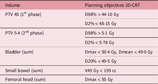
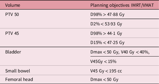
All plans were generated with Eclipse TPS (Eclipse v15·1, Varian Medical Systems, Palo Alto, CA) and with Acuros XB version 15·1 dose calculation algorithm. Dose prescription for 3D-CRT plans was 45 Gy in 25 fractions, in a first phase, and then, 5·4 Gy in three fractions, in a second phase. For VMAT, the prescription was 50·4 Gy in 25 fractions, with integrated boost technique; for this, three full coplanar arcs were used.
Planning objectives for each of the treatment modalities, including dose prescription, coverage and dose limits for organs at risk, are described in Tables 1 and 2, for 3D-CRT and IMRT/VMAT plans, respectively.
Treatment planning objectives used for 3D-CRT plans

Treatment planning objectives used for IMRT/VMAT plans

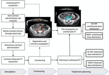
Finally, dose distribution was recalculated on the corresponding unenhanced CT. The same beam weights were kept as for the enhanced CT plans. The entire planning process is shown in Figure 1.
Flow chart showing planning process.

Statistical analysis
In order to compare calculated doses in unenhanced versus enhanced CT plans, a paired t-test was used when the distribution of the difference between groups was normally set apart. Otherwise, the non-parametric Wilcoxon signed-rank test was used. The dose metrics compared were those detailed in planning objectives including coverage, and dose limits for organs at risk, described in Tables 1 and 2, for 3D-CRT and IMRT/VMAT plans, respectively. Furthermore, the gamma index was calculated to compare the IMRT/VMAT unenhanced and enhanced plans.
The gamma passing rate is defined as the percentage of points satisfying the condition gamma index < 1. Reference Low, Harms, Mutic and Purdy17 For evaluation, the following criteria were used: a gamma passing rate of ≥95%, with 2%/2 mm, and ≥90%, with 2%/2 mm global normalisation.
All statistical tests were two-tailed, and a p-value ≤0·05 was considered statistically significant. The analyses were conducted on the Statistical Package for Social Sciences software version 22·0.
Results
Twenty-nine patients fulfilled inclusion criteria. Three patients were excluded due to significant differences in position between the two sets of CTs. Finally, 26 patients were included.
The mean age of the included patients was 62·4 years, (standard deviation (SD) = 12·4). Fifteen patients were women (57·6%), and 11 were men (42·3%). Most of the patients were treated with neoadjuvant intention (80%), and five patients were treated in an adjuvant setting (19·2%). In relation to1 clinical stage, 11 patients had locally advanced rectal cancer (42·3 %), eight patients had early stages (30·7%), and, for seven patients, the clinical stage was not available (26·9%).
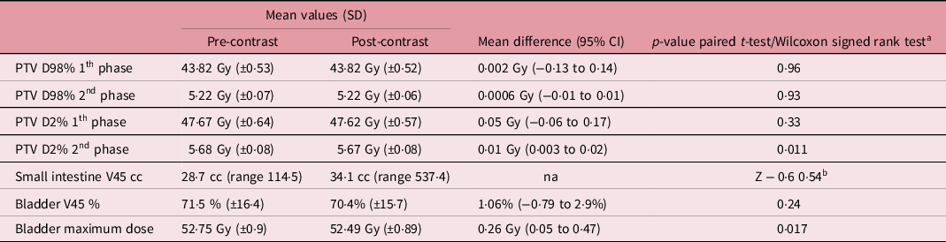
Table 3 shows the differences in mean values of target coverage and dose metrics to organs at risk, as well as the mean differences (MD) when paired sample t-test was done between unenhanced and enhanced 3D-CRT plans. Analysing3D-CRT plans, there were statistically significant differences in PTV D2% from the second phase (MD 0·01 Gy, 95% CI [0·003 to 0·02 Gy] and in maximum doses to bladder (MD 0·26 Gy, 95% CI [0·05 to 0·47 Gy]. There were no statistically significant differences in the other assessed dose metrics.
Mean differences in target coverage and doses to small intestine and bladder in 3D-CRT plans. Dose metrics are reported according with our institutional dose constraints

a Paired t-test was used except data with b.
b Wilcoxon signed-rank test was used.
In the case of VMAT plans, differences in dose metrics between unenhanced and enhanced plans are shown in Table 4. There were statistically significant differences in PTV D98% (MD 0·22 Gy, 95% CI [0·05 to 0·39 Gy]) as well as in PTV D2% (median of 52·52 Gy for non-enhanced and 52·22 Gy for enhanced, Z-4·45 p < 0·0001), volume of small intestine receiving more than 45 Gy (3·1 cc, 95% CI [0·81 to 5·4 cc]), percentage of bladder volume receiving 45 Gy (MD 2·9%, 95%CI [1·4 to 4·3%]) and maximum dose to bladder (MD 0·65 Gy, 95% CI [0·46 Gy to 0·85 Gy]).
Mean differences in target coverage and doses to small intestine and bladder in IMRT/VMAT plans

a Paired t-test was used except data with b.
b Wilcoxon signed-rank test was used.
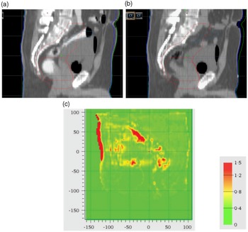
Using the gamma index criteria of 2%/2 mm, the mean passing rate was 95·2% (95% CI [94·1 to 96·36%]) for IMRT/VMAT plans of the entire group. In 25 patients, out of 26, the passing rate was greater than 90%, and, in 15, it was greater than 95%. In Figure 2a, gamma analysis is shown as an example.
Gamma analysis for 2 mm distance to agreement and 2% dose difference criteria. (a) Contrast enhanced CT. (b) Unenhanced CT. (c) Gamma index analysis.

Discussion
The small bowel is one of the most important dose-limiting organs in pelvic radiotherapy. Some studies confirmed that the irradiated bowel volume is closely related to the toxicity caused by pelvic radiotherapy. Reference Baglan, Frazier, Yan, Huang, Martinez and Robertson18–Reference Nuyttens, Robertson, Yan and Martinez20 Herbert et al reported a significant decrease in the incidence of acute and chronic small bowel toxicity using oral contrast as well as a change in the location of the treatment field with the use of small bowel contrast, indicating that planning with contrast leads to changes in the delivery of radiotherapy. Reference Herbert, Curran, Solin, Stafford, Lanciano and Hanks21
In spite of the advantages of using oral contrast, there is concern about possible dosimetric deviations if the dose calculation is performed in the contrast-enhanced CT. Reference Elawadi, Almohsen and Algendy1,Reference Jing, Tian and Tang4 To avoid these theoretical deviations, it is a common practice to obtain two CT scans during the same simulation session: one unenhanced and one enhanced. Reference Hanna, Slevin and Appelt5
However, the promotion of high-value practices in radiation oncology should be a priority because of the increasing costs of cancer care and the increasing expense of ever-advancing technologies. This necessity is more important in countries with difficult access to radiation treatment and very long waiting lists. Reference Smith, Ganz and Bekelman22 This is the reason why this study examines if planning in an enhanced CT causes a dosimetric deviation from an unenhanced plan, which is large enough to justify the practice of performing two CT sets.
Minimal differences were found for the dose metric D2% in the PTV, in the second phase, and for maximum doses to bladder in 3D-CRT plans, although statistically significant. For the remaining parameters, there were no statistically significant differences. In the case of IMRT/VMAT plans, the biggest difference was found in small intestine volume, receiving 45 Gy in enhanced plans, that was 3·1 cc smaller than in non-enhanced, and in maximum doses to bladder with a MD of 0·65 Gy (95%CI [0·46 to 0·85 Gy]), which means a change of 1·6%. There was a statistically significant difference in the dose covering 98% of the PTV of 0·4% of the prescribed dose; however, this difference may not be clinically meaningful.
Our findings are consistent with those reported by other studies that had been conducted in this field. For instance, Elawadi et al conducted a review of 22 studies. Most of the results of these studies suggest a clinically insignificant effect of contrast agents on radiotherapy dose calculations. Anyhow, the majority of these studies assessed the effect of intravenous contrast. Reference Elawadi, Almohsen and Algendy1
Only a few studies investigate the effect of oral contrast in the treatment planification for patients with rectal cancer. Heydarheydari et al reported the results of six patients with rectal cancer, and they found that the relative mean dose and MU (monitor units) differences were less than 2%. Reference Heydarheydari, Farshchian and Haghparast16 Joseph et al examined the effect of oral contrast in 13 patients with rectal cancer, and they reported <0·1% deviation in the dose ratio for all volumes of interest for 3D-CRT plans, while, for IMRT plans, the differences were in the order of 1% for the mean dose. Reference Joseph, Liu and Severin15 Rankine et al reported the impact of oral contrast in three patients and found that dose increase at the isocenter was less than 2·1%. Reference Rankine, Lanzon and Spry23
The study of Jing et al is the only one which concludes that oral contrast agents caused clinically significant changes in the dose calculations for the targets and critical structures, especially for bowel doses. Reference Jing, Tian and Tang4 They reported data from VMAT plans of 33 rectal cancer patients, and they argued that the bowel volume receiving ≥50 Gy was dramatically increased, when oral contrast within the bowel was absent (MD 0·9 cc ± 0·93 cc). Reference Jing, Tian and Tang4 Considering that our study also found differences, even greater than that reported by Jing et al (3·1 cc), additional efforts should be done to determine if this difference could be clinically significant. Jing et al explain these differences in patients in which greater volume of enhanced intestine is overlapping or near to the PTV. They concluded that the larger the volume of the enhanced bowel near the path of the beams, the more significant is the dose underestimation in the calculation. Reference Jing, Tian and Tang4
In our study, using gamma index evaluation, 96% of the included patients satisfied the 2% and 2 mm criteria, in accordance with the study of Elawadi et al, which evaluated the gamma index between unenhanced and enhanced CTs from 226 cancer patients of different locations. Their analysis revealed that 94% of plans satisfied this recommendation, but oral contrast was used just in a few patients. Additionally, they found that bowel movement induced differences in two of three of the included rectal cancer patients. Reference Elawadi, Almohsen and Algendy1
The present study has some strengths. Firstly, unlike all the aforementioned studies, the unenhancement was not simulated. A more comprehensive method was used, using a quasi-experimental approach, in which patients were prospectively included in a protocol designed to scan these patients in the same position twice: once before and once after oral contrast application, trying to avoid differences in positioning. On the other hand, expert radiation oncologists conducted the process of target delineation very carefully, following an institutional protocol. Furthermore, a pilot study was conducted among participating medical physicists, to ensure standardisation of the planning process. Finally, the gamma index evaluation was used as an additional method to evaluate the volumetric dose difference between enhanced and non-enhanced CT.
Some limitations could be mentioned about our study. As oral contrast takes time to reach the intestine, we waited 40 min between unenhanced and enhanced CTs. This fact introduced variations in OARs volumes that could explain some changes in dosimetric parameters. Still, this methodology is more accurate than the methodology used by studies which simulate unenhancement. As the present study is basically dosimetric, it is difficult to decide which differences in doses are clinically meaningful. Studies exploring clinical outcomes may be helpful in this issue; however, this kind of studies has not been done to our knowledge. Finally, in the present study the volume of PTV overlapping with enhanced intestine was not measured, which could be large since five operated patients were included. Some studies have reported that the volume of enhanced intestine within the PTV, or overlapping with the PTV, was significantly correlated with changes in the doses. Reference Jing, Tian and Tang4,Reference Joseph, Liu and Severin15
Conclusion
For most of the assessed outcomes, there were no clinically meaningful differences, between unenhanced and enhanced CT, in target coverage and OARs dose limits for 3D-CRT and IMRT/VMAT plans. The most important difference was found in small bowel dose metrics limits. This difference could lead to an underestimation of the volume of the small intestine receiving 45 Gy when the planning is performed directly on an enhanced CT. However, after assessing concordance of unenhanced and enhanced plans with gamma index, overall, the use of small bowel oral contrast does not significantly influence dose calculations.
These observations indicate that in general, dose calculation performed on an oral contrast-enhanced CT produces reliable plans for most patients, as long as they adhere to protocols with pre-established coverage parameters and OARs dose limits.
Cautions should be taken in specific situations as when a considerable volume of enhanced intestine is overlapping or near to the PTV or when treatment planning dose–volume constraints are very close to being met. In these scenarios, planning in an enhanced CT could be problematic.
Acknowledgements
The authors thank Dr. Luis de los Reyes for his relevant contributions during the conception of this research and Kleber Suárez for his contribution during CT scans acquisition. We also like to thank all physician, physics and radiotherapists from the Radiotherapy department of Hospital SOLCA-Quito for their helpful discussion and advices.
Authors’ Contribution
Nadia Montero-Oleas conceived of, designed, acquired and analysed the data, wrote manuscript drafts and approved them for publication. Andrés Imbaquingo-Cabrera and Cristina Núñez-Silva contributed to design, acquire data, revision of drafts and approval for publication. Alejandro Coloma-Espín, Vladimir Collantes-Cruz and Carlos Molineros contribute to the conception of the study, acquisition, analysis and interpretation of the data, and revision of manuscript. All authors discussed the results and approved the final manuscript.
Funding
This research received no specific grant from any funding agency, commercial or not-for-profit sectors.
Conflict of Interest
None.
Ethical Standards
The authors assert that all procedures contributing to this work comply with the ethical standards of the relevant national guidelines on human experimentation and with the Helsinki Declaration of 1975, as revised in 2008, and have been approved by the institutional committees of the SOLCA-Quito Research Ethics Committee.