


To the best of our knowledge, there are but two reports of patients born with hypoplastic left heart syndrome and a right aortic arch, with none having survived the initial palliative procedure.1, 2 The combination is associated with aberrant origin of the left subclavian artery, which could affect the construction and function of any planned systemic-to-pulmonary arterial shunt. We describe here our recent experience in managing a patient with hypoplastic left heart syndrome and a right aortic arch, who survived the initial palliative surgery, which included placement of a conduit from the right ventricle to the pulmonary arteries.
Case report
The newborn male infant was delivered at 34.5 weeks gestation, weighing 3.2 kilograms at birth. The pregnancy was complicated by poor prenatal care, polyhydramnios, gestational diabetes and cocaine abuse. After delivery, the patient developed respiratory distress, and required endotracheal intubation with ventilatory support. He was transferred to our institution for further evaluation and management of possible congenital cardiac disease.
An echocardiogram demonstrated a hypoplastic left ventricle, with aortic atresia and severe mitral stenosis, as well as left ventricular endocardial fibroelastosis. Additional findings included a right aortic arch, with a left-sided descending thoracic aorta, a left-sided arterial duct, and an aberrant left subclavian artery, which arose from a diverticulum of Kommerell and coursed behind the oesophagus. Magnetic resonance imaging of the chest confirmed the diagnosis (Fig. 1). Except for a horseshoe kidney, no other congenital malformations were detected.
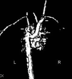
Figure 1. Magnetic resonance image providing a posterior view of the aortic arch, and demonstrating a right aortic arch (¬), with the left subclavian artery (−) arising from a Diverticulum of Kommerell (®). R: right; L: left.
The patient underwent a Damus-Kaye-Stansel procedure, along with placement of a Gore-Tex graft of 5 millimetre diameter between the right ventricle and the pulmonary arteries. His post-operative course was complicated by the need for prolonged mechanical ventilation. Cardiac catheterization at 3 months of age demonstrated moderate stenosis in the ascending aorta distal to the Damus-Kaye-Stansel anastomosis, with a gradient of 40 millimetres of mercury. Subsequently, the patient underwent augmentation of the aortic arch, construction of a bidirectional Glenn shunt, and take-down of the conduit between the right ventricle and the pulmonary arteries. He did well from the cardiovascular standpoint, but remained dependent on the ventilator, requiring a tracheostomy. At 10 months of age, he continues to have a tracheostomy, but is no longer dependent on the ventilator.
Discussion
Abnormalities of the aortic arch are very common in patients with hypoplastic left heart syndrome. Coarctation of the aorta is the most common anomaly, being found in one series with an incidence as high as two in three cases.3 Interruption of the aortic arch is uncommon, but has been well described.4 A right aortic arch, however, although strongly associated with congenital cardiac malformations such as tetralogy of Fallot and common arterial trunk, it is exceedingly rare in patients with hypoplastic left heart syndrome.5–7
As far as we are aware, to date there have been two reports of babies with hypoplastic left heart syndrome and a right aortic arch.1, 2 The first was an infant with VATER syndrome and hypoplastic left heart syndrome due to aortic atresia and mitral stenosis.1 He also had a perimembranous ventricular septal defect, a hypoplastic right aortic arch, and an aberrant left subclavian artery arising from the descending aorta. The patient underwent a balloon atrial septostomy, but died from heart failure before undergoing any surgical procedure. The second report was of an infant who had hypoplastic left heart syndrome with mitral atresia, aortic stenosis, right aortic arch, coarctation of the aorta, large right-sided patent arterial duct, and bilateral superior caval veins.2 The left subclavian artery arose from the left pulmonary artery via a left-sided arterial duct. This patient underwent a Norwood procedure, with a 4 millimetre Gore-Tex shunt placed from the right common carotid artery to the right pulmonary artery, but died on the fifth post-operative day.
Although congenital cardiac disease is more common when the right sided aortic arch branches in mirror-imaged fashion,6, 7 all the cases of hypoplastic left heart syndrome with right aortic arch described thus far have had anomalous origin of the left subclavian artery, albeit that its origin has been variable. As shown in this small number of cases, it can arise directly from the descending aorta or via a diverticulum of Kommerell, or it can arise from the left pulmonary artery via an arterial duct. The embryologic cause of the lesion is abnormal interruption of the initial double arch between the left common carotid artery and the left subclavian artery (Fig. 2).8

Figure 2. Schematic showing the primitive double aortic arch system with the usual site of interruption (A), and the site of interruption in our patient (B). In our case, the result was to produce the aortic arch anomaly as depicted. Ao: Aorta; CCA: common carotid artery, PAD: patent arterial duct; PT: pulmonary trunk; SCA: subclavian artery; VA: vertebral artery.
Interestingly, our patient also had a vascular ring, formed by the right aortic arch with the anomalous left subclavian artery and the patent left-sided arterial duct. This might have contributed to the respiratory failure, since tracheomalacia was documented by flexible bronchoscopy. Ligation and division of the duct provided for relief of the vascular compression on the trachea and oesophagus.
In this population of patients, construction of a systemic-to-pulmonary arterial shunt can be challenging, due to the absence of the left brachiocephalic artery, this being the ideal site for the shunt. Under this circumstance, the origin of the shunt is limited to a smaller right subclavian artery, or the left common carotid artery. We suggest, therefore, that a conduit placed from the right ventricle to the pulmonary arteries offers the best source of flow of blood to the lungs at the time of initial palliation for those with this most unusual variant of hypoplastic left heart syndrome.



