Mammalian fertilisation begins with sperm deposition and migration through the uterus and distal oviduct to the oviductal sperm reservoir, a specialised area where motile spermatozoa transiently bind to the oviductal epithelial surface. Here, the competent spermatozoa transiently slow their metabolism, awaiting the release of the ovum from the ovary. Subsequent to ovulation, spermatozoa undergo capacitation and hyperactivation, resulting in a wave-like pattern of sperm cohort release from the sperm reservoir. Released spermatozoa are guided towards the fertilisation site in the oviductal isthmus by chemotaxis and/or thermotaxis (reviewed in Ref. Reference Suarez and Pacey1). The egg vestments, including the multicellular cumulus oophorus, and the glycoprotein egg coat, the zona pellucida, are the last hurdles encountered by the fertilising spermatozoon prior to its fusion with the oocyte plasma membrane, the oolemma. Several recent reviews discuss progress in research on sperm–egg fusion (Refs Reference Primakoff and Myles2, Reference Inoue3, Reference Vjugina and Evans4). This article addresses this critical step of the fertilisation process in animals and humans, as well as the clinical significance of sperm–oolemma interactions, and their involvement in antipolyspermy defence and oocyte activation.
Sperm surface priming preceding sperm–egg fusion
Prior to fertilisation, the antigens on the sperm head equatorial segment are not exposed for potential interactions with the oolemma. Just like fertilisation itself, sperm priming for fusion with the egg is a stepwise process. During each step, sperm structures are lost or altered irreversibly. Fusogenicity of the sperm plasma membrane is first increased by cholesterol removal and lipid raft formation during sperm capacitation, which probably primes the sperm acrosome for exocytosis (‘the acrosome reaction’) and also enables spermatozoa to detach from the sperm reservoir near the fertilisation site in the oviductal ampullary–isthmic junction (Ref. Reference Suarez and Pacey1). The acrosome reaction results in release of acrosomal enzymes and exposure of a new set of antigens on the sperm head surface. It is thought to be induced by sperm binding to the zona pellucida, although zona-free oocytes can be fertilised in vitro and brought to term (Ref. Reference Wu5), and acrosome removal or vesiculation is observed in pig (Ref. Reference Wu5) and human (Ref. Reference Sathananthan6) spermatozoa coincubated with zona-free oocytes.
In general, acrosomal exocytosis is required for the success of sperm–oolemma fusion. Within the acrosomal segment of the sperm head, proteins coating the inner acrosomal membrane, such as IAM38/ZPBP2, are exposed, possibly to mediate interactions between the sperm head and zona glycoproteins during penetration (Ref. Reference Yu7). On the equatorial segment, thought to be the site of initial sperm binding to the microvilli of the oolemma (Ref. Reference Yanagimachi, Knobil and Neill8), antigens such as equatorin/MN9-antigen are exposed (Ref. Reference Manandhar and Toshimori9). Antibodies against equatorin inhibit mouse fertilisation at the sperm–oolemma fusion step, without affecting sperm motility or sperm penetration of the zona (Ref. Reference Toshimori10). A similar effect was observed after passive immunisation of female mice by intraoviductal injection of anti-MN9 antibody (Ref. Reference Yoshinaga11). Preliminary characterisation of the MN9 antigen reported at a recent meeting suggested that it is a sialoglycoprotein (Ref. Reference Yamatoya12).
Sperm IZUMO is essential for fertility
IZUMO was discovered by proteomic analysis of the antigen recognised by monoclonal antibody OBF13 (Ref. Reference Okabe13) on the equatorial segment of the acrosome-reacted mouse spermatozoa. Male Izumo-null mice are infertile, with an apparent defect of gamete fusion (Ref. Reference Inoue14). IZUMO, named after a fertility shrine in the Shimane prefecture of Japan, is a testis/male-germline-specific member of the immunoglobulin superfamily, a group of cell adhesion molecules (CAMs) best known for their role in the immune response (Ref. Reference Aricescu and Jones15). Of note is the ability of these molecules to bind to identical (homophilic binding) or closely related (pseudohomophilic binding) members of the immunoglobulin superfamily (Ref. Reference Aricescu and Jones15). As IZUMO is testis specific, this opens up the possibility that it could interact with a related CAM protein on the oolemma.
Tetraspanins CD9 and CD81: architects of the oolemma or IZUMO's receptors?
Tetraspanins are hydrophobic integral membrane proteins, characterised by four transmembrane domains and two extracellular loops (Ref. Reference Tarrant16), that serve as organisers of complex plasma membrane protein networks. Common post-translational modifications of tetraspanins include glycosylation and palmitoylation, the latter being important for insertion of tetraspanins into cell membranes. Tetraspanins are enriched in cholesterol-rich microdomains, distinct from lipid rafts, where they organise other proteins such as integrins, kinases, human leukocyte antigens and growth factor receptors (Ref. Reference Lazo17), and can also associate with each other. Such complex associations, referred to as tetraspanin webs, have been implicated in signalling, cell–cell and cell–extracellular-matrix adhesion, and in a variety of specialised functions, in processes such as cell migration/motility, viral infection, immune responses, tumour metastasis and haematopoietic stem cell differentiation (Ref. Reference Yunta and Lazo18). Tetraspanin–tetraspanin interaction appears to be of primary importance within the web; tetraspanin interactions with integrins and other web components appear to be of lesser importance for cell–cell adhesion (Ref. Reference Espenel19). Most of the proteins mentioned above associate with tetraspanins within the cellular membrane (e.g. integrins) or on the inner face of plasma membrane (e.g. protein kinases); little is known about proteins that may bind to the extracellular loops of tetraspanins. In this regard, as discussed below, IZUMO might be one of the few extracellular ligands known to interact with a tetraspanin.
CD9 was first investigated as a potential sperm receptor because of its associations with integrin α6β1, which had previously been implicated in sperm–oolemma binding and fusion. These studies showed that antibodies against CD9 blocked sperm–egg binding and fusion (Ref. Reference Chen20). Independently, a genetic model was developed wherein the female, but not the male, Cd9 −/− mice created to investigate immune responses to pathogens were in fact found to be infertile (Refs Reference Le Naour21, Reference Kaji22, Reference Miyado23). However, mice placed in an extended breeding trial eventually became pregnant at a rate of 60% that of the wild-type controls (Ref. Reference Primakoff and Myles2). The CD9-protein-deficient ova allow for normal zona penetration, but, due to a lack of fusion, spermatozoa accumulate in the perivitelline space as there is no triggering of the zona-pellucida-based antipolyspermy defence (Ref. Reference Inoue3).
Contrary to partial fertility of Cd9 −/− mice, female mice are completely infertile when a related tetraspanin gene, Cd81, is targeted on the Cd9-null background (Ref. Reference Rubinstein24). Infertility of CD9-deficient mice was reversed by injection of Cd9 and Cd81 cDNAs, the latter possibly compensating for the former, in the germinal vesicle stage oocytes (Ref. Reference Kaji25). CD81 is the preferred binding partner of CD9 within the tetraspanin web. Similar to CD9, CD81 interacts with other tetraspanins and with integrins within the tetraspanin web. One known extracellular interaction of CD81 is with hepatitis C viral envelope glycoprotein E2 (Ref. Reference Flint26).
Recently, it was shown that CD81 is also highly expressed on the plasma membrane of cumulus cells surrounding the oocyte (Ref. Reference Tanigawa27). In that study, spermatozoa with a partially retained acrosome were found in the perivitelline space of Cd81-null oocytes that failed to undergo fusion. The authors speculated that spermatozoa could encounter CD81 on the cumulus cells before contacting the zona pellucida and that the lack of such interaction in CD81 animals might impair the fusibility of sperm plasmalemma. However, this does not explain why cumulus- or even zona-free mouse oocytes can be fertilised successfully in vitro.
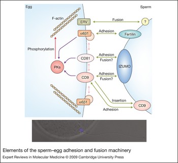
In another recent study, CD9 appeared to be released from the unfertilised ova in the form of membrane vesicles that bind to spermatozoa within the perivitelline space prior to the actual binding of the sperm plasmalemma to the oocyte microvilli (Ref. Reference Miyado28). This unexpected observation was interpreted as a release of exosome-like vesicles that convey the fusogenic factors to sperm plasmalemma. How does IZUMO fit in such a scenario? Could IZUMO be the ligand on the sperm plasmalemma for CD9 (Fig. 1), or does it function independently of CD9/CD81? One of the few ligands that reportedly bind directly to the extracellular loop of CD9 is PSG17 (Ref. Reference Ellerman29), which, similar to IZUMO, is a member of the immunoglobulin superfamily. Possibly, IZUMO could be responsible for binding the oocyte-released vesicles, after which the plasma membrane would become fusogenic based on the acquisition of CD9 molecules from exosomes, which could interact in a homophilic manner with CD9 molecules on the oolemma. A critic could suggest that the authors of the paper describing CD9-containing vesicles simply mistook the cross-sections of oocyte microvilli for vesicles. This is consistent with the authors' observation that there were fewer microvilli on the surface of Cd9-null eggs and with scanning electron microscopy studies showing that microvilli of Cd9 +/+ ova have a higher radius of the curvature of the microvillar tips, compared with Cd9-null ova (Ref. Reference Runge30). In addition, zona-free mouse ova, from which such vesicles would likely disperse away, are easy to fertilise in vitro. It should be noted that scanning electron micrographs of mouse ova do not reveal vesicles on their surface, whereas the oocyte microvilli are very long. However, in support of the vesicle hypothesis, the authors were able to recover CD9-containing vesicles from wild-type ova and use them to restore sperm–oolemma binding in the Cd9-null ova (Ref. Reference Miyado28).
Elements of the sperm–egg adhesion and fusion machinery. Sperm IZUMO may bind to oocyte tetraspanins CD9 and/or CD81 to mediate adhesion and fusion. Within the tetraspanin web on the oolemma, CD9 and C81 may interact with each other and with integrins (e.g. integrin α6β1). Sperm–oolemma adhesion is facilitated by the interactions between oolemma integrins such as α6β1, and sperm disintegrins such as fertilin β. Oolemma-expressed endogenous retroviral envelope proteins (ERVs) could interact with an unidentified sperm plasmalemma component to mediate fusion. Both integrins and tetraspanins could modulate actin polymerisation (F-actin) in the oocyte cortex via signalling pathways involving several protein kinases (PKs), resulting in the formation of the fertilisation cone. Bottom insert shows sperm–oolemma binding during murine fertilisation in vitro. DNA in the sperm head is labelled blue with DAPI (4′,6-diamidino-2-phenylindole).

Integrins and ADAM family proteins might support the binding and fusion process
Integrins bind to extracellular matrix proteins and to a family of snake venom disintegrin proteins termed ADAMs (ADAM stands for ‘a disintegrin and a metalloproteinase domain-containing protein’). Initial observations of integrins on the mammalian oolemma (Refs Reference Bronson and Fusi31, Reference Bronson and Fusi32) were followed by the observation that the mammalian sperm plasma membrane protein fertilin contains an integrin-binding RGD sequence typical of disintegrins (Ref. Reference Blobel33). The involvement of the integrin–disintegrin system in sperm–oolemma fusion is an attractive proposition (Fig. 1) even though integrins are not gamete specific.
Integrin α6β1 was proposed initially as the sperm receptor on the oolemma (Ref. Reference Almeida34), but the mice lacking this integrin were found later to be fertile (Ref. Reference Miller35). Better understanding of the tetraspanin web could explain why mutations or antibody/peptide blocking of integrins in the oolema did have some effect on interactions in vitro. Essentially such interventions, even without having a direct effect on CD9, could alter the architecture of the tetraspanin web on the oolemma, or, if CD9 is indeed a sperm receptor, reduce the accessibility of CD9 to sperm surface ligands. In a similar manner, fertilin subunit β (ADAM2) (Ref. Reference Cho36) and cyritestin (ADAM3) (Refs Reference Nishimura37, Reference Shamsadin38) have been deleted from the mouse genome with little effect on fusion, while effects on sperm–egg interactions (cyritestin) and sperm migration in the female reproductive tract were observed. However, as pointed out in Ref. Reference Primakoff and Myles2, there are more than a dozen male-germline-expressed ADAMs that are yet to be explored for their potential involvement in sperm–oolemma fusion.
CRISP proteins
Epididymal secretory proteins and proteins from seminal plasma could be absorbed onto the sperm surface and interact with the oolemma during fertilisation. Two species of cysteine-rich secretory proteins (CRISPs), both of which are homologues of ion-channel-blocking reptilian and insect toxins (Ref. Reference Roberts39), have been implicated in sperm–oolemma interactions (Ref. Reference Cohen40). Members of the CRISP family are glycoproteins possessing the conserved CAP domain and an overabundance of cysteine residues. Epididymal protein DE (CRISP1) was proposed to have a role in sperm capacitation as well as in sperm–oolemma binding and it is localised on the equatorial segment of the ejaculated, acrosome-reacted rat spermatozoa (Refs Reference Cohen41, Reference Rochwerger and Cuasnicu42). Male and female mice immunised against CRISP1 have reduced fertility (Ref. Reference Ellerman29). Crisp1-null mice are fertile but exhibit reduced zona penetration and sperm–oolemma fusion in vitro (Ref. Reference Da Ros43). The authors suggested that a related CRISP protein could compensate for the absence of CRISP1 in mutant spermatozoa. CRISP2 (TXP2) is an intra-acrosomal protein of testicular origin, antibodies against which caused the accumulation of perivitelline spermatozoa during mouse fertilisation in vitro (Ref. Reference Busso44). In a zona-free fertilisation assay, CRISP2 seemed to compete with CRISP1 for the oolemma binding sites, suggesting that the two proteins bind to complementary/overlapping sites (Ref. Reference Busso44).
Is sperm–oolemma fusion necessary for oocyte activation and antipolyspermy defence?
Mouse oocytes fertilised by intracytoplasmic sperm injection (ICSI) can be refertilised in vitro (Ref. Reference Maleszewski, Kimura and Yanagimachi45). As the fusion step of fertilisation is skipped during the ICSI procedure, it is possible that direct membrane contact or mingling of membrane components alters the oolemma sufficiently to trigger the primary, oolemma-based block to polyspermy. Indeed, a recent study showed that sperm cytosolic factors alone are not sufficient to induce an efficient antipolyspermy defence, while they do trigger other aspects of mouse oocyte activation (Ref. Reference Wortzman-Show46).
Classical studies showed that the fertilising spermatozoon contributes plasma membrane components to the oolemma at fertilisation (Refs Reference Gundersen and Shapiro47, Reference Gabel, Eddy and Shapiro48) and that these sperm-derived membrane components disperse from the site of initial fusion (Ref. Reference Nishioka49). It is not known which sperm plasmalemmal component could be responsible for a change in oolemma receptivity for sperm. The fusion of unfertilised mouse oocytes or parthenogenetically activated eggs with a fertilised mouse oocyte induced a polyspermy block (Ref. Reference Krukowska50). In invertebrates, oolemmal depolarisation is thought to be responsible for the primary block to polyspermy (reviewed in Ref. Reference Yanagimachi, Knobil and Neill8). If that is the case in mammals, parthenogenetic activation or activation by the injection of a spermatozoon or of a sperm extract should trigger an oolemma-based antipolyspermy defence. This is clearly not the case. Consequently, consideration should be given to the possibility that receptor–ligand interactions contribute to oocyte activation and antipolyspermy defence in mammals.
At present, the evidence favours the fusion model of oocyte activation over the ligand–receptor model. In the fusion model, sperm–oolemma fusion enables the solubilisation of the postacrosomal and equatorial sperm perinuclear theca, resulting in the release of a soluble sperm-borne oocyte-activating factor in the ooplasm. This event of natural fertilisation is mimicked by sperm extracts or by whole sperm injection in the ooplasm, yet a contribution of ligand–receptor-based signalling to oocyte activation is still possible. Following the initial studies of the integrin–disintegrin system in mammalian gametes, several papers showed that small RGD-containing peptides can trigger some aspects of oocyte activation (Refs Reference Iwao and Fujimura51, Reference Campbell, Reed and White52, Reference Tatone and Carbone53). Integrins are in fact well suited for a role in oocyte activation, as they are linked to the cytoskeleton and various signalling pathways on the cytoplasmic face of the plasma membrane. In particular, integrins can transmit signals that activate cytoplasmic phospholipases and inositol-trisphosphate-dependent calcium release (Ref. Reference Fenichel and Durand-Clement54), which is generally accepted to be the ooplasmic pathway of the oocyte activation mechanism. Fertilisation-like calcium release has been observed in Xenopus oocytes incubated with RGD peptides (Ref. Reference Iwao and Fujimura51). In bovine oocytes, where calcium oscillations are observed during fertilisation (as opposed to a single calcium transient in Xenopus), RGD peptides induced two transients, but were not able to produce sustained calcium oscillations (Ref. Reference Campbell, Reed and White52). This may explain why a limited cleavage rate was observed in bovine oocytes activated with RGD peptide (~25% in RGD-activated oocytes compared with 60% cleavage in control, ionomycin-activated ones; Ref. Reference Sessions55). Modification of amino acids surrounding the RGD sequence enhanced the ability of RGD-containing peptides to block sperm binding and to induce parthenogenetic activation of bovine oocytes (Ref. Reference Sessions55).
While the interaction of sperm surface ligands with oolemma integrins might not be an essential or sufficient step in oocyte activation, it could contribute to an efficient antipolyspermy defence. In accordance with this ‘secondary’ role of integrins in oocyte activation, coincubation of soluble RGD peptides with the oocyte failed to induce full activation and completion of mouse oocyte meiosis, but elicited cortical granule exocytosis and increased the activity of ooplasmic protein kinase C (Ref. Reference Tatone and Carbone53). It should be noted, however, that the specificity of RGD peptides used to block fertilisation has been questioned (Ref. Reference McLaughlin56).
The role of oocyte cortex in fusion and sperm incorporation
The oocyte cortex is enriched in actin microfilaments and, except for the area overlying the chromosomes, is covered with long, actin-based microvilli (Ref. Reference Longo57). The apex of the microvilli seems to be the point of initial contact with the sperm's equatorial segment and postacrosomal sheath during binding (Ref. Reference Runge30). Disruption of actin microfilaments with cytochalasin B during bovine fertilisation prevents sperm incorporation into the ooplasm, but does not affect sperm–oolemma fusion or oocyte activation (Ref. Reference Sutovsky, Navara and Schatten58). Even a small patch in which sperm plasmalemma and oolemma are fused in such incorporation-arrested zygotes allows for the solubilisation of postacrosomal perinuclear theca and oocyte activation (Ref. Reference Sutovsky59). By electron microscopy, cross-sections of the apical endings of oocyte microvilli can be seen during bull sperm incorporation, inserted between the leaves of sperm head membranes and the perinuclear theca (Ref. Reference Sutovsky60).
The sperm incorporation site in bovine, porcine and other nonrodent mammalian oocytes is relatively small and flat. In contrast, the incorporation of a large murid spermatozoon with its long flagellum and falciform head features the formation of a large surface bulge, termed the incorporation or fertilisation cone (Refs Reference Phillips and Shalgi61, Reference Thompson, Smith and Zamboni62). Despite these morphological differences, microfilament disruptors cytochalasin B, jasplakinolide and latrunculin B reduce the rate of sperm incorporation during in vitro fertilisation of the zona-free mouse ova (Ref. Reference McAvey63). Surprisingly, treatment with a different microfilament disruptor, cytochalasin D, increased the polyspermy rates slightly, possibly by preventing the cytoskeletal rearrangements in the oocyte cortex that are conducive to the polyspermy block (Ref. Reference McAvey63). Neither cytochalasin B nor D had any effect on oocyte activation. Given the method of evaluation (DAPI staining of the nuclei/pronuclei), it is possible that even cytochalasin D prevented the incorporation of the sperm flagellum, but that this was not observable in the absence of flagellar markers or phase contrast microscopy. In a study where sperm tail incorporation was scored (Ref. Reference Terada, Simerly and Schatten64), jasplakinolide did not block sperm head entry, but prevented sperm tail incorporation into the murine ooplasm. This raises the question of whether sperm motility is required for sperm incorporation.
It is generally agreed that flagellar motility is necessary for penetration and that it stops following sperm–oolemma fusion, but exactly at what point of sperm incorporation does the flagellar beating stop? Perhaps motility is necessary to push the sperm plasmalemma and oolemma to close apposition within the fairly wide perivitelline space. As there is a functional plasma membrane enveloping the sperm tail during fusion, it is possible that flagellar movement, although restricted by the fertilisation slit in the zona, could still push the sperm inside the ooplasm. At the same time, oocyte microfilaments, instead of pulling the sperm head in, could simply reorganise the cortex around the sperm head to shore up walls around its perimeter that would eventually become a fertilisation cone. Some studies attributed the inability of human spermatozoa from infertile men to fertilise ova during subzonal sperm injection (SUZI) to the reduced motility of such subfertile spermatozoa (Refs Reference Wolf65, Reference Wolf66).
Oolemma-based block to polyspermy
The above observations suggest a role for cortical microfilaments in the antipolyspermy defence. The mechanism is not clear. There could be a change in the size, density or properties of the oocyte microvilli. Via their link to integrins and the tetraspanin web, microfilaments could guide the reorganisation of fusogenic or sperm-binding membrane domains to suppress sperm-plasmalemma–oolemma binding and fusion once the first fertilising spermatozoon has been incorporated. Cortical granule exocytosis, resulting in zona hardening, could also be affected by cortical microfilaments, although some contradictory results have been reported in rodents (Refs Reference Terada, Simerly and Schatten64, Reference Eliyahu67).
Also highly enriched in murid and ungulate oocyte cortex is the deubiquitinating enzyme UCHL1 (Refs Reference Yi68, Reference Susor69). Female gad mutant mice, which express a mutant form of UCHL1, were found to be subfertile and to have a high rate of polyspermy in vitro (Ref. Reference Sekiguchi70). It is not clear how this enzyme affects the oocyte cortex, but it is possible that it controls the activity and turnover of proteins associated with the cytoplasmic face of tetraspanin web domains by controlling and reversing their ubiquitination. Monoubiquitination and deubiquitination of the cytoplasmic tail is an established mechanism for the endocytosis and degradation of membrane receptors (e.g. Ref. Reference Komada71). Deubiquitination is an early step during the degradation of the ubiquitinated proteins (Ref. Reference Glickman and Ciechanover72). Also, deubiquitination sustains the cellular pool of unconjugated ubiquitin necessary for tagging of substrate proteins prior to their degradation. Shifting the balance of free ubiquitin and substrate-conjugated ubiquitin could alter the dynamics of membrane remodelling that likely occurs in response to oocyte activation. In our preliminary studies, microinjection of antibodies and inhibitors that interfere with UCHL1 significantly altered murine sperm incorporation and in vitro fertilisation rates (Mtango, N., Zhong, Z., Latham, K. and Sutovsky, P., unpublished).
A classical view of the oolemma-based antipolyspermy defence is that of rapid membrane depolarisation following fusion. This mechanism is well documented in invertebrate eggs (e.g. Ref. Reference Longo73), but may not be the primary mechanism of the oolemma-based polyspermy block in mammals (Refs Reference McCulloh, Rexroad and Levitan74, Reference Jaffe, Sharp and Wolf75). Except for a small initial oscillation, the membrane potential of the murine ovum remains constant within one hour following fertilisation, suggesting that membrane block to polyspermy is not voltage sensitive in mammals (Ref. Reference Jaffe, Sharp and Wolf75). Contribution of sperm membrane to the oolemma, rather than the addition of cortical granule content, may result in modification of the oolemma that makes it refractory to fusion with additional sperm following fertilisation in mammals (Ref. Reference Sun76).
Implications for assisted reproduction
There is some evidence of human infertility caused by a failure of sperm–oolemma interactions in patients with autoimmune infertility or sperm flagellar defects (Refs Reference Wolf65, Reference Wolf66). Such infertility is potentially treatable using ICSI. However, given the issue of ICSI not triggering a full oocyte response (e.g. the lack of antipolyspermy defence in mouse ICSI-fertilised zygotes), there is a reason for concern as an incomplete oolemma block to polyspermy could be reflective of reduced developmental potential of ICSI-generated zygotes.
Perhaps of more interest to human reproduction is the targeting of IZUMO by contraceptives. IZUMO is present on the human sperm surface (Ref. Reference Hayasaka77). Recombinant IZUMO was recently tested as an immunocontraceptive in mice. One study showed no substantial effect on fertility despite good circulating titres of antibody against the protein following injection of recombinant IZUMO (Ref. Reference Wang78), while another study reported a 35% reduction in fertility of IZUMO-immunised mice (Ref. Reference Naz79). This may not sound like a complete block, which would be required for a human contraceptive vaccine, but the intricacies of the mouse model should be taken into account, in which a highly efficient immune block of fertilisation is difficult to achieve and a combined, multiantigen vaccine might be necessary (Ref. Reference Naz and Aleem80).
Concluding remarks
Both animal biotechnology and human assisted reproduction could profit from harnessing the mechanism of sperm–egg fusion. Somatic cell nuclear transfer (SCNT/cloning) technology depends upon the success of membrane fusion between donor cell and the recipient ooplast. Typically, fusion is achieved by using a harsh, cell-damaging electrofusion process. Expression of gamete-specific membrane adhesion and fusion proteins in the donor cells used for SCNT could lead to a more natural method of inducing somatic-cell–ooplast fusion. Cloned transgenic animals are an important tool for biomedical research, which in the future may become a xenotransplant source, a food source and a means of producing pharmaceuticals (Ref. Reference Prather81). Identification of autoimmune infertility patients with fusion-incompetent spermatozoa suggests a previously unrecognised aetiology of human male infertility. Such infertility cases will likely continue to be treated by ICSI, yet the examination of autoimmune sera from such patients could provide clues about sperm–egg fusion proteins that have not yet been identified. Such proteins could prove to be suitable contraceptive targets.
IZUMO and CD9 are both essential for sperm–egg interactions, but integrins, disintegrins and other proposed receptors do not appear to be essential. However, their mutations may reduce litter size in controlled mating over an extended period of time and their blocking with competitive peptides and antibodies could result in a block of fertilisation in vitro. Perhaps the importance of a gene for fertility should be judged in multigenerational competitive breeding experiments or mixed insemination experiments with equal numbers of wild-type and mutant gametes. It is possible that while many of these mutants are propagated easily by controlled selective breeding and the short-term reduction of fertility is not observed, they would be eliminated from the genetic pool after several generations of free competitive mating, since their gametes would be inferior in the sperm–egg binding rate compared with wild-type breeders.
For sperm–oolemma fusion, the respective gametes have to first adhere to each other and then the lipid bilayers of their plasma membranes have to fuse. Involvement of both IZUMO and CD9/CD81 in the adhesion step is plausible, but neither protein is highly fusogenic (Ref. Reference Oren-Suissa and Podbilewicz82). So what happens once the binding mediated by IZUMO on sperm plasmalemma and the tetraspanin web proteins on the oolema is achieved? Neither of the above proteins has properties predictive of a membrane-fusion-inducing molecule. Are there membrane fusion proteins on the respective gamete membranes, mediating the actual membrane fusion, similar to viral envelope fusion proteins (Ref. Reference Harrison83) or fusogenic molecules of synaptic vesicles (Ref. Reference Rizo and Rosenmund84)? The means of achieving plasma membrane fusion include zippering- and hairpin-mediated fusion (reviewed in Ref. Reference Oren-Suissa and Podbilewicz82). Expression of endogenous retroviral envelope (ERV) proteins has been found in mouse and human oocytes and the ERV antigen disappeared after fertilisation (Ref. Reference Nilsson85).
Fusogenic, synaptic SNARE proteins are present in spermatozoa, but might be involved primarily in membrane fusion events during acrosomal exocytosis and are lost after its completion (Ref. Reference Ramalho-Santos86). Ferlin (FER1L3), a myoblast fusion protein (Ref. Reference Doherty87), could serve such a purpose in spermatozoa; the Caenorhabditis elegans ferlin (fer-1; a protein closely related to mammalian FER1L3) mutant is infertile because of abnormal membrane fusion during spermatogenesis (Ref. Reference Washington and Ward88). Proteomic analysis of mammalian spermatozoa could reveal other viral and nonviral fusion proteins.
In summary, we have learned a great deal about fusion in the last ten years, yet, in the end, we are still left with this dilemma. That is, at the present level of knowledge, is it possible that IZUMO and/or CD9/CD81 are structural enablers of gamete membrane fusion, rather than actual surface receptors and fusogenic molecules for the opposite gamete? Is the search not yet over?
Acknowledgements and funding
I thank Ms Kathy Craighead for manuscript editing, and my colleagues and collaborators Drs Namdori Mtango, Zhisheng Zhong and Keith Latham for agreeing to disclose our joint unpublished observations. The valuable input from three peer reviewers of this article is much appreciated. Seed funding for my laboratory has been provided by the Food for the 21st Century Program of the University of Missouri.