Introduction
Intracranial aneurysms are an extremely rare cause of spontaneous epistaxis, with few documented cases. The management of such cases is challenging due to the relative anatomical inaccessibility of the bleeding point.
This report documents one such case, and details its management.
Case report
A 72-year-old woman had been admitted to a local emergency department on numerous occasions with significant epistaxis resulting in hypotension. Her only significant past medical history was acute myeloid leukaemia, which was currently in remission.
The patient was seen by the ENT surgeons, who explored her nasal cavity in the operating theatre and performed sphenopalatine artery ablation.
Despite this, she was readmitted with a large epistaxis lasting nearly an hour, again resulting in hypovolaemic shock and requiring intravenous fluid resuscitation.
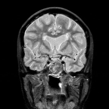
In an attempt to find the cause of the patient's recurrent haemorrhages, a computed tomography angiogram of the internal and external carotid arteries was performed. This demonstrated a large, left-sided intra-cavernous carotid artery false aneurysm, which had eroded through the sphenoid sinuses and was projecting into the nasopharynx. Magnetic resonance imaging demonstrated no evidence of neoplasia around the carotid artery (Figure 1).
T2-weighted, coronal magnetic resonance imaging scan demonstrating the presence of a large intra-cavernous internal carotid artery aneurysm, with erosion into the sphenoid sinus.

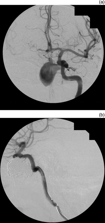
After discussion at the neurovascular multidisciplinary team meeting, the patient underwent angiography with embolisation of the aneurysm. The left internal carotid artery was catheterised, and a non-detachable balloon system was placed as a safety measure in case of aneurysmal rupture. An Echelon microcatheter was deployed into the aneurysm, and multiple endovascular coils were then passed into the aneurysm, achieving satisfactory occlusion of the lumen. There were no immediate complications from this procedure (Figure 2).
(a) Sagittal, pre-operative angiogram demonstrating the large, unprotected aneurysm, prior to endovascular coil placement. (b) The same aneurysm, successfully protected with endovascular coils; note the void of dye within the aneurysm, demonstrating lack of blood flow.

However, one week later the patient experienced a further episode of massive epistaxis, once again resulting in hypovolaemic shock and requiring blood transfusion.
She was taken back to the angiography suite, where recanalisation of the neck of the aneurysm was demonstrated. Therefore, further coils were placed into the aneurysmal remnant. A silk stent was placed from the cavernous carotid up into the supraclinoid carotid, occluding the neck of the aneurysm, to prevent retrograde migration of the coils into the internal carotid artery.
The patient made an uneventful recovery from the procedure and was subsequently discharged.
Despite these measures, the patient went on to have a further episode of massive epistaxis. Her case was again discussed at the neurovascular multidisciplinary team meeting, where consideration was given to angiographic embolisation of the internal carotid artery on the left side, with concurrent embolisation of the facial and internal maxillary arteries bilaterally.
A combination of endovascular and single-photon emission computed tomography (SPECT) imaging was used to demonstrate adequate collateral circulation through both the anterior and posterior communicating arteries in the Circle of Willis, with good perfusion of the left cerebral hemisphere on occlusion of the left internal carotid artery.
Therefore, the decision was made to proceed with left internal carotid artery occlusion. The left internal carotid artery was occluded using Gugliemi detachable coils (GDC) and an Amplatz plug. Again, sufficient collateral flow around the circle of Willis was demonstrated, and stasis of flow across the aneurysm was seen.
After a short post-procedure recovery period, the patient was discharged without neurological deficit, and had no further episodes of epistaxis.
Discussion
Intracranial aneurysms are a rare cause of epistaxis, but should be considered in cases of massive blood loss uncontrolled by conventional surgical measures. In most reported cases, there is erosion of a carotid artery aneurysm through the surrounding carotid canal as it passes through the skull base, with rupture into one of the air-filled facial sinuses. Most reported cases involve a false (rather than true) aneurysm of the internal carotid artery leading to epistaxis.Reference Singh, Thomas, Hoe and Sethi1–Reference Bhatoe, Suryanarayana and Gill4 The reason for this has not been demonstrated, but it is reasonable to suggest that the pressure of blood leaking out around the artery results in slow remodelling of the surrounding bone over time. Eventually, the expanding false aneurysm may erode into a nearby sinus and result in epistaxis.
• Epistaxis as a result of ruptured intracranial aneurysm is extremely rare
• Most cases result from pseudoaneurysms developing after head trauma or skull base surgery
• Traditional treatment techniques involve surgical ligation of feeding vessels
• Endovascular coiling and stenting can be a successful alternative for occluding the aneurysmal lumen, avoiding the risks and complications of open skull base surgery
• In the presented case, conventional endovascular measures resulted in ongoing epistaxis; endovascular ablation of the feeding internal carotid artery was achieved without neurological deficit
In patients experiencing a large volume of epistaxis, conservative management is not an option. The two current methods of treating these aneurysms include surgical ligation of the aneurysm or endovascular coil placement in its lumen. Surgical treatments are valuable for large, complex aneurysms displaying anatomy which prohibits endovascular coil placement. However, in most false aneurysms there is a definable neck, and these would often be best managed through endovascular coil placement, with or without stenting of the feeding vessel to occlude the aneurysm neck.
Surgical ligation of the feeding vessel (in this case the internal carotid artery) may result in permanent neurological deficit if the patient has a deficient circle of Willis and is unable to provide a good collateral circulation. In such cases, a vascular bypass procedure may be undertaken. However, in carefully selected patients endovascular techniques can be valuable in the management of intracranial aneurysms, negating the need for invasive surgery. These techniques are associated with a rapid post-procedure recovery, and avoid the risks and complications of open surgery.