Implications
This study illustrates the potential of an enzyme-based technique to protect polyunsaturated fatty acids (PUFA) against degradation in the rumen. Such a protection technology creates products which potentially increase post-ruminal availability of PUFA which might be of interest both from an animal health perspective as well as to improve the animal product quality. In addition, successful protection of PUFA is ecologically favorable as lesser resources are used. Moreover, this technique can make use of sidestreams from the potato industry.
Introduction
Polyphenol oxidases (PPO) are a group of copper-containing enzymes catalyzing two reactions in the presence of oxygen: the o-hydroxylation of monophenols to o-diphenols and the oxidation of o-diphenols to o-quinones (Mayer, Reference Mayer2006). Quinones are very reactive toward nucleophilic groups, such as the sulfhydryl, amine and imidazole groups of amino acids, and can therefore facilitate protein crosslinking (Zeeb et al., Reference Zeeb, Fischer and Weiss2014). Polyphenol oxidase-mediated protein crosslinking has been studied for several food (Zeeb et al., Reference Zeeb, Fischer and Weiss2014; Isaschar-Ovdat et al., Reference Isaschar-Ovdat, Davidovich-Pinhas and Fishman2016) and neutraceutical (Xu et al., Reference Xu, Teng and Wang2016) applications. In addition, emulsified PUFA were recently protected against rumen biohydrogenation by PPO-mediated protein crosslinking (Gadeyne et al., Reference Gadeyne, Van Ranst, Vlaeminck, Vossen, Van der Meeren and Fievez2015). Protection of PUFA against rumen biohydrogenation increases the post-ruminal absorption of PUFA, which, on the one hand, might be of interest from an animal health perspective, as suboptimal reproduction is an acknowledged problem in intensive dairy farming (Ribeiro, Reference Ribeiro2018) and increased PUFA-intake has been associated with reduced embryo loss (Ambrose et al., Reference Ambrose, Kastelic, Corbett, Pitney, Petit, Small and Zalkovic2006; Elis et al., Reference Elis, Freret, Desmarchais, Maillard, Cognié, Briant, Touzé, Dupont, Faverdin, Chajès, Uzbekova, Monget and Dupont2016). On the other hand, ruminal protection of PUFA could result in meat and milk products with improved nutritional profiles (Lock and Bauman, Reference Lock and Bauman2004; Shingfield et al., Reference Shingfield, Bonnet and Scollan2013), as PUFA are known to promote human health (Palmquist, Reference Palmquist2009; Livingstone et al., Reference Livingstone, Lovegrove and Givens2012).
Without any form of protection, PUFA are extensively biohydrogenated in the rumen when they are included to the feed (Fievez et al., Reference Fievez, Vlaeminck, Jenkins, Enjalbert and Doreau2007; Lanier and Corl, Reference Lanier and Corl2015) or drinking water (Osborne et al., Reference Osborne, Radhakrishnan, Odongo, Hill and McBride2008). These rumen-protected emulsions are digestible post-ruminally, as encapsulated fatty acids were lipolyzed during in vitro abomasal and intestinal simulation (Gadeyne et al., Reference Gadeyne, De Neve, Vlaeminck, Van der Meeren and Fievez2017). Further, recent in vivo research showed the transfer to the milk of dietary trans-10,cis-12 18:2, which had been emulsified and rumen-protected using protein extract from potato tuber peelings in combination with the diphenol 4-methylcatechol (4MC) (Gadeyne et al., Reference Gadeyne, De Neve, Vlaeminck, Van Der Meeren and Fievez2016b).
Despite the promising results, the PPO-mediated protein crosslinking technique also showed serious drawbacks as only a limited amount of oil (2 wt%) could be protected up till now (Gadeyne et al., Reference Gadeyne, Van Ranst, Vlaeminck, Vossen, Van der Meeren and Fievez2015). This is related to the ‘double role’ of the PPO-containing extract, that is emulsification of the PUFA-rich oil and induction of the protein crosslinking. Hence, the PPO-containing protein extract provided both the emulsifying properties and the PPO enzyme. Due to their emulsifying properties, related to their amphiphilic characteristics, proteins tend to adsorb to the oil-water interface, preventing the coalescence of newly formed droplets during emulsification (Dickinson, Reference Dickinson1999). In a previous study by Gadeyne et al. (Reference Gadeyne, De Neve, Vlaeminck, Claeys, Van der Meeren and Fievez2016a), linseed oil was successfully emulsified by a PPO-containing extract from potato peel. However, PPO isoforms were not included in the protein layer covering the emulsions droplets and remained in the bulk phase of the emulsion. Even though no PPO isoforms were adsorbed to the droplet surface, ruminal protection of the linseed PUFA was obtained after exposure to a PPO substrate, inducing protein-crosslinking. This suggests that PPO does not need to be present during emulsion preparation, and other protein emulsifiers can be used to prepare the emulsion. The latter creates perspectives for other protein emulsifiers to replace the emulsifying function of the PPO extract. Obviously, this is of interest as more concentrated emulsions can be prepared when using proteins with strong emulsifying properties.
Accordingly, it was hypothesized that other protein emulsifiers can replace the emulsifying function of the PPO extract (hypothesis A). To test this hypothesis, in a first experiment, five candidate protein emulsifiers were selected for the emulsion preparation, which were crosslinked afterwards through addition of PPO extract and 4MC. The five emulsifiers (sodium caseinate, bovine serum albumin (BSA), gelatin, whey protein isolate (WPI) and soy glycinin) differ in amino acid sequence and structural conformation, which might affect the crosslinking efficiency. To better understand the working mechanism of the rumen protection, the interface of each tested emulsion was characterized by non-reducing sodium dodecyl sulfate polyacrylamide gel electrophoresis (SDS-PAGE) and zymography.
Second, we hypothesized that the findings of the first experiment would allow to protect emulsions containing a considerably higher amount of oil (up to 10 wt%) with less PPO extract, resulting in a considerably greater oil to PPO extract ratio (hypothesis B). To evaluate the latter hypothesis two additional experiments were performed.
Material and methods
Materials
Potato tuber peelings (Solanum tuberosum L.) were collected from different households and frozen at −20°C. Peelings were cooled to −80°C before extraction. Crude linseed oil (40, 194, 169 and 586 mg/g total fatty acids of 18:0, 18:1n-9, 18:2n-6 and 18:3n-3, respectively) was donated by Dumoulin (Kortrijk, Belgium). Whey protein isolate (BiPro JE-099-2-420) was obtained from Davisco Foods International Inc. (Le Sueur, MN, USA). The composition of WPI as stated by the manufacturer was 977 g/kg protein, 3 g/kg fat, 18 g/kg ash (dry weight basis) and 46 g/kg moisture. Gelatin (type A, Type 250 PS 30) was obtained from Rousselot (Ghent, Belgium). Sodium caseinate was purchased from Armor Proteines (Saint-Brice en Conglès, France). Bovine serum albumin (Fraction V) and 4MC were purchased from Sigma-Aldrich (Bornem, Belgium). All chemicals were of analytical grade.
Experimental design
In the first experiment, it was evaluated whether linseed oil can be protected when PPO extract is added after emulsification. Five candidate protein emulsifiers were evaluated: sodium caseinate, BSA, gelatin, WPI and soy glycinin. For this, 10 wt% oil-in-water emulsions were prepared by emulsifying linseed oil in a 1 wt% protein solution, which were diluted fivefold with PPO extract to obtain 2 wt% oil-in-water emulsions. A positive control treatment was included and prepared as described by Gadeyne et al. (Reference Gadeyne, De Neve, Vlaeminck, Claeys, Van der Meeren and Fievez2016a). Emulsions and PPO extract were prepared in triplicate.
In a second experiment, the protection of oil-in-water emulsions with 2, 4, 6, 8 and 10 wt% oil were assessed. The emulsions were prepared starting from an oil-in-water emulsion of 50 wt% linseed oil emulsified in a 1 wt% WPI solution. The proportion of PPO extract was kept constant at 80 wt% in all treatments through dilution with 1 wt% WPI solution.
In experiment 3, the protection of oil-in-water emulsions with 2, 4, 6, 8 and 10 wt% oil were prepared by diluting a 20 wt% oil emulsions with PPO extract, resulting in PPO extract proportions of 90, 80, 70, 60 and 50 wt%, respectively.
For experiments 2 and 3, one batch of concentrated emulsions of 50 and 20 wt% oil were prepared, respectively, while dilution of the emulsions was performed in triplicate.
Enzymatic crosslinking
To induce crosslinking, 4MC was added to the emulsions, which were shaken in open vessels for 24 h. In experiment 1, a final concentration of 40 mM 4MC in the emulsions was used to ensure a high protection efficiency in the control treatment, as reported before by Gadeyne et al. (Reference Gadeyne, De Neve, Vlaeminck, Claeys, Van der Meeren and Fievez2016a). For each treatment, a negative control was prepared, replacing 4MC with 0.01 M phosphate buffer (PB, pH 7), without 4MC.
In experiment 2 and 3, the concentration of 4MC was chosen to obtain a minimum amount of 0.16 mmol 4MC/m2 interfacial area, based on experiment 1 and the observation of Gadeyne et al. (Reference Gadeyne, Van Ranst, Vlaeminck, Vossen, Van der Meeren and Fievez2015). Therefore, 4MC concentration varied in function of interfacial area. In experiment 2, final 4MC concentrations of 20, 50 75, 100 and 125 mM were used for emulsions containing 2, 4, 6, 8 and 10 wt% oil, respectively. In experiment 3, final 4MC concentrations of 50, 100, 125, 175 and 200 mM were used, respectively. Emulsions of 2 and 10 wt% oil without 4MC served as a negative control for both experiments.
Isolation of soy glycinin from extruded soybean meal
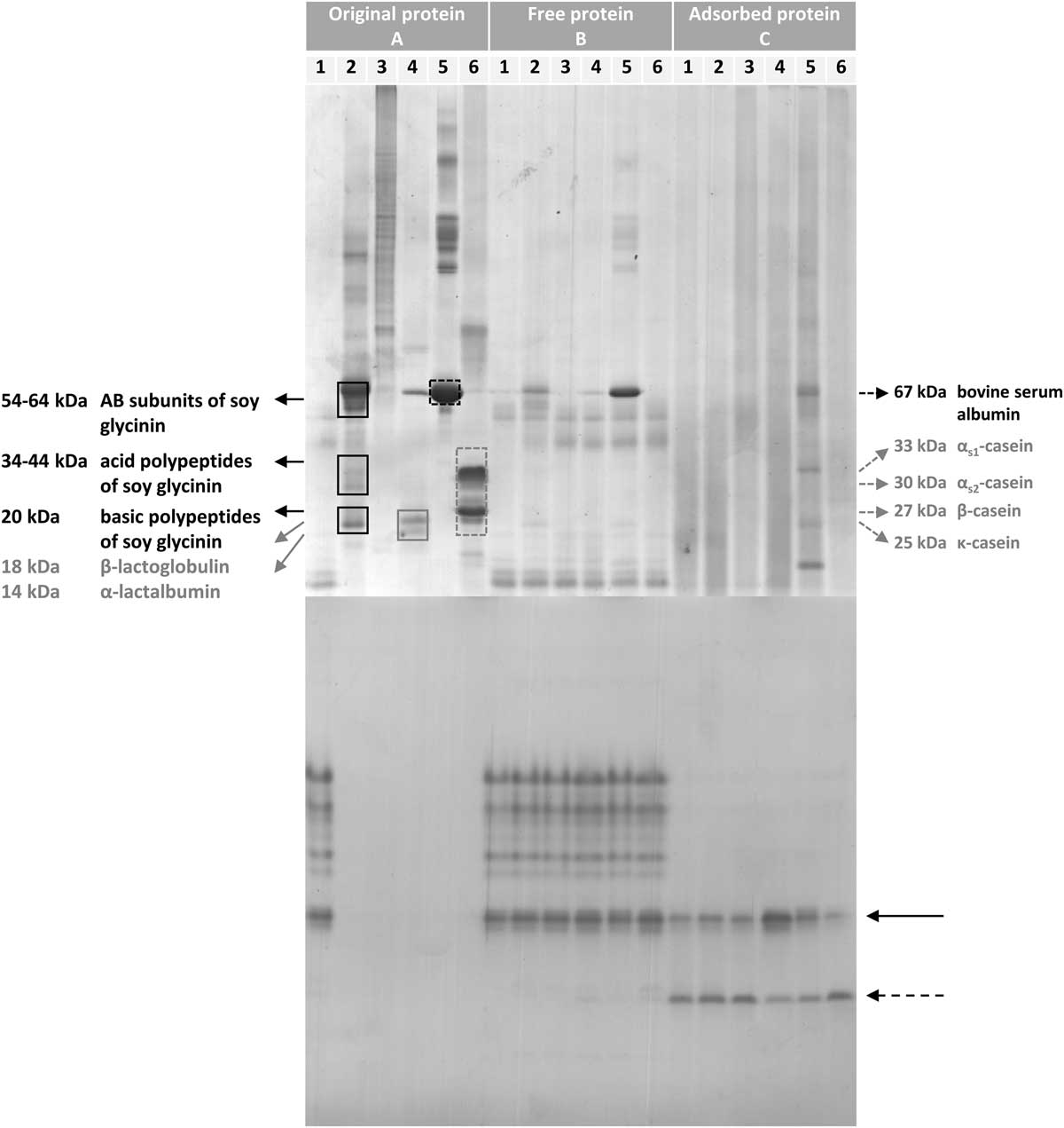
Soy glycinin was isolated from extruded soybean meal (49/3.5 HiPro from AR Coproducten; Didam, the Netherlands). The isolation protocol was adapted from Isaschar-Ovdat et al. (Reference Isaschar-Ovdat, Rosenberg, Lesmes and Fishman2015) with small modifications (Supplementary Material S1). The resulting protein pattern obtained by non-reducing SDS-PAGE is shown in Figure 1 (lane 2A).
Sodium dodecyl sulfate polyacrylamide gel electrophoresis (SDS-PAGE) (top) and zymogram (bottom) of the original protein solutions (A), the free protein fraction (B) and the protein adsorbed to the droplet surface of the emulsions (C) of experiment 1. Emulsions were prepared with polyphenol oxidase extract (1; control) or with soy glycinin (2), gelatin (3), whey protein isolate (4), bovine serum albumin (5) or sodium caseinate (6) and diluted (4 : 1 w/w) with polyphenol oxidase (PPO) extract before analysis. Protein bands were identified by comparison with data reported in the literature (Wolf, Reference Wolf1993; Ercili Cura et al., Reference Ercili Cura, Lille, Partanen, Kruus, Buchert and Lantto2010; Ma et al., Reference Ma, Forssell, Partanen, Buchert and Boer2011; Macierzanka et al., Reference Macierzanka, Bordron, Rigby, Mills, Lille, Poutanen and Mackie2011). The arrows in the zymogram indicate the appearance of a new protein band (dashed arrow) upon adsorption to the interface, presumably originating from the splitting of a PPO dimer (full-line arrow).

Crude polyphenol oxidase extract: preparation and characterization
Polyphenol oxidase extract was prepared as described by Gadeyne et al. (Reference Gadeyne, De Neve, Vlaeminck, Claeys, Van der Meeren and Fievez2016a). The extract was analyzed for protein content according to Winters and Minchin (Reference Winters and Minchin2005) using BSA as a standard (100% pure, Calbiochem, San Diego, CA, USA). Apparent PPO activity of the extract was evaluated by addition of 4MC, a diphenolic PPO substrate. The enzymatic characteristics are the results of several isoenzymes present in the extract and may be influenced by other compounds present in the PPO extract, therefore the characteristics are described as ‘apparent’ quantities. The oxidation reaction was started by addition of 750 μl 0.04 M 4MC to 50 μl extract diluted with 2.2 ml PB (pH 7). Formation of the corresponding quinone 4-methylquinone (4MQ) was monitored for 1 min by measuring the absorbance at 400 nm using an UV-VIS spectrophotometer (Genesys 10 S UV-VIS; Thermo Scientific, Madison, WI, USA). The apparent PPO activity in katal was calculated from the slope of the initial linear part of the curve based on a standard series of 4MQ solutions, prepared through chemical oxidation of 4MC using sodium periodate. Analyses for protein content and apparent PPO activity were executed in triplicate (analytical replicates). In experiment 1, the PPO extract contained 3.82±0.33 mg protein/ml extract and had an apparent PPO activity of 154±13 nkatal/mg protein. Experiment 2 and 3 were performed using PPO extract containing 3.03±0.25 mg protein/ml extract with an activity of 144±33 nkatal/mg protein.
Emulsion preparation
Emulsions were prepared by premixing linseed oil in a protein solution with a high-speed Ultra Turrax (S25N-10G dispersing tool; T25 Basic, Ika Werke, Staufen, Germany) until a homogeneous mixture was obtained. Fine emulsions were obtained by passing the premix through a Microfluidizer five times (M110S; Microfluidics Corp., Newton, MA, USA) at 21 MPa.
Emulsion characterization
Emulsions were characterized before dilution with PPO extract and after crosslinking in presence of 4MC. Droplet size distributions were determined by laser diffraction (Malvern Mastersizer S; Malvern Instruments, Malvern, UK) whereby the refraction indices were set at 1.5295+0.1i for the oil and 1.33 for the continuous phase. Emulsions were ~20 times diluted in a 1% (w/w) SDS solution before analysis. A few drops of the diluted sample were added to the MS-17 wet sample dispersion unit, filled with deionized water, until an obscuration level between 10% and 30% was obtained. The analysis was performed in triplicate and the emulsion droplets were characterized in terms of volume-weighted mean diameter (D 4,3) and surface-weighted mean diameter (D 3,2). Results were confirmed by observations using an optical microscope (Olympus CX40; Olympus Optical Co., Tokyo, Japan) equipped with a AxioCam ERc 5s video camera (Carl Zeiss Microimaging GmbH, Göttingen, Germany). A droplet of the diluted sample (±20 times in a 1% (w/w) SDS solution) was placed on a microscope slide using a capillary and carefully covered with a coverslip. A drop of immersion oil (Merck Co., Darmstadt, Germany) was placed on the coverslip before characterization with 1000-fold magnification.
Separation of free and adsorbed protein
The free and adsorbed protein fractions of the emulsions in experiment 1 were collected before crosslinking through a procedure described previously by Gadeyne et al. (Reference Gadeyne, De Neve, Vlaeminck, Claeys, Van der Meeren and Fievez2016a). In brief, after an aliquot (25 ml) of the emulsion was centrifuged (30 min; 30 000×g; 4°C), a sample of 5 ml of the serum phase was filtered through a 0.20 μm syringe filter (Chromafil RC-20/25; Macherey-Nagel GmbH & Co. KG, Düren, Germany) and collected as the free protein fraction. Another aliquot (15 ml) of the serum phase was removed from the emulsion and 20 ml 0.01 M PB (pH 7) was added (replacing the original serum phase). The cream phase was re-dispersed by thorough vortexing. This washing step, centrifugation and replacement of 20 ml of the serum phase with 0.01 M PB (pH 7), was repeated. After an additional centrifugation step, 20 ml of the serum phase was replaced by 1 wt% SDS solution in 0.1 M PB (pH 7). The sample was vortexed thoroughly to re-disperse the cream phase and stirred gently overnight to allow the SDS molecules to replace the adsorbed protein on the emulsion droplets. After a final centrifugation step, the adsorbed protein was collected by filtering an aliquot of the serum phase through a 0.20 μm syringe filter (Chromafil RC-20/25). The protein concentration of both the free and the adsorbed protein were determined as described for the PPO extract. The collected protein fractions, and the original 1 wt% protein solutions were diluted to obtain similar protein concentrations before loading on gels for SDS-PAGE and zymography.
Gel electrophoresis and zymography
The free and adsorbed protein fractions of emulsions in experiment 1 were analyzed by SDS-PAGE and PPO zymography to examine the partitioning of PPO isoforms between the bulk phase and the droplet surface of the emulsions according to Gadeyne et al. (Reference Gadeyne, De Neve, Vlaeminck, Claeys, Van der Meeren and Fievez2016a). The visualization of the bands during zymography is initiated by the oxidation of 4-methylcatechol to its corresponding ortho-quinone, which is catalyzed by the PPO isoforms. The ortho-quinone, readily reacts with 4-amino-N,N’-diethylaniline sulfate forming a colored adduct, visualizing the protein as a purple band.
Evaluation of ruminal protection efficiency
Batch in vitro incubations were carried out to assess ruminal protection of PUFA in emulsions as described previously (Gadeyne et al., Reference Gadeyne, Van Ranst, Vlaeminck, Vossen, Van der Meeren and Fievez2015). In short, incubation flasks contained 1 ml 2 wt% oil-in-water emulsion, 250 mg hay and 24 ml buffer/rumen fluid solution (3.8 : 1 ratio), which are appropriate amounts to study ruminal biohydrogenation (BH) in vitro (Fievez et al., Reference Fievez, Vlaeminck, Jenkins, Enjalbert and Doreau2007). Correspondingly, emulsions with higher concentrations were diluted to 2 wt% oil with distilled water before addition of 1 ml emulsion to the incubation flask. The buffer consisted of 3.58 g Na2HPO4.12H2O, 1.55 g KH2PO4, 0.124 g MgCl2.6H2O, 8.74 g NaHCO3 and 1 g NH4HCO3 per liter of distilled water. Rumen contents were collected from three rumen-fistulated sheep before morning feeding. The animals were fed grass hay ad libitum combined with a grain-based concentrate (200 g/day) distributed twice daily to cover their requirements. They had free access to drinking water. Fistulation was approved by the ethical commission (file number 241, 2014) of the Institute for Agricultural and Fisheries Research (ILVO, Belgium). Rumen contents of the three sheep were combined and sieved (1 mm mesh) before incubation. Flasks were flushed with CO2 to obtain anaerobic conditions and incubated under intermittent shaking at 39°C in a batch culture incubator (Edmund Bühler GmbH, Hechingen, Germany). After 24 h, flasks were transferred to an ice bath to stop microbial activity. To monitor microbial activity in the incubations, gas production (Hassim et al., Reference Hassim, Lourenço, Goel, Vlaeminck, Goh and Fievez2010), pH (Hanna Instruments, Temse, Belgium) and volatile fatty acid formation were assessed (Gadeyne et al., Reference Gadeyne, De Ruyck, Van Ranst, De Neve, Vlaeminck and Fievez2016c). Gas composition was converted to absolute gas production rates based on the pressure accumulation in the flask (Infield 7 C handheld read-out device equipped with a T1 Stitch-Tensiometer; UMS GmbH, München, Germany). These incubation characteristics were not reported as no major differences were observed between treatments, indicating changes in the extent of BH were not due to changes in microbial activity. The emulsions were incubated in duplicate or triplicate (analytical replicates).
The extent of BH was calculated based on fatty acid analysis of the samples before and after incubation. A 5 ml aliquot incubation fluid was freeze-dried and analyzed after direct transesterification, using a gas chromatograph (Gadeyne et al., Reference Gadeyne, Van Ranst, Vlaeminck, Vossen, Van der Meeren and Fievez2015). Peaks were identified based on their retention times, compared with external standards as a reference (GLC463; Nu-Check Prep Inc., Elysion, ML, USA). Quantification of fatty acid methyl esters was based on the area of the internal standard (C13:0) and on the conversion of peak areas to the weight of the fatty acid (FA) by a theoretical response factor for each FA (Ackman and Sipos, Reference Ackman and Sipos1964; Wolff et al., Reference Wolff, Bayard and Fabien1995). Finally, BH (%) and protection efficiency (%) was calculated according to Gadeyne et al. (Reference Gadeyne, Van Ranst, Vlaeminck, Vossen, Van der Meeren and Fievez2015) as follows:
 $$ \eqalignno{&{\rm BH(\%){\,\equals\,}}\cr &{{{\rm (mg}\,{\rm 18\colon\!3n{\hbox{-}}3}\,{\rm per}\,{\rm mg}\,{\rm total}\,{\rm C18}\,{\rm FA)}_{{{\rm 0}\,{\rm h}}} {\rm \minus (mg}\,{\rm 18\colon\!3n{\hbox{-}}3}\,{\rm per}\,{\rm mg}\,{\rm total}\,{\rm C18}\,{\rm FA)}_{{{\rm 24}\,{\rm h}}} } \over {{\rm (mg}\,{\rm 18\colon\!3n{\hbox{-}}3}\,{\rm per}\,{\rm mg}\,{\rm total}\,{\rm C18}\,{\rm FA)}_{{{\rm 0}\,{\rm h}}} }}{\rm {\times}100} \cr\cr& {{\rm Protection}\,{\rm efficiency}\,{\rm (\%){\equals}}{{{\rm BH}_{{{\rm without}\,{\rm 4MC}}} {\rm \minus BH}_{{{\rm with}\,{\rm 4MC}}} } \over {{\rm BH}_{{{\rm without}\,{\rm 4MC }}} }}{\rm {\times}100}} $$
Statistics
In experiment 1, emulsion and concomitant PPO extract preparation and characterization were executed in triplicate. Results were analyzed by the MIXED procedure of SAS (SAS Enterprise Guide 7; SAS Institute Inc., Cary, NC, USA). Analytical replicates were averaged before analysis. For the emulsion characteristics in experiment 1, the following model was used: Y ijk =μ+R i +Pr j +CL k +Pr i ×T k +ε ijk , with Y ijk the variable of interest, μ the overall mean, R i the random effect of statistical replicate (i=1, 2, 3), Pr j the fixed effect between subjects of protein emulsifier (j=polyphenol oxidase, soy glycinin, WPI, gelatin, bovine serum albumin or sodium caseinate), CL k the fixed effect within subjects of enzymatic crosslinking (k=before or after enzymatic crosslinking of the emulsions) and ε ijk the residual error. Enzymatic crosslinking was considered as repeated measures. BH and protection efficiency were analyzed by the following model: Y ij =μ+R i +Pr j +ε ij , with Y ij the variable of interest, μ the overall mean, R i the random effect of statistical replicate (i=1, 2, 3), Prj the fixed effect of protein emulsifier (j=polyphenol oxidase extract (as positive control), soy glycinin, WPI, gelatin, BSA or sodium caseinate) and ε ij the residual error. Data of experiment 2 and 3 were analyzed according to the following model: Y ij =μ+R i +T j +ε ij with Y ij the variable of interest, μ the overall mean, R i the random effect of statistical replicate (i=1, 2, 3), T j the fixed effect of treatment (j=2, 4, 6, 8 or 10 wt% oil) and ε ij the residual error. A post hoc Tukey test was used to determine pair-wise differences between means at a significance level of P<0.05.
Results
Experiment 1
Before crosslinking, no pronounced differences in emulsion characteristics were observed between the treatments (Table 1). The emulsions were interfacially characterized by separating and analyzing the free and adsorbed fractions of the emulsions by SDS-PAGE and zymography. The protein fractions were compared with the protein solutions originally used for emulsion preparation (Figure 1). When comparing the protein bands of the original protein solutions (lanes A), the bands of the PPO extract (lane A1) are remarkably lighter than the protein bands in the protein emulsifier solutions (lanes A2 to A6) and the protein patterns of the protein emulsifier solutions greatly differed in both molecular weight as well as number of protein bands. In lane A2, major bands between 54 and 64 kDa were identified as the AB subunits of soy glycinin, the subunits each consisting of an acid (A) and a basic (B) polypeptide linked by a disulfide bond (Wolf, Reference Wolf1993). Only minor bands of the soy glycinin polypeptides were found. Gelatin migrated as a smear of polypeptides (lane A3). The WPI solution contained α-lactalbumin, β-lactoglobulin and a substantial amount of BSA (lane A4). In lane A5, BSA predominantly migrated as a band with a molecular weight of 67 kDa (Ma et al., Reference Ma, Forssell, Partanen, Buchert and Boer2011), as well as some minor high-molecular-weight bands and one lower-molecular-weight band. In lane A6, the typical migration pattern of casein subunits (25 to 33 kDa) (Ercili Cura et al., Reference Ercili Cura, Lille, Partanen, Kruus, Buchert and Lantto2010; Macierzanka et al., Reference Macierzanka, Bordron, Rigby, Mills, Lille, Poutanen and Mackie2011) was obtained with some minor bands in the higher-molecular-weight region. The free protein fraction of all emulsions (lanes B1 to B6) contained crude PPO extract protein bands (lane B1), which can be found in every lane at the bottom of the gel. Only proteins abundantly present in the original protein solutions were detected in the free protein fraction of the emulsion; that is the AB subunits of soy glycinin (lane B2) and BSA (in lanes B4 and B5). Sodium dodecyl sulfate polyacrylamide gel electrophoresis of the adsorbed protein fractions mainly resulted in a smear of bands (lanes C1 to C4, and lane C6). In contrast to the free fraction, no major band of soy glycinin was found in lane C2. The major band of BSA is visible in lane C5, while several lower-molecular-weight bands also appeared.
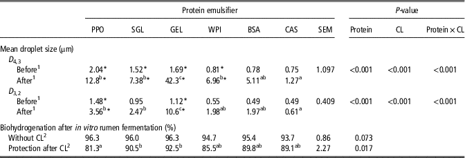
Effect of protein emulsifier and enzymatic crosslinking (CL) on emulsion characteristics and biohydrogenation parameters determined by in vitro ruminal fermentation of emulsions containing polyphenol oxidase (PPO) prepared with different protein emulsifiers (experiment 1)

PPO=polyphenol oxidase; SGL=soy glycinin; GEL=gelatin; WPI=whey protein isolate; BSA=bovine serum albumin; CAS=sodium caseinate; SEM=standard error of the mean; CL=enzymatic crosslinking; D 4,3=volume-weighted mean diameter; D 3,2=surface-weighted mean diameter.
Entries followed by ‘*’ indicate a significant difference with the measurement before enzymatic crosslinking.
a,b,cEntries with different superscripts differ significantly within the row.
1 Characterization before or after enzymatic crosslinking.
2 Non-crosslinked emulsions (without 4-methylcatechol as PPO substrate) served as negative control to calculate the protection efficiency against biohydrogenation of the crosslinked emulsions.
The zymogram (Figure 1, bottom) visualized the PPO isoforms in the different fractions of the emulsions. In the original protein solutions (A) only the PPO extract showed PPO activity, generated by multiple isoforms (lane A1). All isoforms of the PPO extract could be detected in the free protein fraction of each emulsion (lanes B1 to B6), while in the adsorbed protein fraction of the emulsions only PPO isoforms with a lower molecular weight were detected (lane C1 to C6, indicated by the arrows). In addition, a new (low-molecular-weight) PPO isoform (lanes C1 to C6 in comparison with lane A1) appeared (indicated by the dashed arrow). In contrast with the free protein fraction (lanes B1 to B6), some differences in band intensity could be observed between the adsorbed protein fractions (lanes C1 to C6), where the two adsorbed PPO isoforms (indicated by the arrows) seemed inversely correlated in intensity. The appearance of new PPO isoforms was not observed in every replicate of the experiment (Supplementary Material Figure S1).
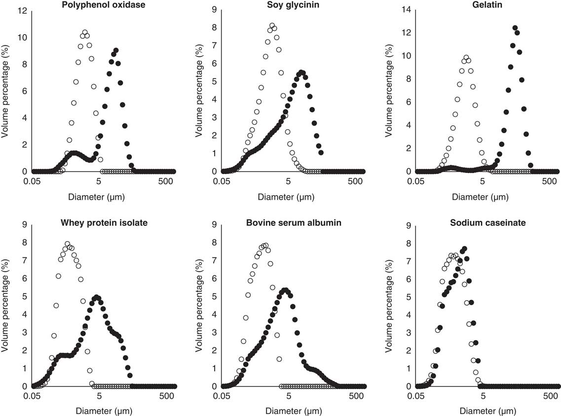
After dilution of the 10 wt% emulsions with PPO extract, interfacial crosslinking was induced by incubating the emulsions with 40 mM 4MC. Interfacial crosslinking caused the droplet size distributions to shift to larger droplet sizes for all emulsions (Figure 2). The largest shift occurred for gelatin emulsions, where the volume-weighted mean diameter increased 25 times following protein crosslinking and the surface-weighted diameter increased more than nine times. Volume- and/or surface-weighted mean diameter significantly increased after interfacial protein crosslinking (Table 1), except for the sodium caseinate- and BSA-stabilized emulsions. For the latter treatments only a small shift toward larger droplet sizes occurred (Figure 2).
Droplet size distributions of emulsions before (open circles) and after (full circles) enzymatic crosslinking of interfacial protein in experiment 1. Emulsions were prepared with polyphenol oxidase extract (control) or with soy glycinin, gelatin, whey protein isolate, bovine serum albumin or sodium caseinate and diluted (4 : 1 w/w) with polyphenol oxidase extract after emulsification.

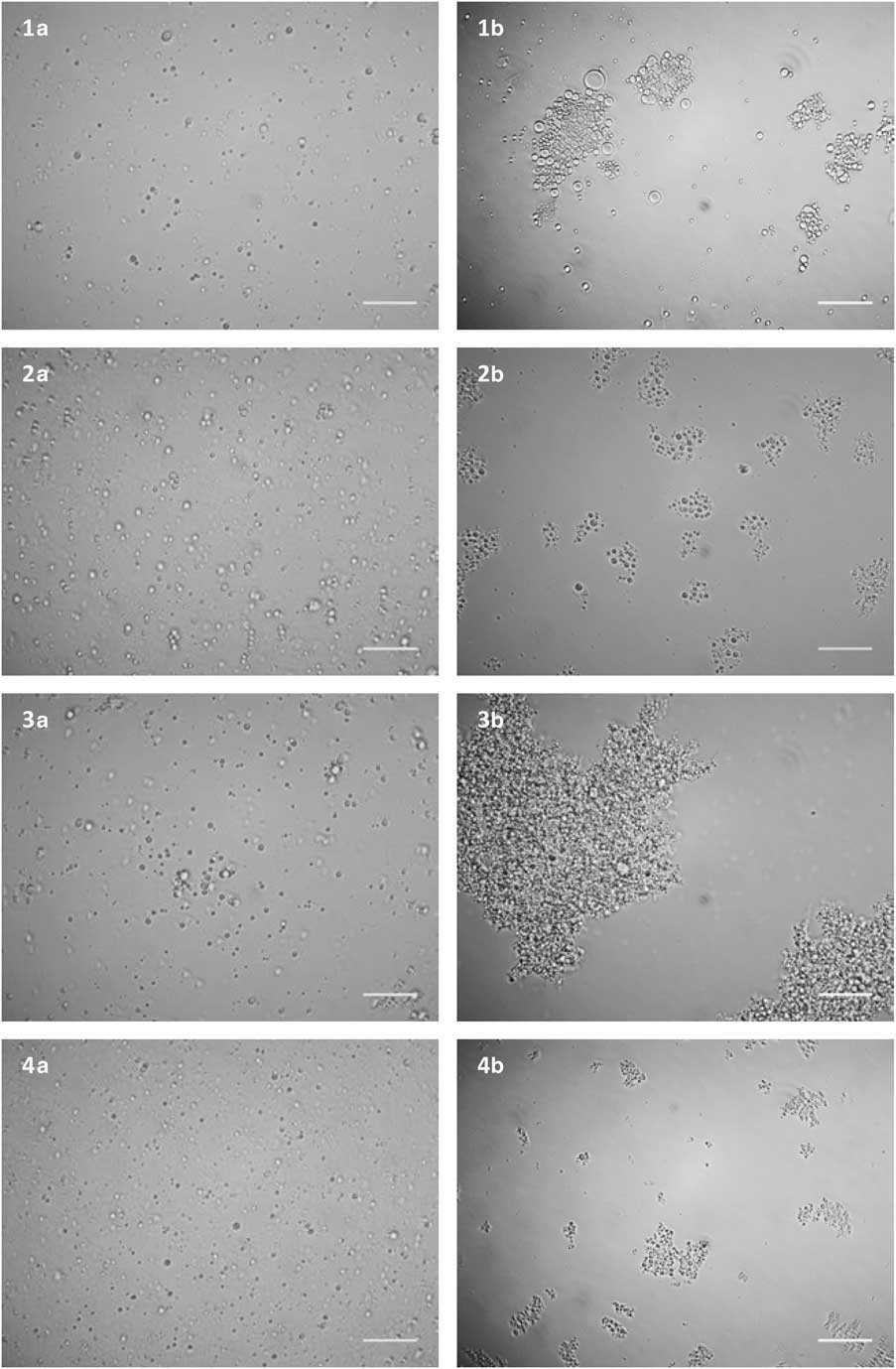
Indeed, after interfacial crosslinking aggregate formation was observed in the microscopic images (Figure 3). The largest aggregates were visible in the gelatin emulsions, which corresponds to the major shift in the droplet size distribution.
Microscopic observations of emulsions before (a) and after (b) enzymatic crosslinking of interfacial protein show formation of aggregates in experiment 1. Emulsions were stabilized by polyphenol oxidase extract (1), soy glycinin (2), gelatin (3) or whey protein isolate (4). Scale bars indicate a distance of 20 μm.

The protection efficiency of the crosslinked emulsions was evaluated in an in vitro batch simulation of the rumen environment. The PUFA, 18:3n-3, was almost completely biohydrogenated in the negative control emulsion (Table 1). Protection efficiencies of emulsions treated with 40 mM 4MC exceeded 80% for all treatments (Table 1). The lowest protection efficiency was obtained for the control treatment. For gelatin (P=0.015) and soy glycinin (P=0.045) a significantly higher efficiency was found compared with the control treatment, while other protein emulsifiers resulted in intermediate protection efficiencies (86% to 90%).
Experiment 2
In this experiment, the potential to increase the oil concentration was explored while the proportion of the PPO extract was kept the same as in the test treatments of experiment 1 (i.e. 80 wt% PPO extract). A range of emulsions with 2, 4, 6, 8 and 10 wt% oil was prepared by diluting a 50% oil emulsion. The crosslinked 2 wt% oil emulsions served as positive control emulsion.
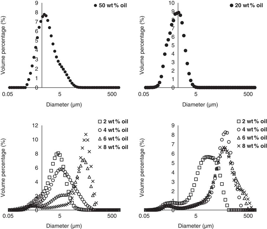
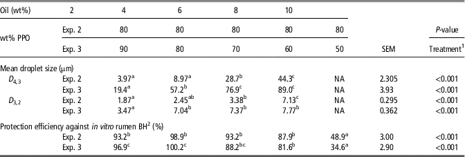
The 50 wt% oil emulsion was characterized by a right-skewed droplet size distribution (Figure 4) with a D 4,3 of 2.10 μm and mode of 1.17 μm. After crosslinking, increasing droplet aggregation was found for increasing oil levels (Table 2). The emulsions with the highest oil concentration, 10 wt% oil, were not stable during the crosslinking reaction, as oiling off was observed after 24 h incubation with 4MC. In this case the droplet size distribution was not determined, but formation of large aggregates was visible, even with the unaided eye.
Droplet size distributions of emulsions in experiment 2 (left) and experiment (3) before (top) and after (bottom) crosslinking. Emulsions of 50 wt% oil and 20 wt% oil were prepared with a 1 wt% whey protein isolate (WPI) solution and diluted with polyphenol oxidase (PPO) extract and 1 wt% WPI solution (experiment 2) or with PPO extract alone (experiment 3) to obtain emulsions of 2, 4, 6, 8 and 10 wt% oil. Droplet size distribution of 10 wt% oil emulsions were not determined due to oiling-off.

Effect of variation in oil content, or both oil content and amount of crude polyphenol oxidase (PPO) extract, on emulsion characteristics and protection efficiency against in vitro biohydrogenation, for emulsions of experiments 2 and 3, determined after enzymatic crosslinking

Exp.=experiment; D 4,3=volume-weighted mean diameter; D 3,2=surface-weighted mean diameter; NA=not available, no measurement due to oiling-off; wt% PPO=proportion of PPO extract in emulsion; SEM= standard error of the mean.
a,b,cEntries within a row with different superscripts differ significantly.
1 Treatment: Variation of oil concentration (experiment 2) or both oil concentration and proportion of PPO extract.
2 Non-crosslinked emulsion of 2 and 10 wt% oil (incubated without 4-methylcatechol as PPO substrate) served as negative control.
To determine the protection efficiency non-crosslinked emulsions of 2 and 10 wt% oil, showing 96.4±0.2% biohydrogenation of 18:3n-3, served as a negative control. When emulsions were crosslinked with 4MC high ruminal protection efficiencies (87%) were obtained, except for the 10 wt% emulsion, for which a protection of only 35% was found.
Experiment 3
Here, it was assessed whether emulsions with higher oil concentrations could also be protected when less PPO was used for enzymatic crosslinking. Therefore, a range of emulsions with 2, 4, 6, 8 and 10 wt% oil were prepared by diluting a 20 wt% oil emulsion with PPO extract. The emulsions contained 90, 80, 70, 60 and 50 wt% PPO extract, respectively.
The 20 wt% oil emulsion was characterized by a monomodal distribution with a D 4,3 of 0.93 μm (Figure 4). Crosslinking of the emulsions led to droplet aggregation, with larger aggregates for higher oil concentration (Table 2). Similar to experiment 2, oiling off was observed for the emulsions containing 10 wt% oil. Compared with emulsions of the same oil level of experiment 2, larger aggregates were formed after crosslinking but similar protection efficiencies were obtained.
Protection efficiencies were calculated based on biohydrogenation of 18:3n-3 in the negative control (the average biohydrogenation of 2 and 10 wt% oil emulsions which were incubated without 4MC) which equaled 96.4±0.2%. Within experiment 3, the highest protection was found for emulsions with 2, 4 and 6 wt% oil. In the 8 wt% oil emulsions a slightly lower but still effective protection was found, exceeding 80%. The 10 wt% oil emulsions was considered ineffectively protected (<50%).
Discussion
Polyphenol oxidase-mediated protein crosslinking
In order to protect PUFA from ruminal metabolism, the potato tuber peel PPO should cause extensive crosslinking of the adsorbed protein, a reaction typically characterized by browning (Friedman, Reference Friedman1997). The PPO-mediated crosslinking of the protein emulsifiers tested in the current study (soy glycinin, gelatin, WPI, BSA and sodium caseinate) in the presence of a phenolic substrate has been reported previously. However, none of these studies used PPO extracted from potato tuber peelings to induce the crosslinking of these protein nor used 4MC as phenolic substrate. Le Roes-Hill et al. (Reference Le Roes-Hill, Palmer, Rohland, Kirby and Burton2015) reported covalent crosslinking of casein, gelatin and BSA in the presence of l-3,4-dihydroxyphenylalanine and l-tyrosine, by bacterial PPO isolated from Streptomyces. Soy glycinin could be crosslinked by Bacillus megaterium PPO in the presence of caffeic acid, P-coumaric acid and chlorogenic acid (Isaschar-Ovdat et al., Reference Isaschar-Ovdat, Rosenberg, Lesmes and Fishman2015). Crosslinking of the whey proteins α-lactalbumin and β-lactoglobulin was achieved using mushroom PPO in the presence of caffeic acid (Thalmann and Lötzbeyer, Reference Thalmann and Lötzbeyer2002).
Crosslinking of the interfacial protein did not only lead to browning of the emulsions but also caused aggregation of the emulsion droplets. The emulsions stabilized by soy glycinin and WPI showed aggregate formations of similar size, while emulsions stabilized by PPO extract and especially the gelatin-stabilized emulsions showed the formation of large aggregates, up to 100-fold of the original droplet size. Caution needs to be taken when comparing the size of these aggregates, keeping in mind the results obtained by laser diffraction analysis relies on the assumption of perfectly round isotropic particles, while, this is not the case when aggregation takes place. Aggregation during enzymatic crosslinking of protein in emulsions can be ascribed to interdroplet crosslinks that are formed as a consequence of collisions during the crosslinking reaction as observed by Isaschar-Ovdat et al. (Reference Isaschar-Ovdat, Rosenberg, Lesmes and Fishman2015). Several studies report the promotion of aggregate formation and stabilization by covalently crosslinking interfacial protein, mainly by transglutaminase (Yang et al., Reference Yang, Liu and Tang2013; Zeeb et al., Reference Zeeb, Beicht, Eisele, Gibis, Fischer and Weiss2013), but also by laccase (Maier et al., Reference Maier, Oechsle and Weiss2015) or the chemical crosslinker glutaraldehyde (Maier et al., Reference Maier, Ensenberger, Irmscher and Weiss2016). In the sodium caseinate- and BSA-stabilized emulsions of the current study, crosslinking mainly seemed to occur between protein adsorbed to the surface of the same droplet, as the emulsion characteristics did not change after crosslinking. Also, Sato et al. (Reference Sato, Perrechil, Costa, Santana and Cunha2015) and Ma et al. (Reference Ma, Forssell, Partanen, Buchert and Boer2011), who studied laccase-mediated crosslinking of protein in sodium caseinate- and WPI-stabilized emulsions, respectively, reported no change of mean droplet size after enzymatic crosslinking.
Ruminal protection efficiency
A plethora of studies describe the ruminal biohydrogenation of PUFA in oils supplemented to the rumen (Fievez et al., Reference Fievez, Vlaeminck, Jenkins, Enjalbert and Doreau2007; Osborne et al., Reference Osborne, Radhakrishnan, Odongo, Hill and McBride2008; Lanier and Corl, Reference Lanier and Corl2015), demonstrating the need for ruminal protection. Nevertheless, (nano-)emulsions added to the diet or drinking water were hypothesized not to require protection due to rapid passage through, or bypassing, the rumen with the liquid phase. Recently, El-Sherbiny et al. (Reference El-Sherbiny, Cieślak, Szczechowiak, Kołodziejski, Szulc and Szumacher-Strabel2016) simulated the addition of soybean and fish oil to the drinking water in the form of nanoemulsion and the results showed a reduction in biohydrogenation of C18:3n-3 of more than 70% (calculated from the proportion in C18 fatty acids, compared with the control) in a RUSITEC system. However, when soybean oil was administered by continuous infusion of an emulsion in the in vivo study of Baumann et al. Reference Baumann, Chouinard, Lebeuf, Rico and Gervais2016), numerous biohydrogenation intermediates and limited increase in 18:2n-6 in the milk fatty acid profile indicated extensive ruminal biohydrogenation, suggesting emulsification itself does not effectively prevent biohydrogenation. The high biohydrogenation levels found in the non-crosslinked emulsions in the current study confirms the latter statement.
Previous studies showed the potential for PPO-induced crosslinking to effectively protect 2 wt% emulsions of PUFA-rich oil stabilized by a crude potato peel extract against ruminal BH (Gadeyne et al., Reference Gadeyne, Van Ranst, Vlaeminck, Vossen, Van der Meeren and Fievez2015 and Reference Gadeyne, De Neve, Vlaeminck, Claeys, Van der Meeren and Fievez2016a), where the extract functioned both as emulsifier as well as the source of PPO. In the current study, a new approach was evaluated, where the potato peel extract was added only after emulsification by use of external protein emulsifiers. The use of other emulsifiers than the protein present in the protein extract is of interest as, on the one hand, emulsifiers can be selected with excellent emulsifying properties to facilitate the protection of more concentrated emulsions, and on the other hand, this gives the opportunity to reduce the amount of potato peel extract. The new protocol led to slightly higher protection efficiencies in comparison with the control treatment, where PPO was present from the start. Despite the difference in composition and size between the emulsifying proteins, protections were high for all treatments and no major differences in protection were observed. It appears, in the presence of 4MC, all tested protein emulsifiers can be used to obtain effective protection of 2 wt% linseed oil emulsions. Therefore, it can be concluded that, as hypothesized (hypothesis A), the emulsifier function of the PPO extract can be replaced by other protein emulsifiers.
As 10 wt% oil emulsions couldn’t be successfully protected, hypothesis B only could be partially retained: emulsions containing up to 8 wt% oil were successfully protected using only 60 wt% PPO extract. Thus, in comparison with the original protocol (2 wt% oil and 98 wt% PPO extract), the new protocol resulted in an improvement of more than 650% of the oil to PPO extract ratio (w/w). Emulsion stability seemed to be the limiting factor in the 10% oil emulsions, as these emulsions showed oiling-off after incubation with 4MC. The stirring during the incubation with 4MC probably increased the number of droplet collisions resulting in more coalescence and oiling-off.
For a ruminal protection system to be effective, it should not only ensure bypassing the rumen but also post-ruminal release. Gadeyne et al. (Reference Gadeyne, De Neve, Vlaeminck, Van der Meeren and Fievez2017) showed complete hydrolysis of fatty acids from PPO-mediated rumen-protected emulsions through in vitro abomasal and intestinal simulation. The mechanism of this release has not been studied before and to elucidate how the fatty acids become available for lipolysis more research is needed in this respect. Two different mechanisms could be hypothesized to provoke the fatty acid release: (i) either the protein-bound phenols – which are created by reaction of quinones with nucleophilic groups of the proteins – are dissociated from the protein under the acidic conditions of the abomasum, or (ii) the acidic conditions of the abomasum change the conformation of the protein in the protective matrix in such way that the binding sites of the pepsin and other proteases in the digestive tract become available and hence initiate digestion. From those two mechanisms, the former seems to be less likely, as this reaction would progress via a non-aromatic transition state.
Adsorption behavior of polyphenol oxidase isoforms
Sodium dodecyl sulfate polyacrylamide gel electrophoresis of the original protein solutions in experiment 1 resulted in remarkably less intense protein bands in the control compared with the other protein solutions (Figure 1, top; lane A1 v. lanes A2 to A6). This was mainly attributed to a higher variety in protein and polypeptide bands. Good separations of the protein bands were obtained for the different protein solutions, except for gelatin. As the aim of the analysis was mainly to locate the PPO isoforms and their activity, the protocol was not adjusted for the separation of the gelatin subunits.
The zymogram (Figure 1, bottom) showed multiple bands with PPO activity in the PPO extract, in accordance to the observations of Gadeyne et al. (Reference Gadeyne, De Neve, Vlaeminck, Claeys, Van der Meeren and Fievez2016a). As no staining was observed in the region of the PPO isoforms on the Coomassie-stained gel, the results showed only a low concentration of enzyme was present in the extract. Polyphenol oxidase isoforms were detected in both the free and adsorbed fractions in all emulsions. However, in the adsorbed fractions only low-molecular-weight isoforms were detected (Figure 1, lanes C1 to C6). The PPO isoform in the adsorbed fraction with the lowest molecular weight (indicated with the black dashed arrow) was not detected in the PPO extract (lane A1). This new protein band could be explained by splitting of a PPO dimer, of which the reactive site most probably became available for proteases in the PPO extract upon unfolding during adsorption. Higher susceptibility of proteins toward proteolytic enzymes upon adsorption to the oil/water interface was indeed reported before (Marengo et al., Reference Marengo, Miriani, Ferranti, Bonomi, Iametti and Barbiroli2016). Minor bands in the PPO isoform pattern differed slightly between replicates (Supplementary Material Figure S1) but generally one PPO isoform and/or (presumably) its dimer could be found in all adsorbed protein fractions. Hence, the zymogram showed similar partitioning of the PPO isoforms in all treatments.
In conclusion, in the current study a new approach was shown to optimize the use of a potato tuber peelings extract for the PPO-mediated protection of linseed oil against rumen biohydrogenation. The described protection technology, based on the use of a natural enzyme complex, provides a tool to increase the PUFA outflow from the rumen, promoting both animal (reproductive) health, as well as the production of healthier animal products, enriched in PUFA. The current technology was optimized by replacing the emulsifying function of the PPO-containing extract with another protein emulsifier resulting in an improvement of >650% of the PPO extract to oil ratio. These findings have a great impact on the practical feasibility of future in vivo trials, needed to further validate this new technology. Further optimization of the technology should be possible by tackling the destabilization of concentrated emulsions during protein-crosslinking through adjustment of the emulsion formulation and/or the crosslinking procedure.
Acknowledgments
Nympha De Neve received a PhD grant from the government agency Flanders Innovation & Entrepreneurship (VLAIO-Belgium, grant number 131632). The authors thank Sieglinde Debruyne and Alexis Ruiz Gonzalez who reviewed this manuscript with great attention. They greatly appreciate technical assistance from the staff of LANUPRO, in particular, Charlotte Melis.
Declaration of interest
The authors declare no potential conflict of interest.
Ethics statement
The fistulation of the animals from which the rumen fluid was sampled was approved by the ethical commission of the Institute for Agricultural and Fisheries Research (ILVO, Belgium; file number 241, 2014).
Software and data repository resources
Data were not deposited in an official repository.
Supplementary material
To view supplementary material for this article, please visit https:/doi.org/10.1017/S1751731118000423